Biology Unt 1 (B3 Tissue structures and function)
1/49
There's no tags or description
Looks like no tags are added yet.
Name | Mastery | Learn | Test | Matching | Spaced |
|---|
No study sessions yet.
50 Terms
What is an epithelial tissue?
Covers all the surfaces of the body
Lines all the tubes
This group of tissues forms a protective barrier.
(They are found inside of the lungs)
What is a muscle tissue?
Allows body parts to move through contraction
What is a connective tissue?
Connective tissue supports, connects, and protects other tissues and organs.
What is a nervous tissue?
Nervous tissue transmits information throughout the body.
What are the 2 types of epithelial tissues?
Columnar and squamous
What is the role of the ciliated epithelial cell? ( within the columnar epithelium)
Hair like structures on their cell membrane called cilia- moves mucus containing trapped pathogens away from the lungs.
found in trachea and bronchi
what is the role of the goblet cell? ( within the columnar epithelium)
Produces a protein called mucin
which traps pathogens that have been inhaled in.
found in trachea and bronchi
Where in the lungs are squamous epithelial tissues found?
Alveoli
What is the shape of squamous epithelial cells?
Flat and thin- often one cell thick
How are the gases in the alveoli wall adapted to travel into and out of the bloodstream?
Large surface area- more surface available for the diffusion of o2 to occur
Squamous epithelium (SEC) is one cell thick- short diffusion pathway as oxygen molecules have to pass through one thin SEC to get to capillaries
Rich network of capillaries- keeps a strong oxygen gradient because oxygenated blood moves away and is replaced by deoxygenated blood.
Which factors increase the risk of developing chronic bronchitis?
smoking, silica dust and air pollution
what happens to the structures of the lungs when a person suffers from chronic bronchitis?
Inflammation of bronchi and narrows airway
increased number of mucus in the gland
airway is obstructed by thick mucus
makes it harder for air to move in and out of lungs, leading to coughing and breathing difficulties.
Emphysema is caused by smoking. Describe what happens to the alveoli when a person suffers from emphysema and give symptoms
smoke damages cilia
pathogens get trapped in the mucus
macrophages move to lungs to kill bacteria
macrophages release enzymes to breakdown alveoli
surface area decreases and diffusion is slower
SYMPTOMS: easily tired, fatigue or breathlessness when exercising
Explain why a person suffering from emphysema may feel tired
has a smaller surface area (lung tissue)
reduction in oxygen uptake
leads to more anaerobic respiration
Differences of epithelium and endothelium tissues
Epithelium- is a lining tissue that covers OUTSIDE surfaces
Endothelium- is a lining tissue that covers INSIDE surfaces
What is the role of the endothelium inside arteries and veins?
Lines the inside of the blood vessels
These cells are smooth and flat
Which reduces resistance for blood flowing through them
Explain the role of the endothelium in blood capillaries
capillaries are one cell thick
this layer of single cells are endothelial cells
thin to provide short diffusion pathways
Atherosclerosis- Explain the problems caused if cholesterol accumulates in the walls of the arteries
causes an inflammatory response which gets bigger
so narrows lumen of the artery
and decreases blood flow through the lumen of the artery
These deposits are called plaque and fatty deposit
To try and help the body, what engulfs the cholesterol and what do they become
Explain how atheroma leads to blood clots
A type of phagocyte called macrophage which become foam cells
(2)
Endo of artery gets damaged
cholesterol is deposited behind endo + launches immune response
macrophages engulf cholesterol + form foam cells
This combination of macrophage and cholesterol forms a plaque
lumen of artery gets narrow and increases BP more, atherome can burst
causes blood clots as it stops the flow of blood and o2 to cells for respiration
If this occurs in coronary artery, can lead to heart attack
Explain the risk factors that can lead to atherosclerosis/cardiovascular disease
Smoking (carbon monoxide and nicotine)-
carbon monoxide binds to haemoglobin and reduces o2 transport
BP and heart rate increase, greater risk of damaged endothelium
Nicotine increases platelets stickiness so higher risk of blood clots
Diet-
Raises blood cholesterol levels
more cholesterol available for macrophages to engulf, so more foam cells and plaque formation
salt increases blood volume which raises BP
High blood pressure-
Increased BP damages the endothelium
damage triggers an inflammatory response
macrophages engulf cholesterol, forming foam cells
build up of plaque behind the endothelium
Describe the structure of the myelin sheath
made up of schwann cells which wrap around the axon
cell membrane is made up of mostly lipids
lipids are electrical insulators
What is the purpose of myelin?
myelin sheath acts as an insulator
lipids prevents the loss of ions from motor neurones
increases the speed of electrical impulse
The resting potential of neurones is -65mV. Explain what this means
inside of axon is more negative that the outside
creates negative resting potential of -65mV
There are more positive ions outside the axon than inside
Explain how the resting potential is set up and maintained
The sodium–potassium pump moves 3 Na⁺ out of the neurone and 2 K⁺ in, using ATP.
This makes Na⁺ more concentrated outside the neurone.
It also makes K⁺ more concentrated inside the neurone.
K⁺ diffuses out through open potassium channels because of its concentration gradient.
Na⁺ channels remain closed, preventing Na⁺ from re-entering.
Because more positive ions leave than enter, the inside becomes negatively charged compared to the outside, producing the resting potential of about –65 mV.
Describe the events of the action potential
Depolarisation Occurs & Na+ channels open, permeability to Na+ ion increases & enter axon by diffusion
Threshold- increased membrane potential -70mV to -50mV and threshold reached (all or nothing law)
Voltage-gated Na+ ion channels open & Na+ channels close at +30mV
Action potential occurs
Repolarisation occurs- K+ ion channel opens, permeability to K+ ion increases & leave axon by diffusion.
Hyperpolarisation occurs at -90mV, reference to refractory period
What is the purpose of hyperpolarisation?
prevents neurone from being re stimulated instantly
The period of time when no new action potential can be generated is called the refractory period
This is important as it helps ensure action potential travels in one direction down a neurone
What is the term for when the action potential jumps from node to node?
Saltatory conduction
How does saltatory conduction affect conduction and why?
Schwann cells wrap around the axon to form a myelin sheath
This increases the speed of nerve conduction
Myelin acts as an insulator preventing ion loss
Action potentials only occur at the nodes of ranvier
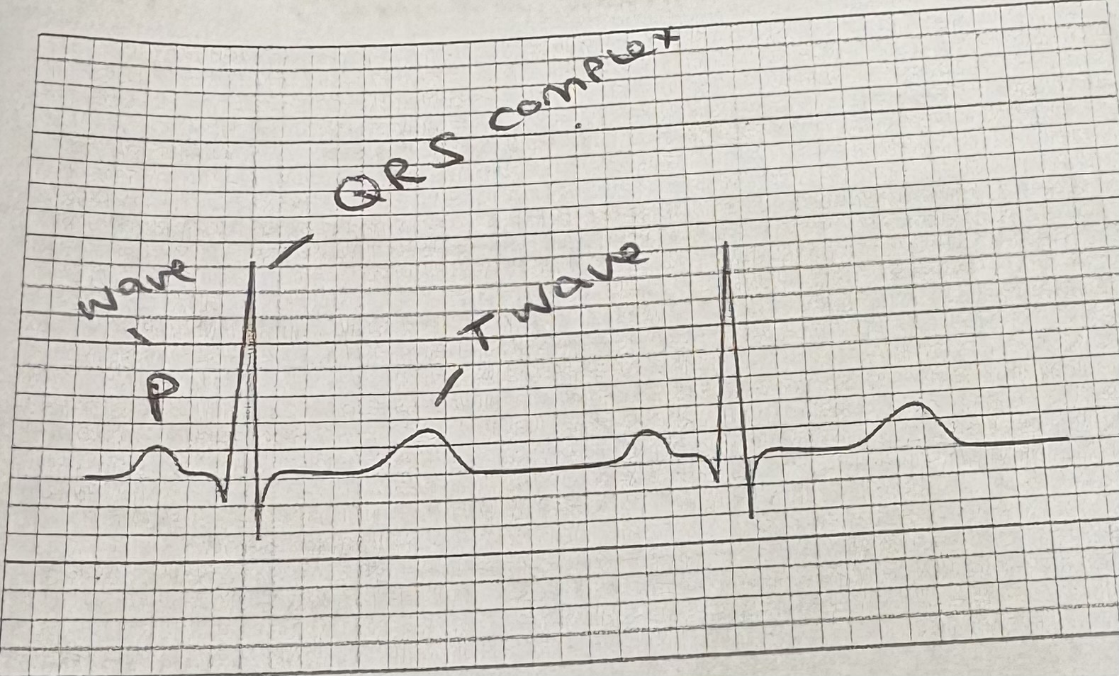
What factors would affect this normal ECG?
increase of heart rate caused by exercise, then QRS complex would be closer together
Describe the events of the synapse
Action potential reaches the presynaptic neurone.
Voltage gated channels open; Ca²⁺ diffuses into presynaptic neurone
Vesicles move to and fuse with the presynaptic membrane.
Neurotransmitters are released into the synaptic cleft- (exocytosis)
Neurotransmitters diffuse across the cleft.
They bind to receptors on the postsynaptic membrane.
Na+ channels open; Na⁺ diffuses in the postsynaptic neurone.
A new action potential is generated in post synaptic membrane
What happens to neurotransmitters after depolarisation?
Neurotransmitters are removed from receptors.
They are broken down by enzymes in the synaptic cleft.
Breakdown products are reabsorbed into the presynaptic neuron.
They are repackaged into vesicles to make more neurotransmitter.
Neurotransmitters are attached to postsynaptic membrane after depolarisation. Why is this important?
Allows postsynaptic neurone to rest
Na+ ion channels close after depolarisation
Neurotransmitter are reused in presynaptic neurone
What is an inhibitory neurotransmitter?
Decreases the likelihood of an action potential.
closes Na⁺ channels or opening Cl⁻ channels.
Makes the membrane more negative → harder to reach threshold.
What is an excitatory neurotransmitter?
Increases the likelihood of an action potential.
Works by opening Na⁺ channels.
Makes the membrane less negative → easier to reach threshold.
Explain how agonists work
Chemicals with a similar shape to neurotransmitters.
Bind to the same receptors.
Produce a similar response to the natural neurotransmitter.
Examples: nicotine, muscarine.
Explain why nicotine and muscarine are classed as agonists
They both mimic acetylcholine (neurotransmitter)
Increases the activity of receptors
Binds to neurotransmitter receptor of postsynaptic neurone
Causes Na+ ion channel to open
Explain how antagonists work
Chemicals have a similar shape to the normal neurotransmitter.
They bind to the same receptor sites.
They do not trigger a response.
They block the normal neurotransmitter from working.
Examples: atropine and curare.
Explain why atropine and curare are classed as antagonists
They have opposite effects on the neurotransmitter, they are similar to
Binds to the neurotransmitter receptor of postsynaptic neurone
Causes opposite response
What is parkinson’s disease?
What is the role of dopamine in the body? Link to number one.
A disease caused by the death of some neurones in the brain that release dopamine
This decreases the amount of dopamine being released at synapse and affects movement.
In parkinson’s, there is a ? supply of dopamine releases at synapses in the brain
Higher
If there is a reduces amount of dopamine, explain why it may lead to uncontrolled jerky movements
Less dopamine (neurotransmitter) released into synaptic cleft from pre-synaptic neurone
Less dopamine diffuses across synaptic cleft to post-synaptic neurone
Less dopamine attaches to its complementary receptor on membrane of post-synaptic neurone
Less ion channels open so there’s less depolarisation of post-synaptic neurones
Explain how L-dopa works
L-dopa is a precursor for dopamine
It can enter the brain and convert into dopamine & increases amount of dopamine in brain
More dopamine is released into + diffuses across synaptic cleft
More dopamine binds to receptor on post-synaptic neurone + depolarises neurone
How would a dopamine agonist work?
It would have a similar shape as dopamine neurotransmitters
The agonist would bind to receptors on post-synaptic membrane + have similar affect to dopamine
What is the role of serotonin in the body?
In some cases of depression, there is a ? supply of serotonin released at synapses in the brain
Its a neurotransmitter that regulates mood
Lower
If there is a reduced supply of serotonin, explain why it may lead to a low mood
Less serotonin (neurotransmitter) released into synaptic cleft from pre-synaptic neurone
Less serotonin diffuses across synaptic cleft to post-synaptic neurone
Less serotonin attaches to its complementary receptor on membrane of post-synaptic neurone
Less ion channels open so there’s less depolarisation of post-synaptic neurones
Why do muscle fibres contain many mitochondria?
site of aerobic respiration
molecules need atp for contraction of muscles
Arrange the following from smallest to largest- myofilaments, muscle fibres, myofibrils and muscles
How are they related to one another?
muscle, muscle fibres, myofibrils and myofilaments
(2)
muscles are like ropes
muscle fibres are strands inside the rope
myofibrils are tiny threads inside each strand
myofilaments such as actin & myosin made the threads work
COMPARE SLOW TWITCH FIBRES AND FAST TWITCH FIBRES
ST has slower speed, FT has rapid speed
ST has lots of blood vessels as they need big supply of o2, FT use less blood vessels
ST use aerobic respiration, FT uses anaerobic respiration
ST has many mitochondria for long lasting energy, FT has fewer mitochondria
ST has lots of myoglobin as it stores o2, FT uses less myoglobin
ST are red due to lots of myoglobin, FT are white due to less myoglobin
Explain 2 advantages of fast muscle fibres having lots of glycogen
Quick energy supply- glycogen can be broken down quickly, which gives them energy almost immediately
Works without oxygen- uses anaerobic r that needs glycogen so lots stored helps produce energy when no o2 is available.
why do ST use less glycogen?
How powerfully do they both contract?
How quickly do they fatigue?
ST mainly uses o2, not large stores of glycogen for long duration activities.
ST not very powerful as they’re built for endurance, FT very powerful as its built for quick strong bursts of force
ST slow to fatigue due to using o2 efficiently and less build up of lactic acid, FT fast to fatigue as anaerobic r produces lactic acid that tires them