muscle physiology flashcards
1/23
There's no tags or description
Looks like no tags are added yet.
Name | Mastery | Learn | Test | Matching | Spaced |
|---|
No study sessions yet.
24 Terms
epimysium
a fibrous connective tissue sheath that surrounds the entire muscle
fascicle
a bundle of skeletal muscles
endomysium
a connective tissue sheath surrounding each muscle fiber that contains capillaries and nerves
perimysium
a connective tissue sheath surrounding several muscle fibers
motor unit
comprised of a single motor neuron and all of the muscle fibers it innervates
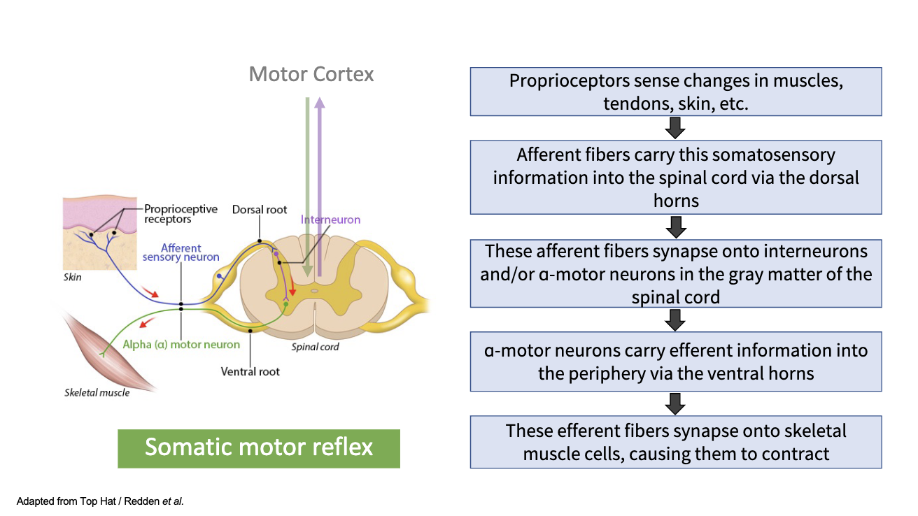
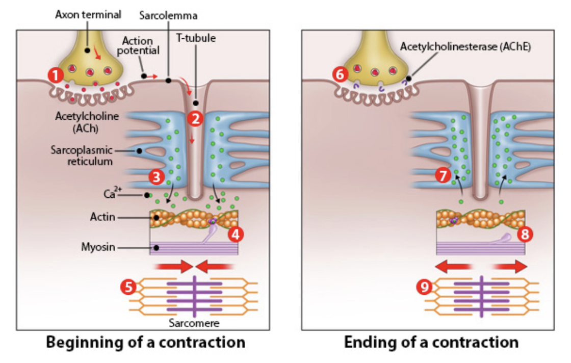
neuromuscular junction (NMJ)
specialized synapse between the somatic (α) motor neuron’s nerve terminal and the motor end plate of the skeletal muscle cell

excitation/contraction (E/C) coupling
transformation of this electrical signal (in the muscle cell) into contraction of sarcomeres
muscle action potentials sensed in t-tubules trigger Ca2+ release from the SR and cause muscle contraction
process:
action potential travels along sarcolemma of muscle fiber
action potential reaches t-tubules, allowing the action potential to rapidly penetrate into the interior of the muscle fiber
voltage-gated calcium channels in the SR membrane open in response to action potential, causing a rapid influx of calcium ions from the SR into the cytoplasm of the muscle fiber
the released calcium ions bind to troponin, a protein located on the actin filaments within the muscle fiber
troponin binding causes a conformation change in the troponin-tropomyosin complex, exposing active sites on the actin filaments
myosin heads bind to the exposed active sites on the actin filaments, forming cross-bridges
the myosin heads undergo a power stroke, causing thin filaments to slide over the thick filaments, shortening the sarcomere and resulting in muscle contraction
troponin
protein located on the actin filaments within the muscle fiber
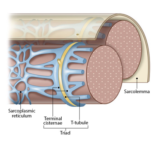
t-tubule
invaginations of the sarcolemma that extend deep into muscle fiber and allows rapid transmission of action potentials from the surface of the muscle fiber to the interior
sarcoplasm
cytoplasm of the muscle cell
sarcolemma
plasma membrane of the muscle cell
actin
protein that produces thin contractile filaments within muscle cells
myosin
protein that produces dense contractile filaments within muscle cells
steps from motor neuron action potentials to skeletal muscle contractions
acetylcholine binds to skeletal muscle fibers (nicotinic receptors)
action potentials moves into t-tubules
Ca2+ release by sarcoplasmic reticulum (trigger for contraction)
Ca2+ binds to troponin; active cross-bridge cycling
skeletal muscle contraction
acetylcholinesterase in cleft of NMJ degrades acetylcholine
Ca2+ pumped into extracellular fluid and SR
tropomyosin blocks cross-bridge formation
skeletal muscle contraction is prevented
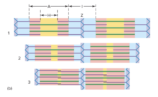
sarcomere
the repeating structural unit of myofibrils in striated muscle; area between consecutive z-lines, consisting of a dark band in the middle and a light band on either side; smallest contracile unit of a muscle cell
thin filaments: actin
thick filaments: myosin
sarcomeres shorten during muscle contraction due to thick and thin filaments sliding past one another
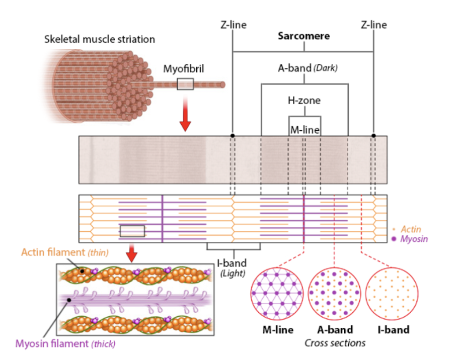
parts of a sarcomere
z-discs (z-lines): borders between neighboring sarcomeres; appears as a dark line in the middle of the I-band
a-band: length of thick filament; dark area of striation and identifies location of myosin filaments
m-line: center of sarcomere
i-band: region of thin filaments without any overlapping thick filaments; only consists of actin
h-zone: region of thick filaments without any overlapping thin filaments, only consists of myosin

muscle contraction (filaments)
shortening of sarcomeres via sliding of thin filaments (actin) towards m-lines
thick and thin filament lengths are constant but overlap incrases
a-bands maintain their length
i-bands and h-zones narrow
→shortening of myofibrils → shortening of muscle cell → muscle contraction
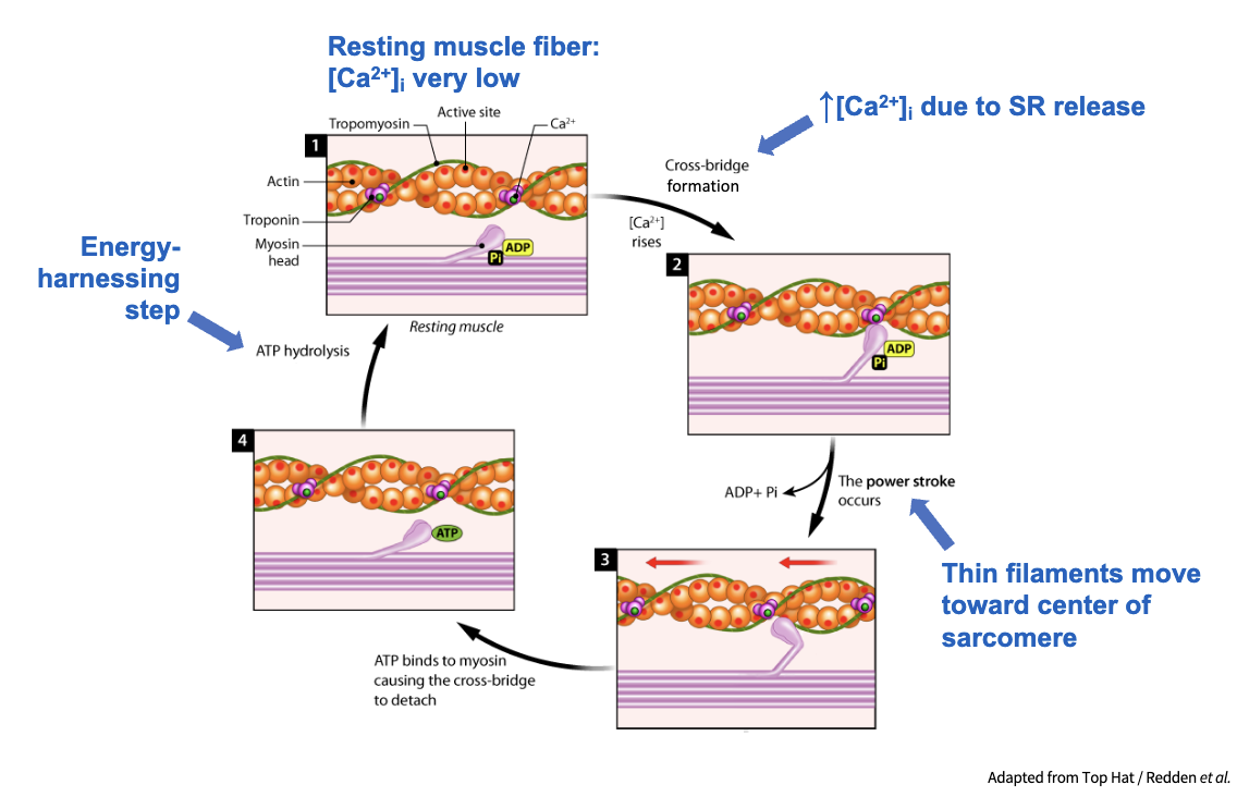
cross-bridge cycle
each myosin head binds to an exposed active site on the actin filament, forming a cross bridge
powerstroke: after cross-bridge formation, myosin head undergoes a conformational change, pivoting towards the center of the sarcomere, called a power stroke, which causes the actin filament to slide relative to the myosin filament and pull the thin filament towards the center of the sarcomere
during the power stroke, ADP and inorganic phosphate are released from the myosin head
the myosin head remains bound to the actin filament until a new ATP molecule binds to it
ATP binds, causing the myosin head to detach from the actin filament
binding of ATP provides energy necessary for myosin head to return to its high-energy cocked state
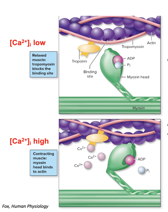
Ca2+ dependence of cross-bridge attachment
in relaxed muscle: the myosin/actin interaction is inhibited; tropomyosin lies in the grooves of the actin filaments → cross-bridge attachments are blocked
in stimulated muscle: active cross-bridge cycling causes muscle to contract; Ca2+ binds to troponin → tropomyosin moves → cross-bridge attachments can occur
twitch
the response of a muscle to a single action potential
↑ action potential frequency → summation of muscle response
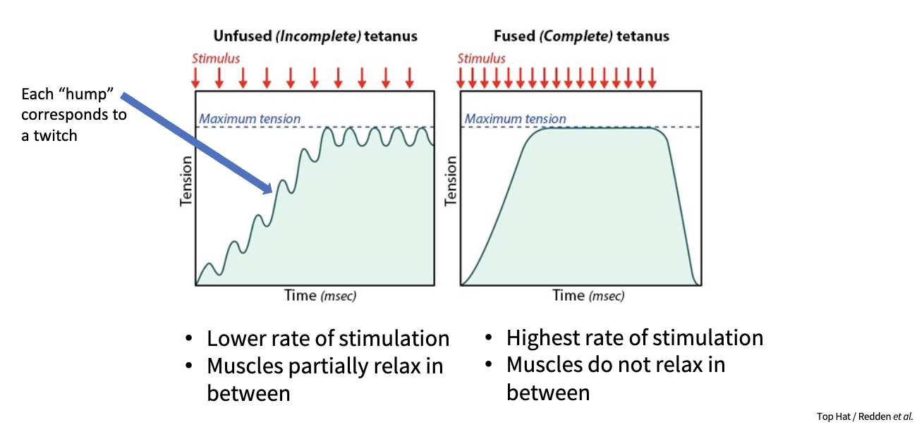
skeletal muscle behavior
contraction strength increases as:
more motor units are recruited
number of myofibrils increases
frequency of stimulation increases
myofibril thickness increases
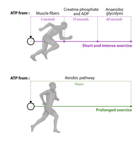
aerobic vs anaerobic ATP
aerobic ATP production:
during low to moderate intensity, sustained activities
dominant energy system for activities lasting several minutes or longer/endurance exercises (long distance running, cycling, swimming)
processL with oxygen present, glucose or fatty acids are metabolized in the mitochondria through glycolysis, krebs cycle, and ETC
anaerobic ATP production:
during high-intensity, short-duration activities where atp demand is very high
without oxygen, glucose broken down into pyruvate, converted to lactate which can lead to muscle fatigue
top down control
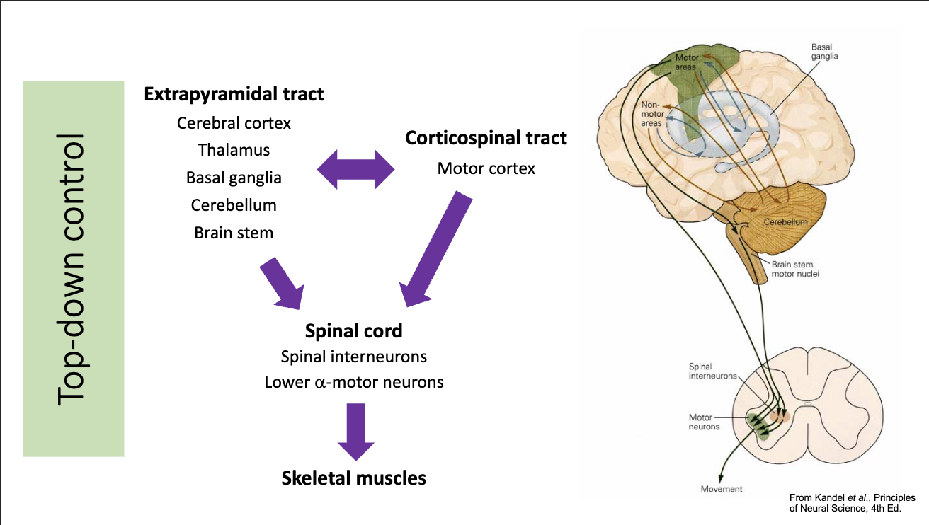
bottom up control