Topic 2 - Unit 2- Fundamentals of sensory physiology
1/7
There's no tags or description
Looks like no tags are added yet.
Name | Mastery | Learn | Test | Matching | Spaced | Call with Kai |
|---|
No study sessions yet.
8 Terms
Neuron structure and function
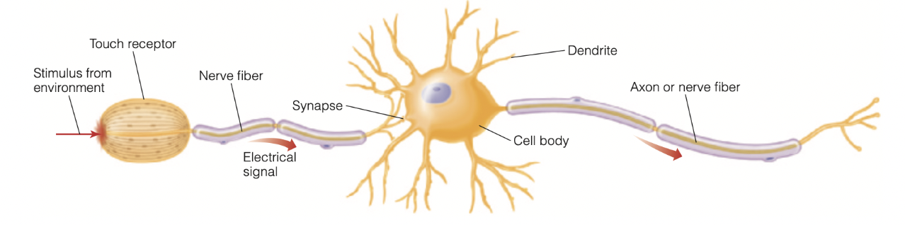
The neuron on the right consists of a cell body, dendrites, and an axon, or nerve fiber. The neuron on the left that receives stimuli from the environment has a receptor in place of the cell body.
Neuron Components: Cell body (keeps cell alive), dendrites (receive signals from other neurons), and axon/nerve fiber (conducts electrical signals). Sensory receptors are specialized neurons that respond to environmental stimuli.
Neural Processing: Neurons are interconnected in complex networks—signals don't travel in straight lines but through interconnected pathways where they meet and are affected by other signals. This neural processing creates neurons that respond to specific features like slanted lines, faces, movement direction, or tastes.
How Electrical Signals in Neurons Are Studied:
Electrical signals are recorded from the axons (or nerve fibers) of neurons using small electrodes to pick up the signals.
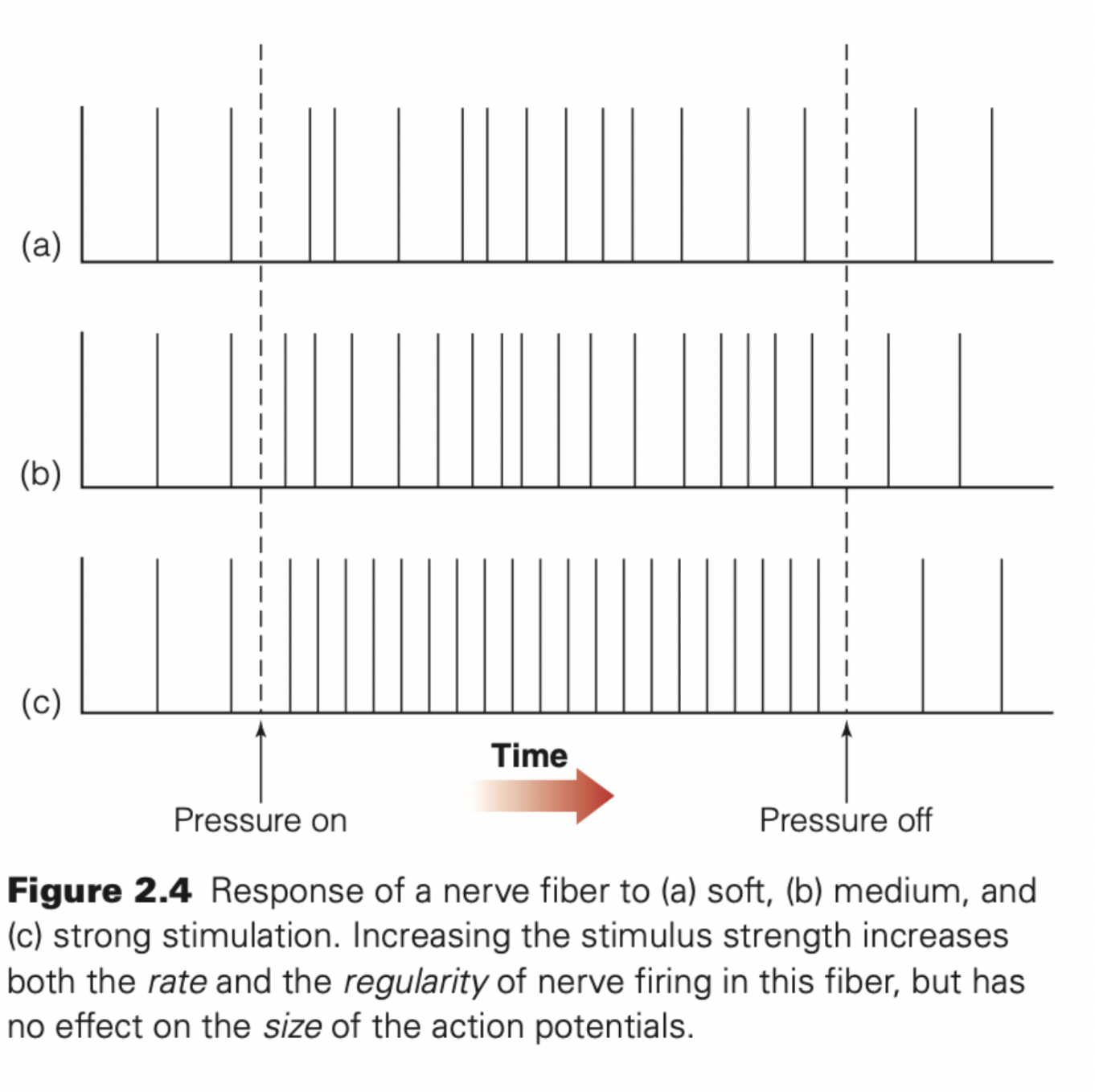
The same neuron is stimulated at different intensities
Researchers record its electrical activity using electrodes
Rate of firing:
Meaning
Stronger stimulation → more action potentials per second
Weak stimulation → fewer action potentials
Example:
Soft touch → slow firing
Strong pressure → rapid firing
Regularity of firing:
Meaning
Stronger stimuli produce:
More consistent
More regular firing patterns
Weak stimuli:
Produce irregular or sporadic firing
Action potentials
Resting State (–70 mV)
What this means
The inside of the neuron is more negative than the outside.
This difference in electrical charge is called the resting membrane potential.
–70 mV is normal for most neurons.
Why this happens
Different ions (Na⁺, K⁺) are unevenly distributed
The membrane is more permeable to K⁺
The sodium–potassium pump maintains this imbalance
Measuring the Charge (Electrode)
Meaning
An electrode is placed near or inside the neuron.
It measures the voltage difference between inside and outside.
At rest, it reads –70 mV.
3. Rising Phase (Depolarization)
What’s happening
A stimulus triggers the neuron
Voltage-gated sodium (Na⁺) channels open
Sodium rushes into the neuron
Result
Inside becomes less negative, then positive
This is called depolarization
Appears as the rising phase on the graph
4. Falling Phase (Repolarization)
What’s happening
Sodium channels close
Potassium (K⁺) channels open
Potassium flows out of the neuron
Result
The inside becomes negative again
This is called repolarization
Appears as the falling phase of the action potential
5. Return to Resting State:
What happens next
The neuron may briefly become more negative than –70 mV (hyperpolarization)
The sodium–potassium pump restores normal ion distribution
The neuron is ready for another signal
Synaptic transmission
Action potentials can't jump the synapse (gap between neurons). Instead:
Action potential triggers release of neurotransmitters from synaptic vesicles
Neurotransmitters flow across synapse
Bind to matching receptor sites on receiving neuron (like key in lock)
Cause voltage change in receiving neuron
Two Response Types
Excitatory: Causes depolarization (more positive inside), increases firing likelihood
Inhibitory: Causes hyperpolarization (more negative inside), decreases firing likelihood
Multiple inputs combine to determine if receiving neuron fires. Both excitation and inhibition are essential for neural processing.
Sensory coding: how neurons represent information
Specificity Coding
One neuron represents one specific stimulus/concept (e.g., "grandmother cell" fires only for your grandmother). Quiroga et al. found neurons responding specifically to Steve Carell or Halle Berry from multiple views and representations. However, limited recording time means these neurons might respond to other stimuli if tested further—grandmother cells likely don't exist.
Sparse Coding
A small group of neurons with overlapping responses represents each stimulus through their firing pattern. Most neurons remain silent. Evidence supports this for visual objects, auditory tones, and odors.
Population Coding
Large groups of neurons create unique patterns for each stimulus. Allows representation of huge numbers of stimuli through different patterns. Evidence exists across all senses.
Brain representation and modularity
Historical Context
Phrenology (Gall & Spurzheim, 18th century) wrongly claimed skull bumps revealed mental faculties but correctly proposed different brain areas serve different functions.
Modularity Evidence
Neuropsychology: Broca's area (frontal lobe) = speech production; Wernicke's area (temporal lobe) = speech comprehension
fMRI studies: Superior temporal sulcus (STS) = "voice area" responds specifically to vocal sounds vs. environmental sounds
fMRI Method
Measures blood flow changes in brain voxels (2-3 mm cubes containing many neurons). Active areas consume more oxygen, changing hemoglobin's magnetic properties. Colors indicate activation levels during tasks vs. baseline.
Distributed representation
Perceptions involve networks of brain areas working together, not just isolated modules.
Examples:
Pain: Activates multiple areas for sensory (location, intensity), emotional (unpleasantness), and motor (reflexive) components
Object perception: Houses, faces, chairs show maximum activity in separate areas BUT also activate widespread cortical regions
Connectivity between brain areas
Structural Connectivity
Physical fiber pathways connecting brain regions (the "road map")
Functional Connectivity
Neural activity patterns flowing through networks during specific functions (the "traffic patterns")