Looks like no one added any tags here yet for you.
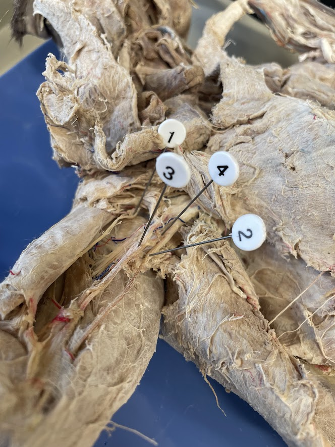
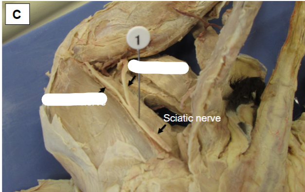
1 (smallest and most anterior, on the posterior surface of the biceps brachii)
musculocutaneous nerve (cat)
2 (most posterior and goes towards the elbow joint)
ulnar nerve (cat)
3 (found between the musculocutaneous and ulnar nerves and is superficial; it is usually bundled in connective tissue with a brachial artery (red) and brachial vein (blue))
median nerve (cat)
4 (found between the musculocutaneous and ulnar nerves, but is thicker, and is found near the proximal end of the arm)
radial nerve (cat)
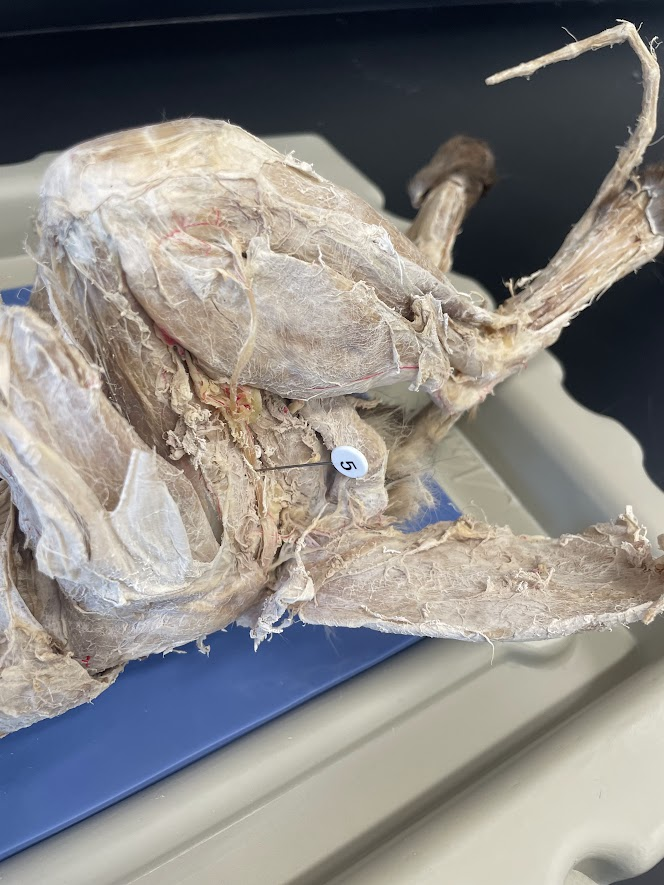
5 (only large nerve on the dorsal side of the thigh)
sciatic nerve (cat)
just remember that 1-4 are near the shoulder, and 5 is from the lower back to leg area
just remember :)
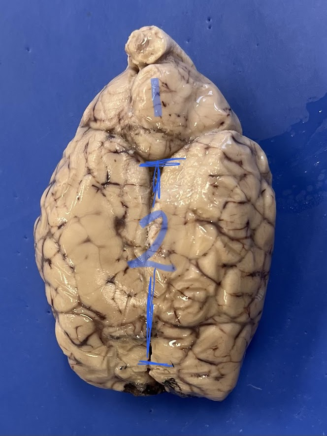
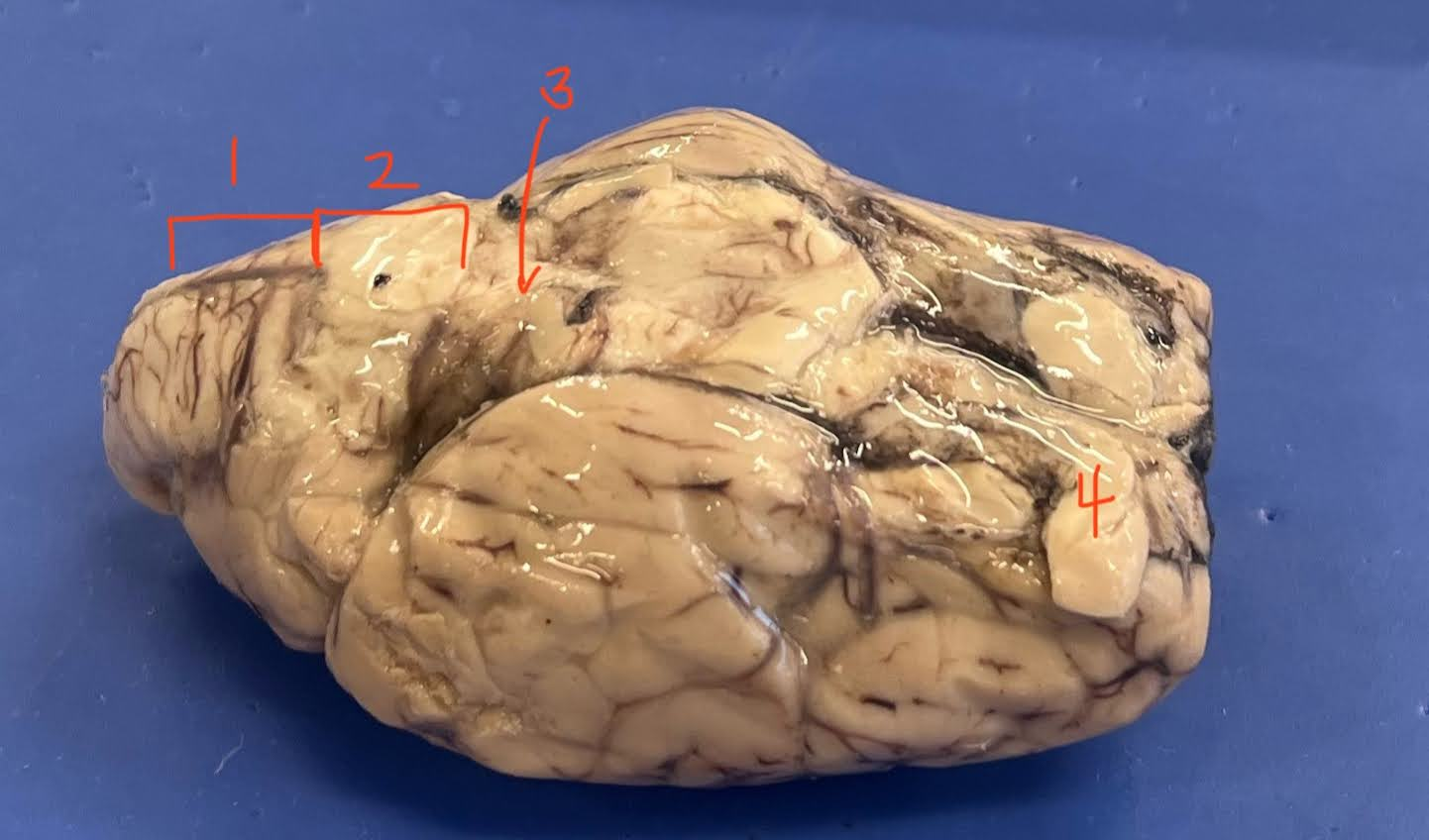
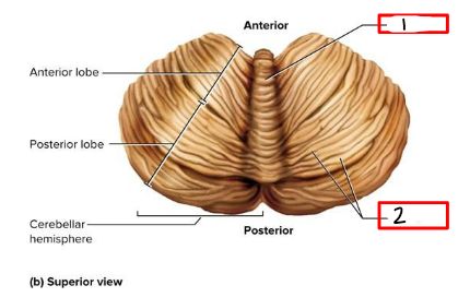
1
cerebellum (sheep brain)
2
longitudinal fissure (sheep brain)
1
medulla oblongata (sheep brain)
2
pons (sheep brain)
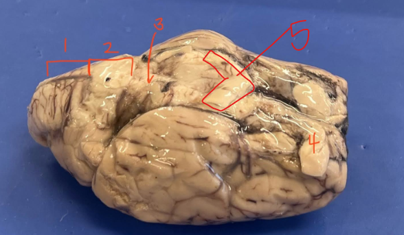
5
optic chiasm (sheep brain)
4
remnants of olfactory bulb (sheep brain)
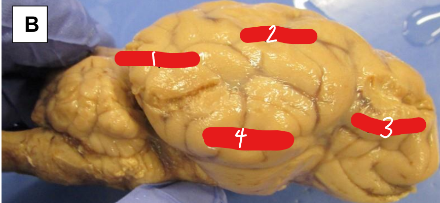
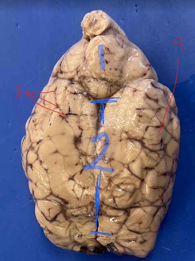
1
occipital lobe (sheep brain)
2
parietal lobe (sheep brain)
3
frontal lobe (sheep brain)
4
temporal lobe (sheep brain)
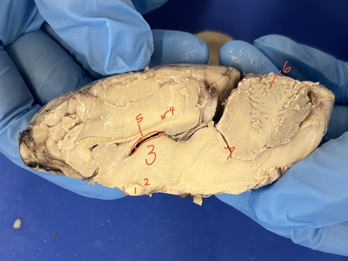
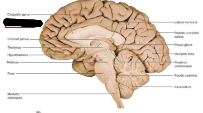
1
optic chiasm (sheep brain)
2
hypothalamus (sheep brain)
3
thalamus (sheep brain)
4
corpus callosum (sheep brain)
5
lateral ventricle (sheep brain)
6
arbor vitae (sheep brain)
7
fourth ventricle (sheep brain)
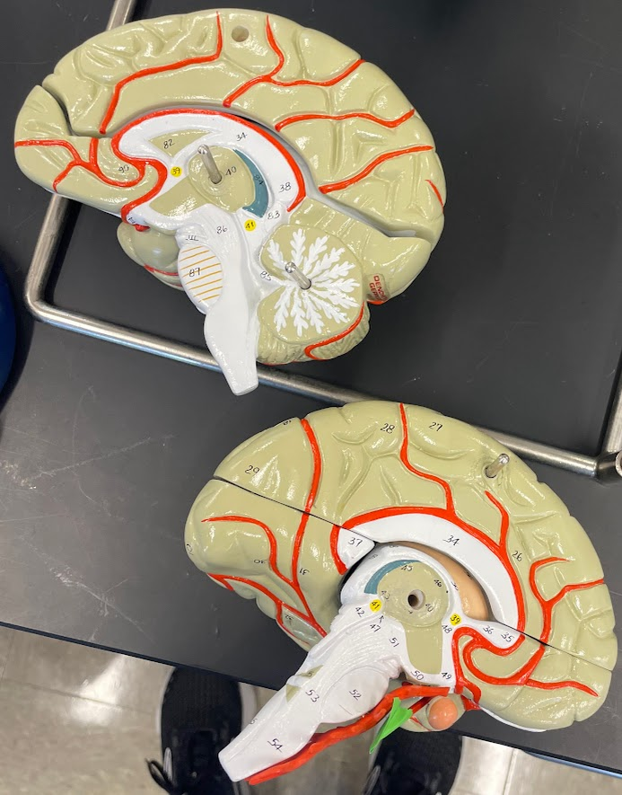
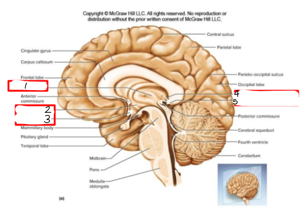
40
thalamus (model)
1
thalamus (ppt)
2
hypothalamus (ppt)
3
optic chiasm (ppt)
4
part of the epithalamus (habenula) (ppt)
5
part of the epithalamus (pineal gland) (ppt)
corpus callosum (ppt)
what makes up the dienchephalon
thalamus, hypothalamus, epithalamus, optic chiasm, and parts of cranial nerve 2 (CN II) (ppt)
______________________ nerves arise when the sciatic nerve splits in the distal part of the thigh
tibial and fibular (cat)
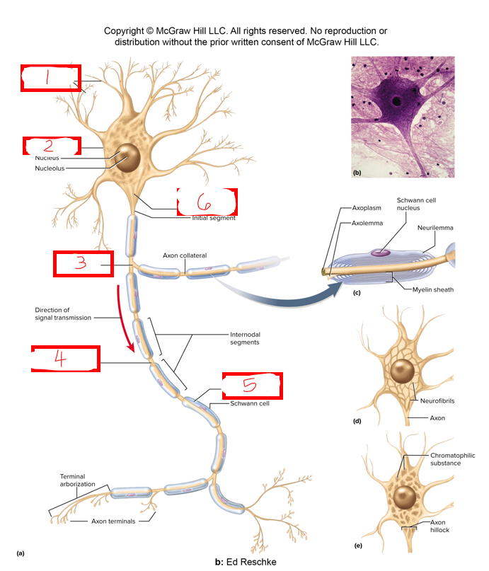
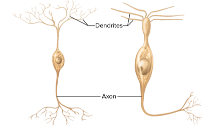
1
dendrites (ppt)
2
neurosoma/soma/neurons cell body (ppt)
3
axon (ppt)
4
myelin sheath gap/nodes of ranvier (ppt)
5
myelin sheath (ppt)
6
axon hillock (part of trigger zone) (ppt)
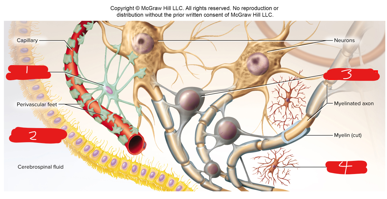
1
astrocyte (ppt)
2
ependymal cell (ppt)
3
oligodendrocyte (ppt)
4
microglia (ppt)
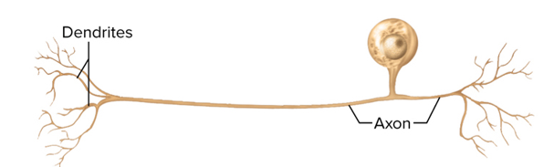
what type of neuron is this
unipolar
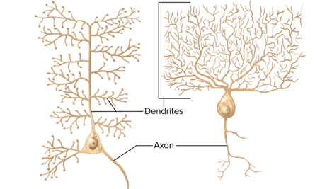
what type of neuron is this
multipolar
what type of neuron is this
bipolar
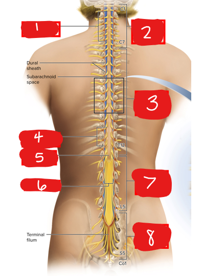
1
cervical enlargement (ppt)
2
8 cervical spinal nerves (ppt)
3
thoracic spinal nerves (ppt)
4
lumbosacral enlargement (ppt)
5
medullary cone (ppt)
6
cauda equina (ppt)
7
lumbar spinal nerves (ppt)
8
sacral spinal nerves (ppt)
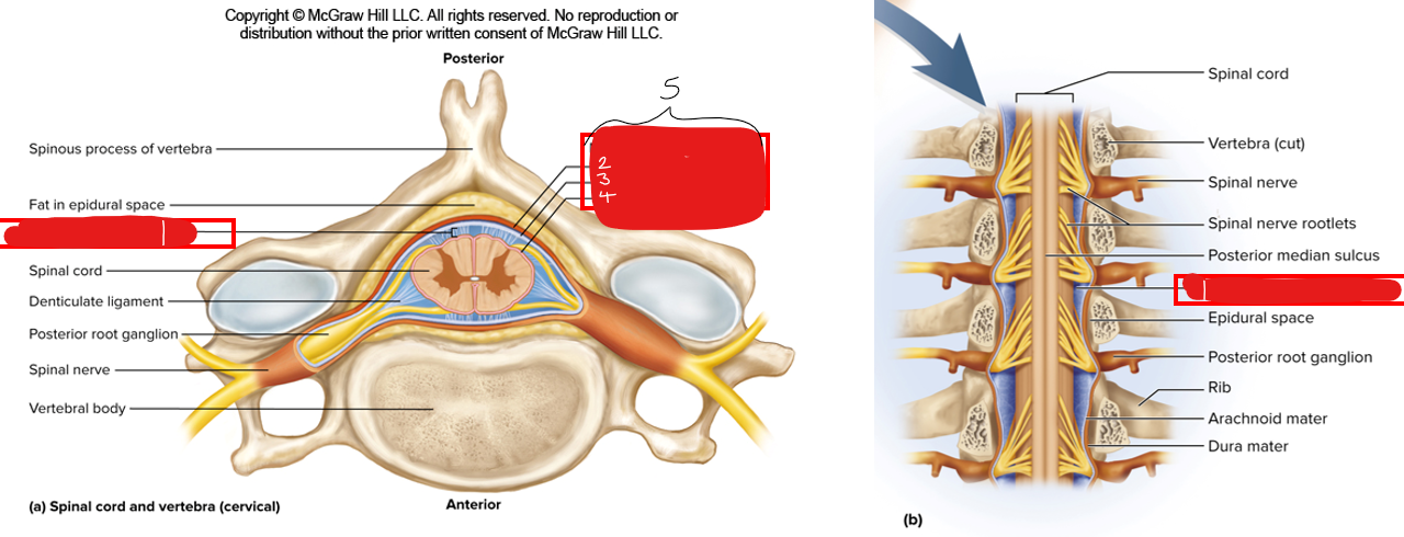
1
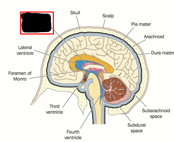
subarachnoid space (where csf circulates) (ppt)
2
dura mater (ppt)
3
arachnoid mater (ppt)
4
pia mater (ppt)
5
meninges (ppt)
1
spinal cord (ppt)
2
dorsal/posterior root ganglion (ppt)
3
anterior root (ppt)
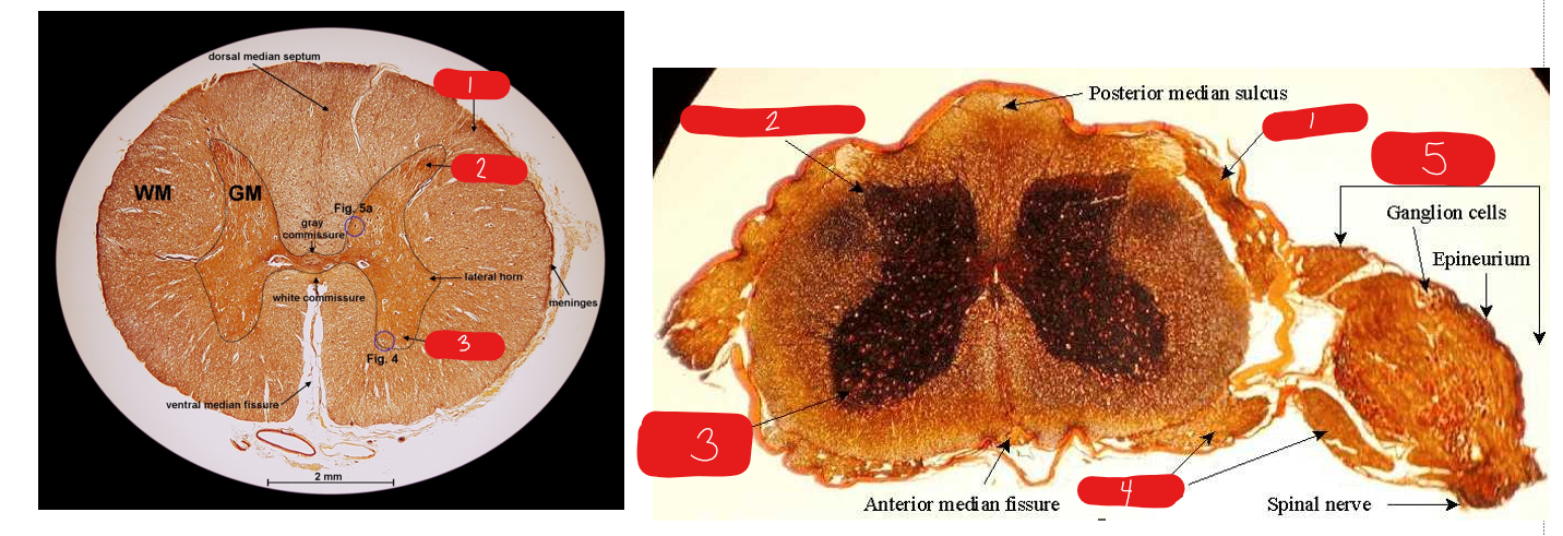
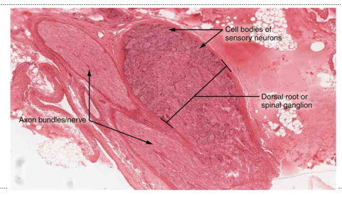
1
dorsal root (histology)
2
posterior/dorsal horn (histology)
3
anterior/ventral horn (histology)
4
ventral root (histology)
5
dorsal root ganglion (histology)
what is the histology image of
spinal cord cross section (histology)
what is the histology image of
dorsal root ganglion (histology)
what is a neurite made of
dendrites(s) and axon(s)
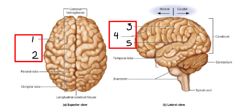
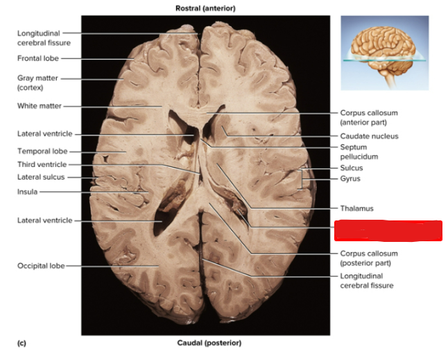
1
frontal lobe (ppt)
2 and 3
central sulcus/sulci (ppt)
4
gyri (ppt)
5
lateral sulcus
transverse cerebral fissure (ppt)
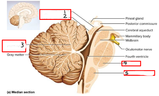
1
superior colliculus (ppt)
2
inferior colliculus (ppt)
3
arbor vitae (ppt)
4
pons (ppt)
5
medulla oblongata (ppt)
1
vermis (ppt)
2
folia (ppt)
3 (grooves)
sulci (sheep brain)
4 (lifted fold between 2 grooves)
gyri (sheep brain)
thin _____ mater directly covers the brain
pia
choroid plexus (ppt)
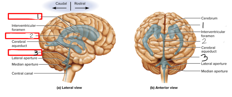
1
lateral ventricles (ppt)
2
third ventricle (ppt)
3
fourth ventricle (ppt)
choroid plexus CSF production (ppt)
a
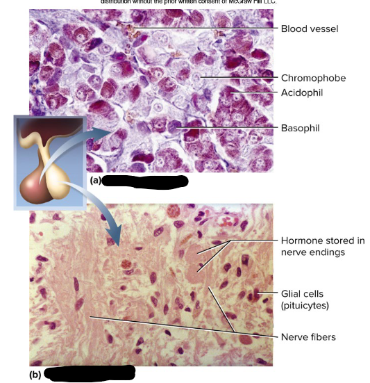
anterior pituitary/adenohypophysis (histology)
b
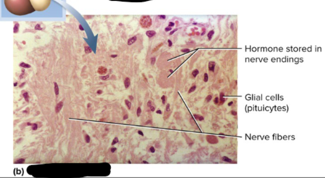
posterior pituitary/neurohypophysis (histology)
what is a hormone produced here (in the posterior pituitary)?
oxytocin (OT)
what is a hormone produced in the anterior pituitary/adenohypophysis?
thyroid stimulating hormone (TSH)
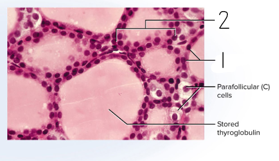
what is this slide a picture of
thyroid (histology)
1
follicular cells of thyroid (histology)
2
follicles of thyroid (histology)
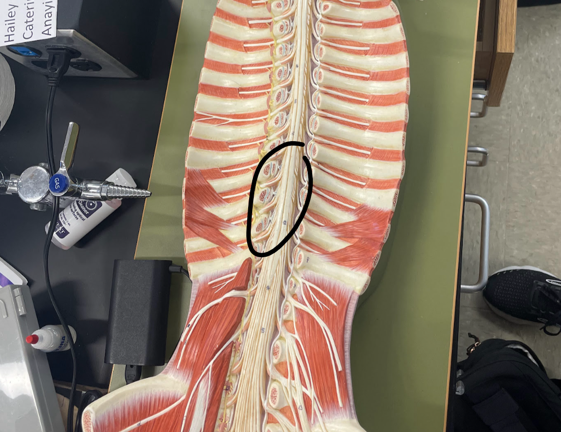
what general area is circled
cervical enlargement (model)
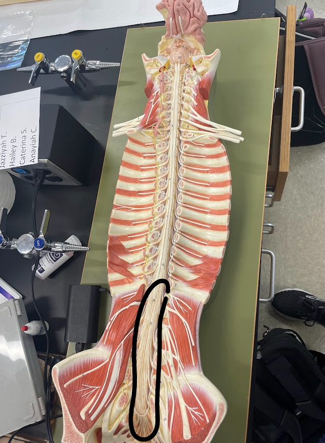
what general area is circled
lumbar enlargement
cauda equina (model)
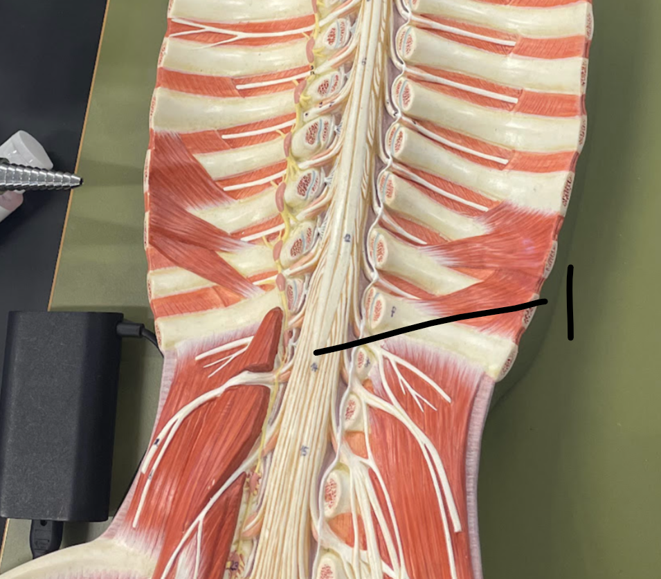
1
medullary cone (model)