Prepare for flag race
1/137
Earn XP
Description and Tags
Week 1 - skull anatomy, Week 3 - bones and bony landmarks, Week 4 - Appendicular Muscles, Week 5 - Axial muscles
Name | Mastery | Learn | Test | Matching | Spaced | Call with Kai |
|---|
No analytics yet
Send a link to your students to track their progress
138 Terms
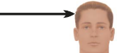
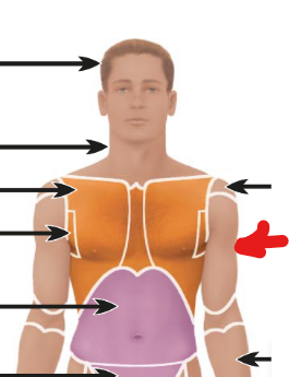
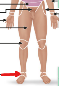
Cephalic
The head
Cervical
The neck

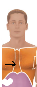
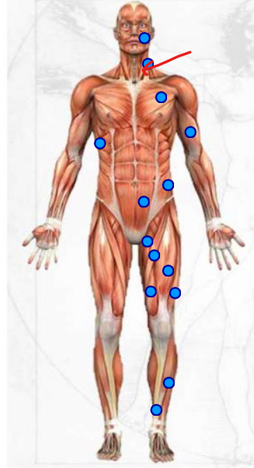
Thoracic
the chest area but above the abdomen
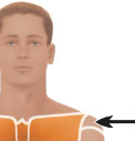
Acromial
the shoulders
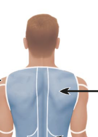
Scapular
the shoulder blades
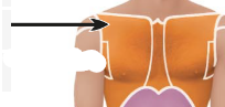
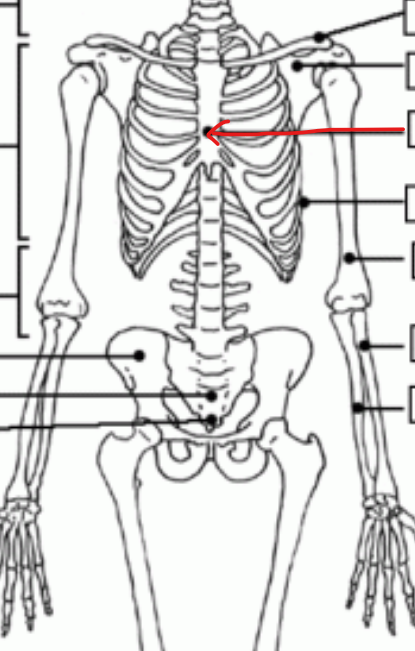
Sternal
middle of the chest area
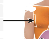
Axillary
the armpit area
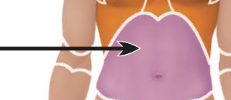
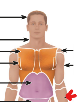
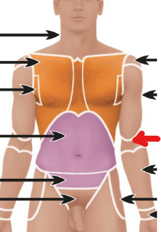
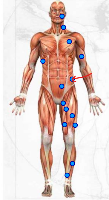
Abdominal
the whole stomach area below the thoracic area
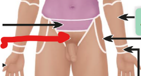
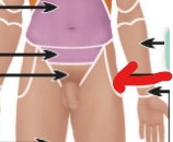
Pelvic
between the abdominal area and inguinal area

Inguinal
right above the pubic and is the groin
Brachial
upper arm area
Antebrachial
lower arm area
Antecubital
the elbow
Coxal
the hip
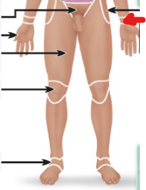
Pollex
the thumb
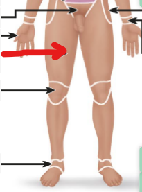
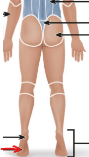
Femoral
the upper leg area
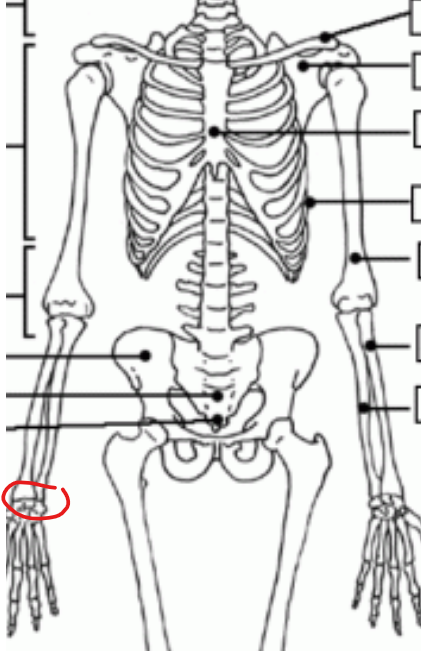
Carpal
the wrist
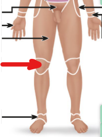
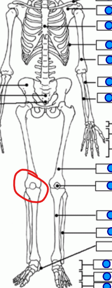
Patella
the knee (anterior view)
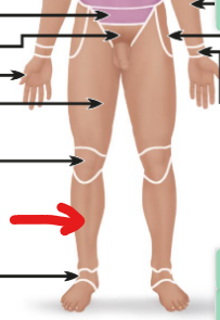
Fibular
lower leg area
Tarsal (region)
the ankle
Vertebral
spine in the dorsal cavity (the back)
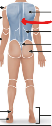
Lumbar
the lower back centre area
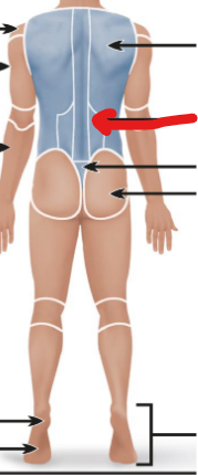
Sacral
below the lumbar vertebral and between the upper gluteal
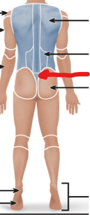
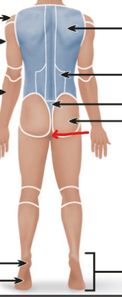
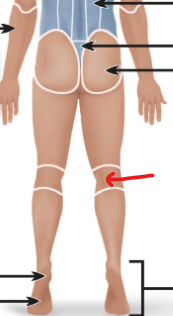
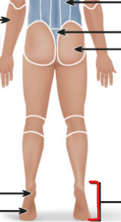
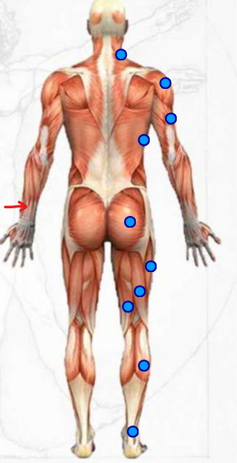
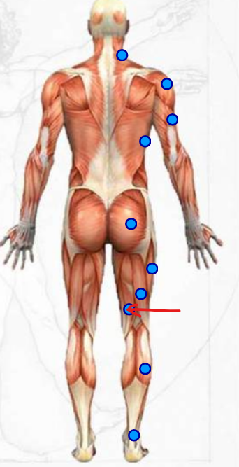
Gluteal
the buttcheeks
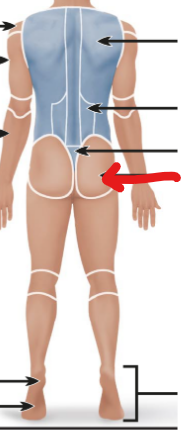
Perineal
the butthole
Popliteal
the knee (posterior view)
Calcaneal
the heel
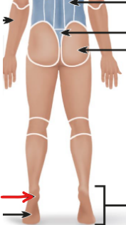
Plantar
the sole of the feet below the heel
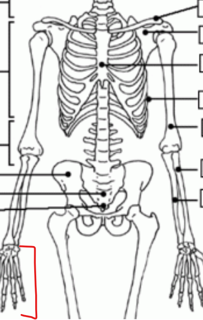
Pedal
the whole foot region
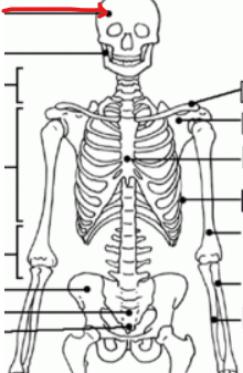
Cranium
bone of the head
Cervical vertebrae
spine in the neck area
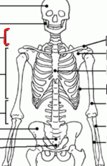
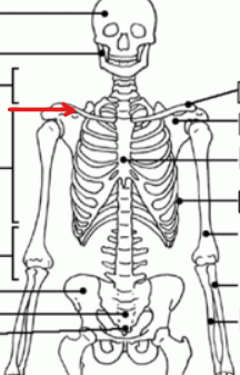
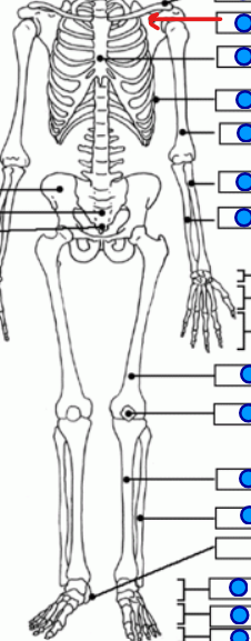
Clavicle
the collarbone
Humerus
the upper arm bone
Lumbar vertebrae
spine in the lumbar cavity S
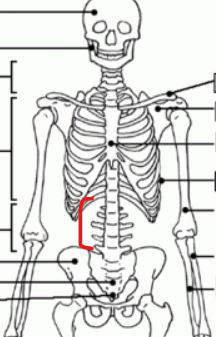
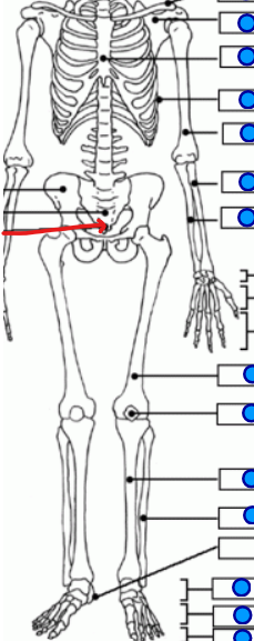
Sacrum
triangular bone below the lumbar vertebrae and above the coccyx
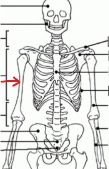
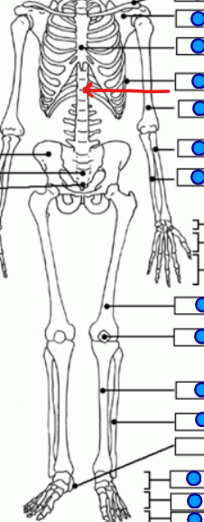
Sternum
the bone on the spine area superior to the lumbar vertebral
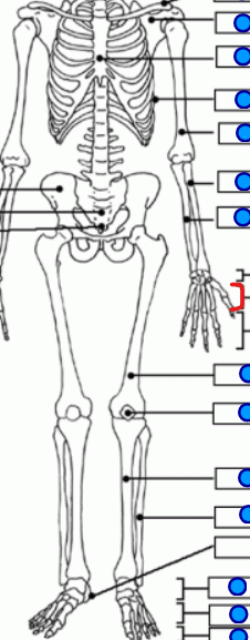
Carpals
the wrist
Phalanges
bones making up the upper fingers and bones in toes
Patella
bones in the knees
Scapular
triangular bone making the shoulder blades
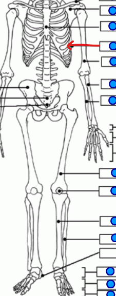
Ribs
curved bones forming the thoracic region
Thoracic vertebrae
spine in the thoracic region
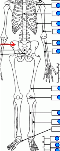
Pelvis
Outer triangular bone in the pelvic region, surrounding the sacrum
Radius
later lower arm bone
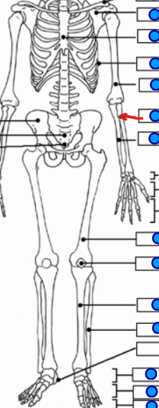
Ulna
the medial lower arm bone
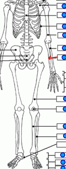
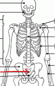
Coccyx
triangular bone at the very bottom under the sacrum
Metacarpals
lower finger bones between the carpals and the phalanges
Femur
upper leg bone
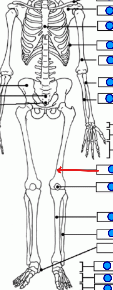
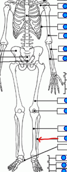
Tibia
medial lower leg bone
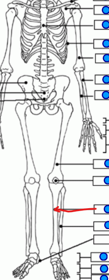
Fibula
lateral lower leg bone
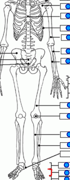
Tarsals (skeletal)
7 short bones right inferior to the ankle

Metatarsals
bones between the tarsals and the phalanges
Temporalis
the temple muscles
Masseter
muscle on the sides of the lower face (right next to the ear)

Buccinator
muscle in the cheek on face
Trapezius
triangular muscle next to the neck

Deltoid
triangular muscle around the shoulders
Biceps brachii
muscle on the upper arm
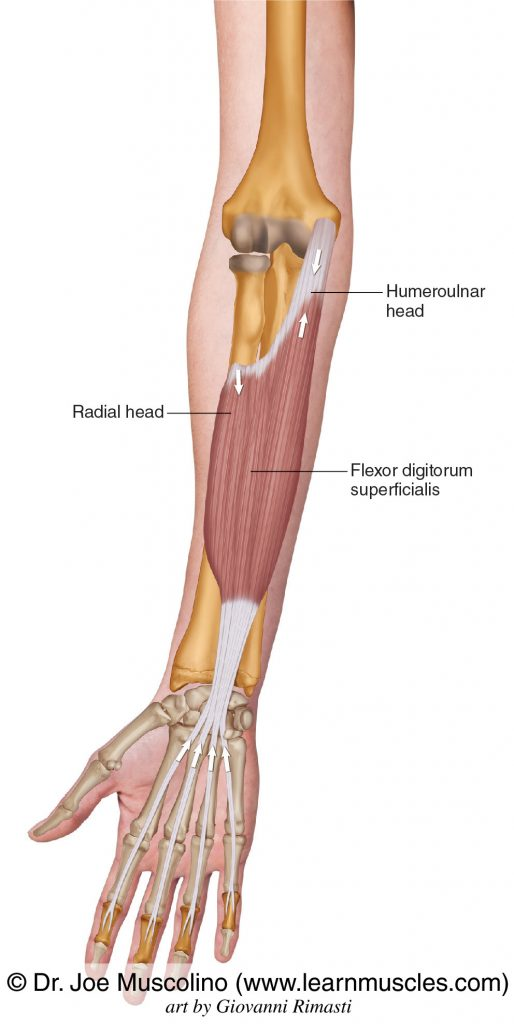
Flexor digitorum superficialis
lower arm inner muscle
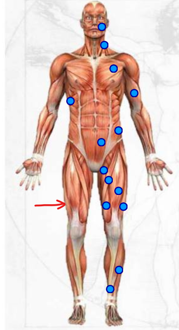
Rectus femoris
upper leg muscle

Vastus lateralis
outside muscle on the upper leg area
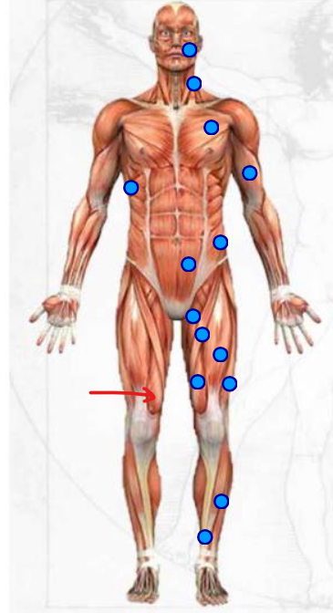
Vastus medialis
inner muscle right above the knee
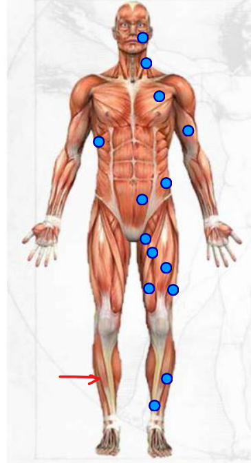
Tibialis anterior
outer muscle in the lower leg area
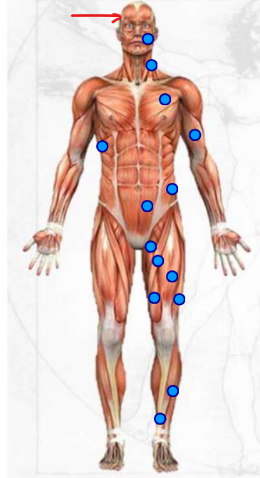
Orbicularis oculi
muscle in the eye

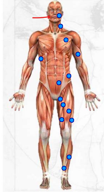
Orbicularis oris
muscle surrounding the lips, making up the mouth

Sternocleidomastoid
muscle making up the neck area
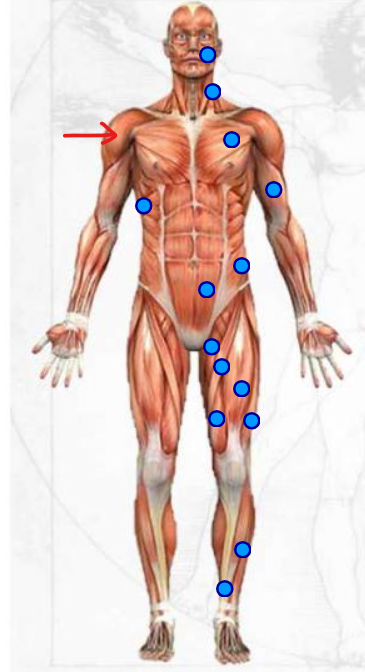
Pectoralis major
The chest pecks

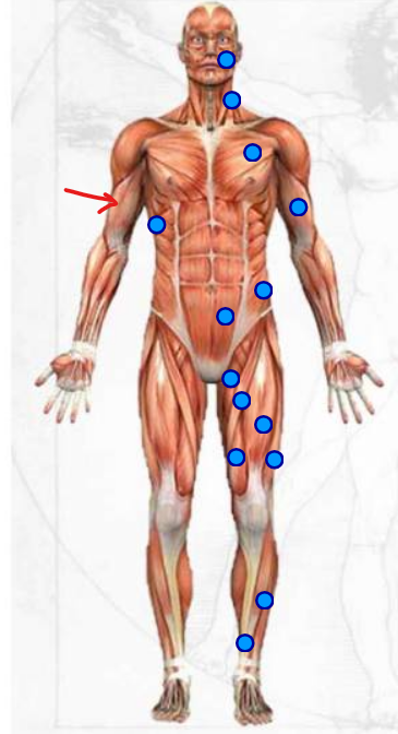
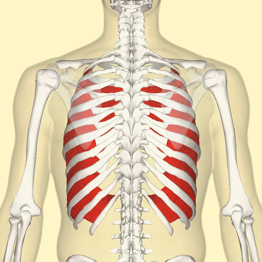
Internal intercostals
inner muscles between the ribs
Rectus abdominis
the abs

Transverse abdominis
very internal muscle acting like a corset
Vastus intermedius
muscle directly underneath the rectus femoris
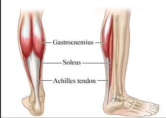
Gastrocnemius
inner muscle on the lower leg area

Soleus
muscle in the calf area (kinda in the middle of the lower leg)
Triceps Brachii
upper arm muscle in the posterior view

Extensor digitorum
muscle in the lower arm region
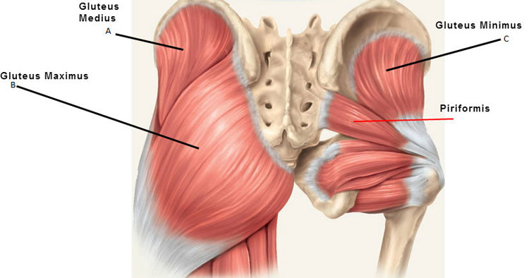
Gluteus minimus
inner upper muscle on the buttcheeks
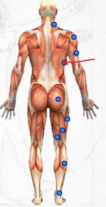
Latissimus dorsi
triangular lower side muscles int he dorsal cavity
Gluteus medius
muscle in the upper right corner of the buttcheeks

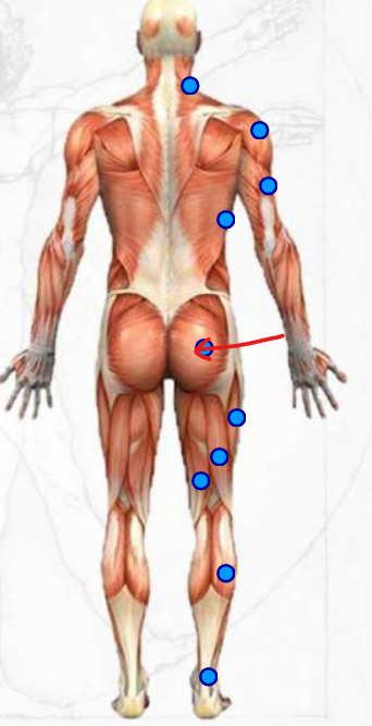
Gluteal maximus
main part of the buttcheeks
Biceps femoris
main inner upper leg muscle

Semitendinosus
the muscle to the left of the biceps femoris in the upper leg
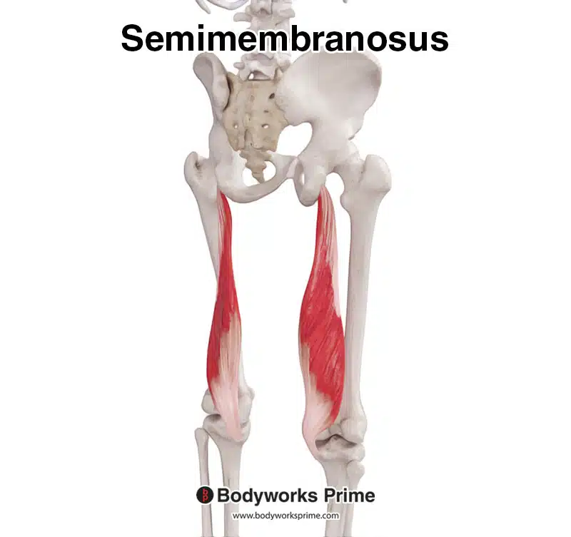
Semimembranosus
inner leg muscle from the posterior view in the upper leg region
‘Epi’
prefix meaning ‘on top of;
Tuberosity
round and roughened patch of bon that slightly protrudes, serving as an attachment point for muscles
Foramen
a hole through a bone
Origin
attachment site for muscles on a bone which don’t move when muscle contracts
Insertion
attachment point for a muscle on a bone which move when the muscle contracts
Articulation
the join point of 2 or more bones
Condyle
rounded articular projection
Frontal bone
the forehead
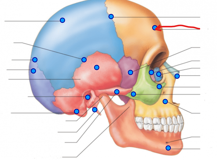
Parietal bone
on the top sides of the skull

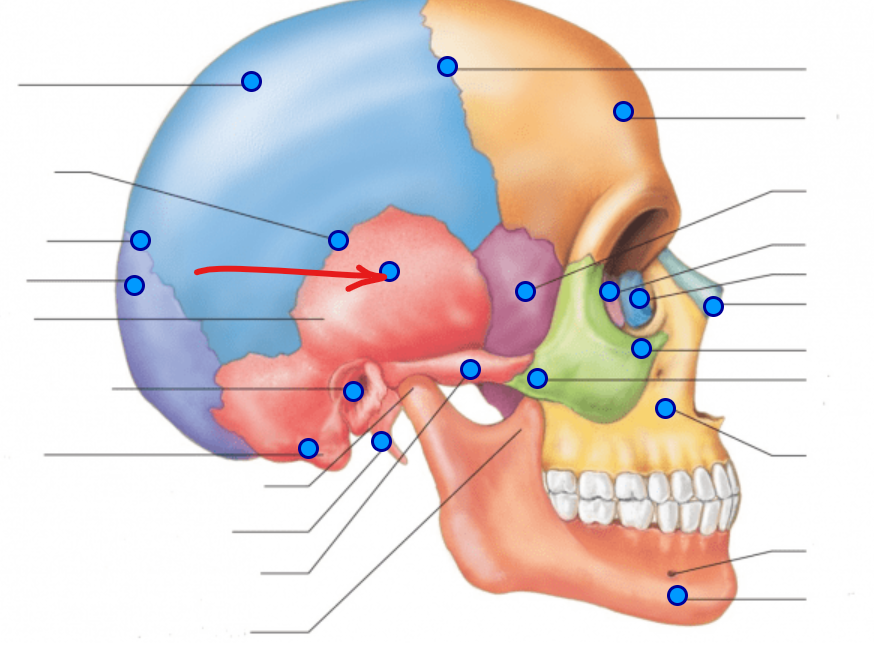
Temporal bone
At the temporalis
Landmark: external acoustic meatus (ear canal → hole into the ear)
Occipital bone
The back of the skull
Landmark: occipital condyles (sticking out of occipital region) and foramen magnum (hole in the middle of the skull)
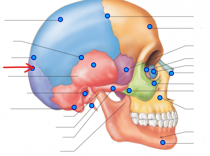
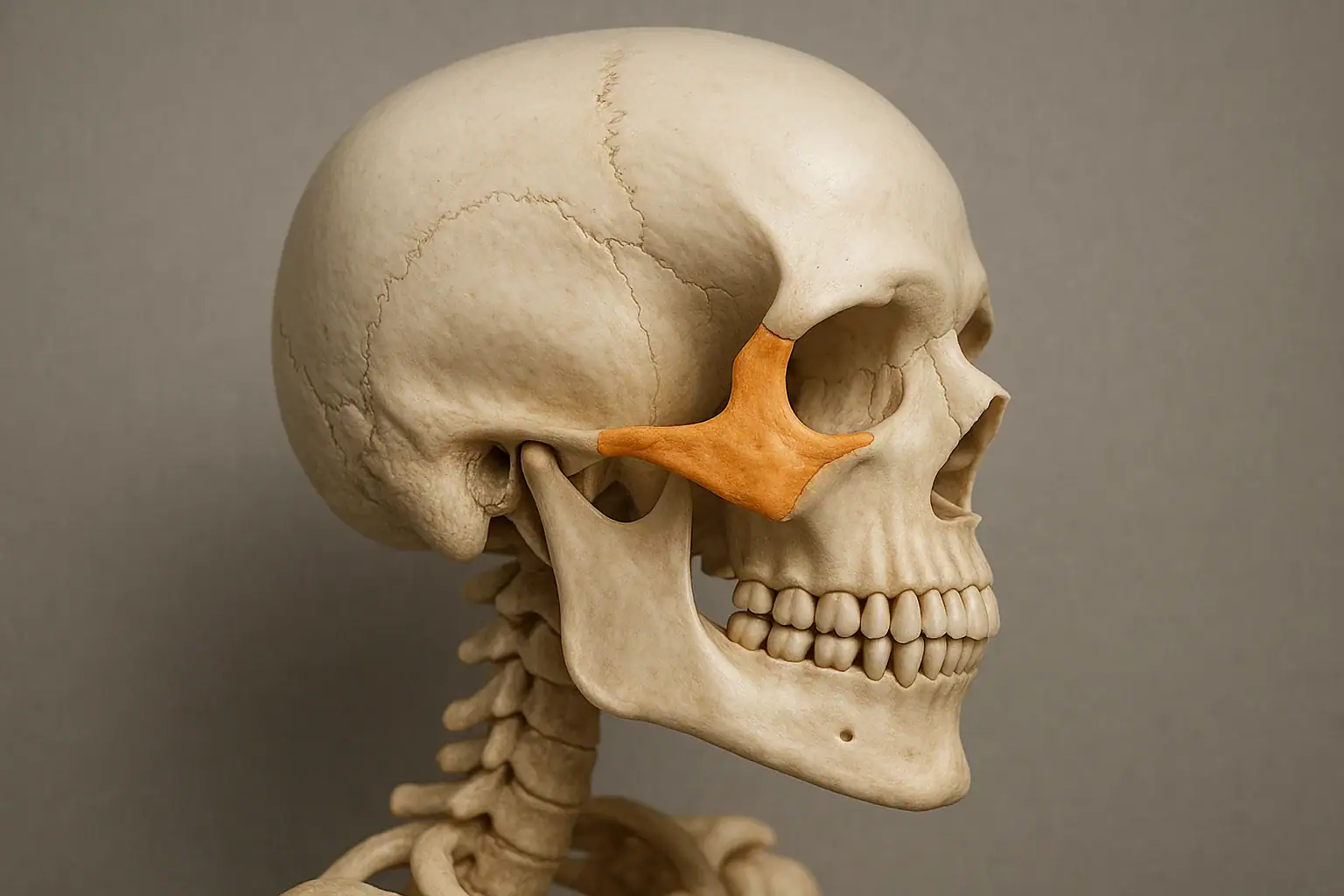
Zygomatic bones
bone of the cheekbones
Landmarks: Zygomatic process (Zygomatic arch - pointy part of the cheekbone)
Maxilla
bone of the upper jaw/mouth

Mandible
bone of the lower jaw
Landmark: mandibular condyle (temporomandibular joint)

Mastoid process
prominent bone behind the ear
Sutures
joints between 2 bones of the skull
Coronal
suture across the middle of the skull
Sagittal
suture going from the front to the back of the skull