Bones of the Proximal Forelimb and the Shoulder Joint
1/29
There's no tags or description
Looks like no tags are added yet.
Name | Mastery | Learn | Test | Matching | Spaced |
|---|
No study sessions yet.
30 Terms
Evolutionarily, what has happened to the pectoral skeleton and the limbs in domestic species compared to humans? (Cursorial Adaptations)
Cursorial Adaptations - speed or endurance. Most cursorials are either predators or medium - large sized herbivores.
1.Reduced pectoral skeleton - narrower and deeper
More cranial/ caudal movement
Clavicle absent / vestigial (Present in cats)
Deep, narrow chest
2.Elongation at proximal end of limb
Scapula lies lateral and vertical
3.Elongation at distal end of limb
long metacarpals, walk on toes or
hooves (increased leg length)
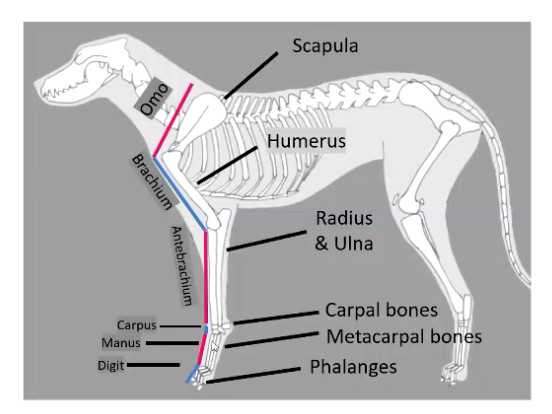
Describe the main regions of the forelimb.
Omo (shoulder)
Brachium (arm) - from shoulder to elbow
Antebrachium (forearm) - from elbow to carpus
Carpus (wrist)
Manus (hand) - distal to carpus
Digit (toe)
How is the scapula oriented in domestics?
Why is it orientated this way?
How is it attached to the rest of the musculoskeletal system?
Vertical orientation
Shoulder joint relatively lower in limb
Increases limb length → increasing stride length → more ground covered
Restricted lateral movement (abduction/adduction)
• Absent / vestigial clavicle
Muscular attachment between forelimb & trunk
What lateral surfaces of the scapula are palpable?
Lateral surface:
• Dorsal border - palpable
• Cranial border - palpable
• Caudal border - difficult to palpate / not palpable
beneath muscle
• Scapular spine - palpable, divides scapula into two portions
Supraspinous fossa
Infraspinous fossa
• Acromion process - palpable
Guide for location of shoulder joint
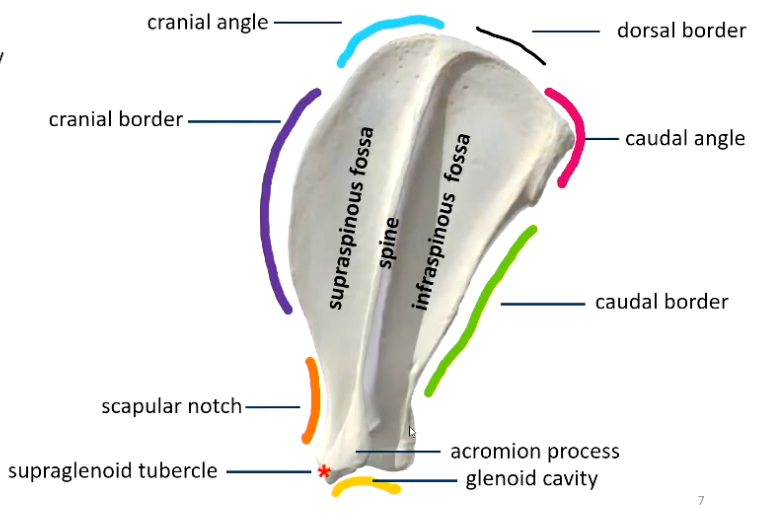
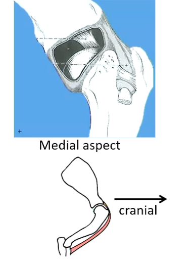
What key features and attachment sites are found on the ventral and lateral aspects of the scapula?
Ventral surface:
Glenoid cavity :
Concave
smooth subchondral / articular surface for shoulder joint
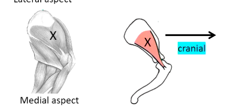
• Supraglenoid tubercle
Attachment of biceps brachii muscle
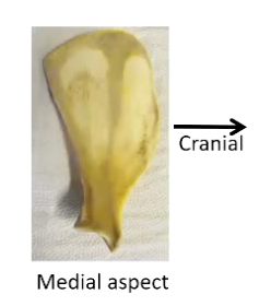
What structures are found on the medial / costal surface of the scapula?
Flat - scapular glide (Scapula gliding across the surface of the rib-cage)
Rough bone - muscle attachment
Serratus ventralis muscle (dorsal)
Subscapularis muscle (ventral)
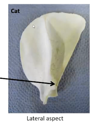
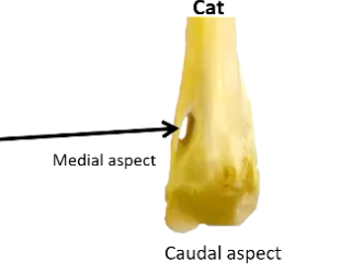
How does the scapula in a cat compare to a dog?
Cats:
• More rounded cranial angle than dog
• Suprahamate process
proximal to acromion process
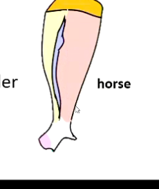
How does the scapula appear in large species?
Large species:
• Tuber on spine
• Cartilage extension to dorsal border
Horse specifically does not have acromnion process
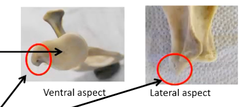
What are the centers of ossification in the scapula?
What potential issue may result?
• 2 centres of ossification:
Body
Supraglenoid tubercle
Potential issue:
- origin of tendon of biceps brachii m.
- Physis weak point = avulsion fracture - as it is near the growth plates
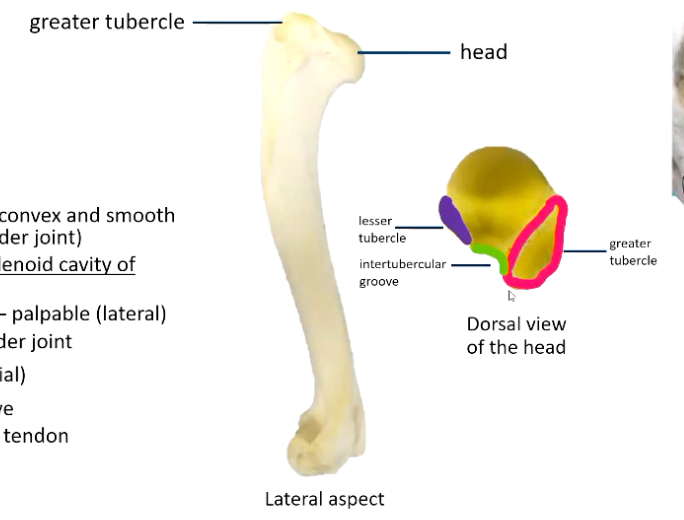
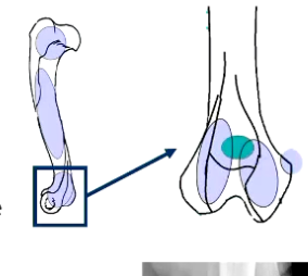
What are key features of the proximal end of the humerus?
Describe the surface of the head and any tubercules
• Head
articular surface, convex and smooth surface (for shoulder joint)
articulates with glenoid cavity of scapula
Greater tubercle - palpable (lateral)
Location of shoulder joint, palpation can help locate
• Lesser tubercle (medial)
• Intertubercular groove (Between the tubercles)
Passage of biceps tendon through it
Smooth surface
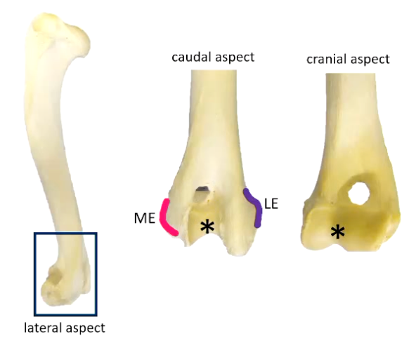
What structures are found on the distal end of the humerus?
• Distal end called the condyle
• Medial epicondyle (ME) - palpable
• Lateral epicondyle (LE)- palpable
• Trochlea *
Is a central depression
Is a smooth articular surface for elbow joint
• Ulnar / olecranon fossa
Caudal aspect
• Radial fossa
Cranial aspect
• Supratrochlear foramen:
Nothing passes through, it is a mechanical foramen, in some dogs it provides a hook or stability to elbow joint
May not be present in short legged (chondrodystrophic) dogs
Absent in larger species eg. horse
Is the supratrochlear foramen present in cats?
NO
• No supratrochlear foramen, instead have Supracondyloid foramen
• Brachial artery & median nerve pass through
• Vulnerable in fractures
What are the areas of ossification in the humerus?
Development:
• 5 Centres of ossification:
• Proximal epiphyses
• Body
• Condyle:
Medial epicondyle
Medial half
Lateral half
Describe the anatomic location of the shoulder joint capsule or bicipital bursa.
What ligaments holds the tendon of the bicep in place
• Forms part of joint capsule extends into intertubercular groove which envelopes the tendon, contains fluid and protects it from wear and tear
between greater & lesser tubercles
• Wraps around tendon of origin of Biceps brachii
(originates on supraglenoid tubercle)
• Tendon held in place by Transverse ligament
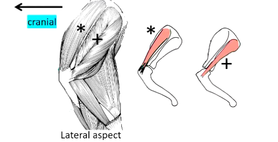
What are the main lateral support muscles in the shoulder joint?
Note Origin
Insertion
Nerve supply
• Lateral support:
• Supraspinatous muscle *
O= Supraspinous fossa
• Infraspinatous muscle +
O = Infraspinous fossa
• | = proximal lateral humerus
• Nerve supply = suprascapular nerve
What are the main medial support muscles in the shoulder joint?
Note Origin
Insertion
Nerve supply
• Medial support:
• Subscapularis muscle X
O = Subscapular fossa
I = proximal medial humerus
• Nerve supply = subscapular nerve
How is skeletal muscle arranged?
How do they usually originate and insert?
• Fibres arranged in parallel bundles
• Encased in fibrous tissue
• Attached to rigid structures - usually bone
Origin - usually proximal
Insertion - usually distal
How do muscles attach to bones?
Attachments:
• Directly to bone
Rough surface area
Bony bumps / tubercles
• Via aponeurosis
Sheets of fibrous tissue
Areas of restricted access
• Via tendons
What are the functions of muscle, how do they work when movement occurs?
• Muscle fibres contract - muscle belly shortens
• Points of attachment pulled closer together
• Effect depends on location of origin & insertion
What does antagonistic pairs mean?
opposite actions
Action of muscles depends on what factors?
• Action depends on:
Specific joints) crossed
Aspect of joints) crossed
• Note: a muscle has no effect on a joint if it doesn't cross it!
Reminder** Where do extrinsic muscles originate and insert?
What four movements of the limb relative to the trunk can occur?
• Origin on axial skeleton
• Insertion in appendicular skeleton
• Movement of limb relative to trunk:
Protraction (limb moves cranially)
Retraction (limb moves caudally)
Adduction (medially)
Abduction (laterally)
Reminder** Where do intrinsic muscles originate and insert?
What movements can occur here?
• Origin & insertion within appendicular skeleton
• Movement limited to within limb
Flexion / Extension
Rotation
• Flexion - reduced flexor angle
non weight bearing position
• Extension - increased flexor angle
weight bearing position
What is the origin and insertion of the brachiocephalic muscle?
What is its movement function?
• Origin = cervical vertebrae & skull
• Insertion = humerus
• Located cranial to the limb
Forelimb PROTRACTOR
Shoulder EXTENSOR
What species is the clavicle present in and what does it allow in a diagnostic setting?
• Clavicle
Bony remnant in muscle cranial to shoulder
Present in cats
Absent in dogs
Allows differentiation on radiographs
What is the origin and insertion of the latissimus dorsi?
• Origin = thoracic vertebrae
• Insertion = humerus
• Located caudal to the limb
• Function:
Forelimb RETRACTOR
Also propulsion
Shoulder FLEXOR

What are the origins and insertions of the serratus ventralis muscle?
As well as its functions
• Origins:
• Thoracic wall (ribs)
• Cervical vertebrae
Insertions
• proximal scapula (medial aspect) - proximal to pivotal point, which is the region where the scapula can rotate
• Located between forelimb and trunk
• Function (relating to locomotion):
• Cranial portion (black arrows)- Protracts
• Caudal portion (orange arrows) - Retracts
What are the origins and insertions of the trapezius muscle?
As well as its functions
• Origin = cervical & thoracic vertebrae
• Insertion = proximal scapular spine
• 2 parts - cranial and caudal
• Located proximal to limb
• Proximal to pivotal point
• Function :
ABDUCTION of the limb
( Also protraction)
What are the origin and insertion points of the pectoral muscles?
What about the function?
Pectoral muscles
• Origin = sternum
• Insertion = humerus
• Two heads: Deep & superficial groups
• Located medial to limb
• Distal to pivotal point
• Function:
• ADDUCTION of the limb
Which forelimb muscles are innervated by the brachial plexus?
Which are not?
NOT = brachiocephalic and trapezius
Are innervated
Latissimus dorsi
Serratus ventralis
Pectoralis