GI Embryology I and II
1/23
There's no tags or description
Looks like no tags are added yet.
Name | Mastery | Learn | Test | Matching | Spaced |
|---|
No study sessions yet.
24 Terms
What are the abdominopelvic quadrants
The gut is subdivided into four quadrants based on the location of the umbilicus or the belly button
→ RUQ
→ LUQ
→ RLQ
→ LLQ
What occurs during the 2nd week of pregnancy?
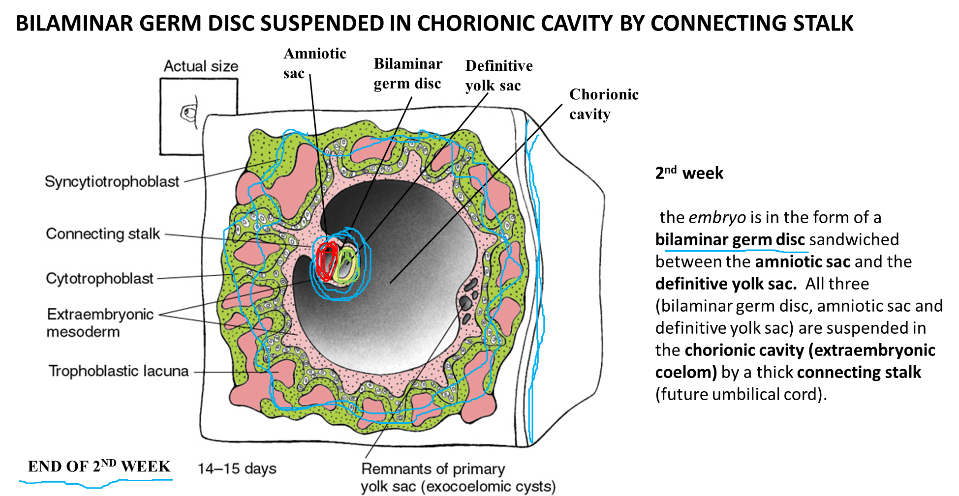
Following fertilization in the 2nd week the embryo will form the bilaminar germ disc
→ the bilaminar germ disc is sandwiched between the amniotic sac and the yolk sac
→ both of the above structures are suspended in the chorionic cavity by a thick connecting stalk that will act as the future umbilical cord
What happens to the amniotic sac during development?
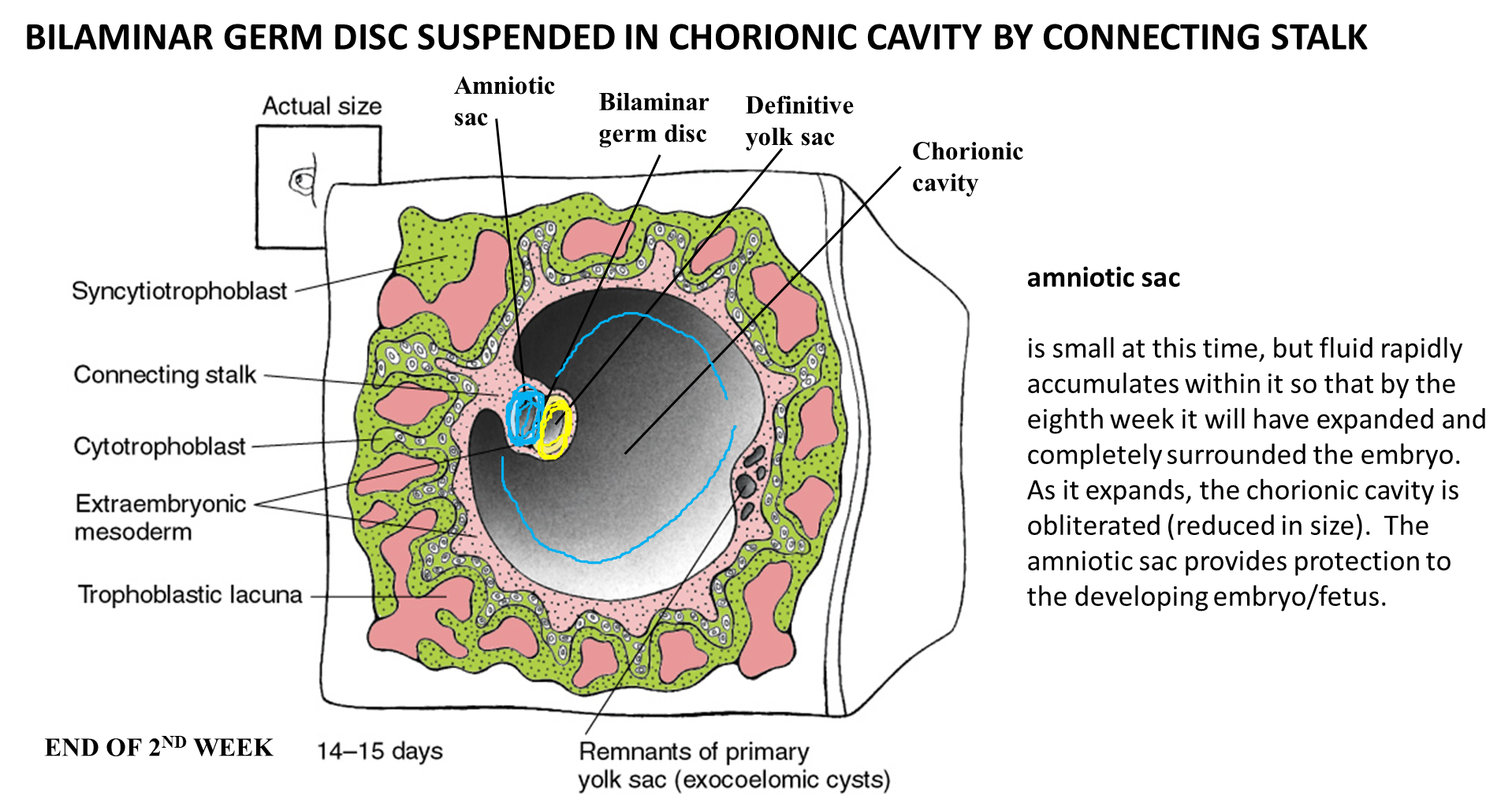
The amniotic sac will rapidly fill with fluid, so that by the eighth week the sac will have expanded and surrounded the entirety of the embryo
→ as the amniotic sac expands, the chorionic cavity reduces in size
What occurs during the third week of development?
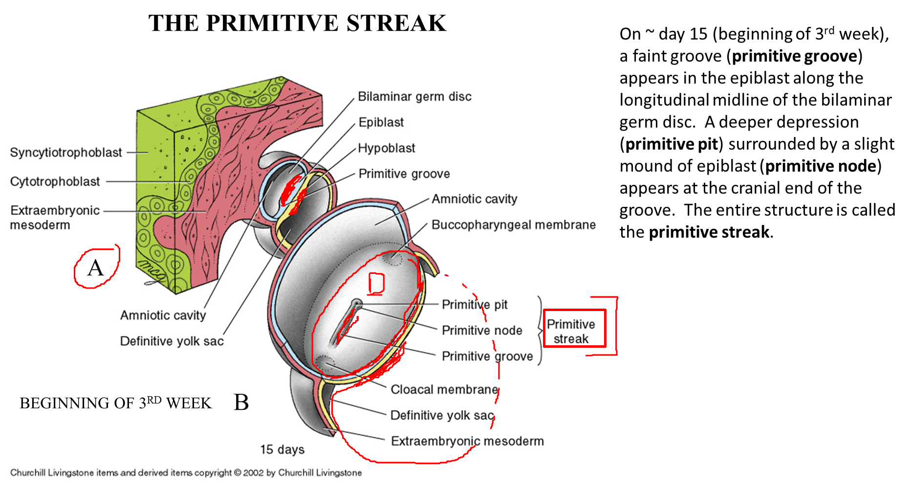
Gastrulation occurs during the third week of development where the epiblast will form the three primary germ layers
→ this occurs when the epiblast cells located in the primitive streak will move downward and form the three germ layers
What happens to the Lateral Plate Mesoderm
Lateral Plate Mesoderm
→ this subdivision of the mesoderm will subdivide into a somatic layer which lines the body wall and a visceral/splanchnic layer which lies on the organs themselves
→ in between these two growing layers is the intraembryonic coelom which will eventually become the body cavity
How does the gut tube develop?
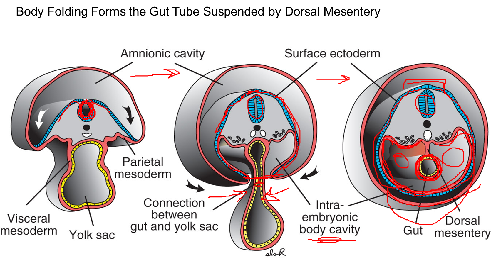
During the fourth week of development
→ as the neural tube begins to close, the surface ectoderm will begin to wrap around the sides of the developing embryo, eventually becoming a continuous circle after fusion on the ventral midline
→ as the lateral walls of the embryo grow around, it will pinch off the yolk sac forming the gut tube which is lined by lateral plate mesoderm
at the same time, cranial caudal folding is occurring as well
How does the mesentery develop in the embryo?
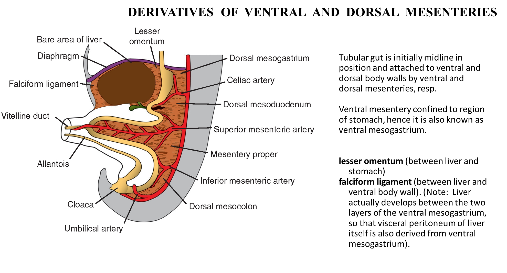
The endodermal tubular gut is initially midline and is attached to both the ventral and dorsal body walls via the dorsal and ventral mesenteries
1) the mesenteries are derived from mesoderm
→ in an adult, the dorsal mesentery is where the majority of our mature mesentery is derived from
→ in an adult, the ventral mesentery will be more modified and disappears becoming the lesser omentum and falciform ligament
What does the dorsal mesentery in the embryo go on to form?
1) Dorsal Mesentery
In the stomach the dorsal mesogastrium will go on to form two derivatives
→ Greater Omentum
→ Splenorenal Ligament
In the intestine, the dorsal mesentery will form four major structures
→ The mesentery - supporting structure of the ileum and jejunum
→ Mesoappendix - mesentery for the appendix
→ Transverse and Sigmoid mesocolon
2) importantly, in the intestinal tract only the transverse and sigmoid colon develop mesentery because the mesentery of the ascending and descending mesocolons disappear as we develop
What is the difference between intraperitoneal, retroperitoneal, and secondarily retroperitoneal
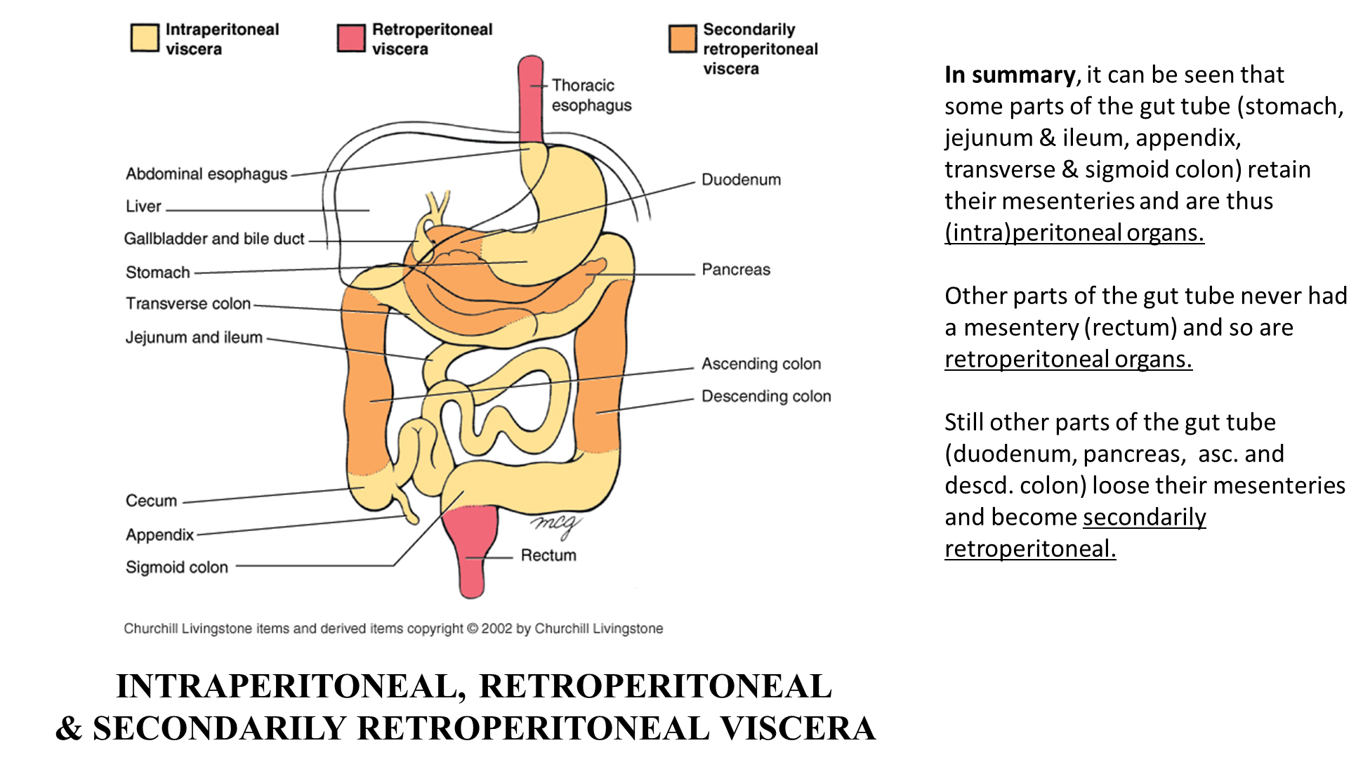
1) Retroperitoneal Organs are organs that have never had a mesentery attached
2) Intraperitoneal organs will still have their mesentery attached
2) if an organ is secondarily retroperitoneal, it means that these structures have lost their mesentery
→ duodenum, pancreas, ascending and descending colon
How does the stomach develop in the embryo?
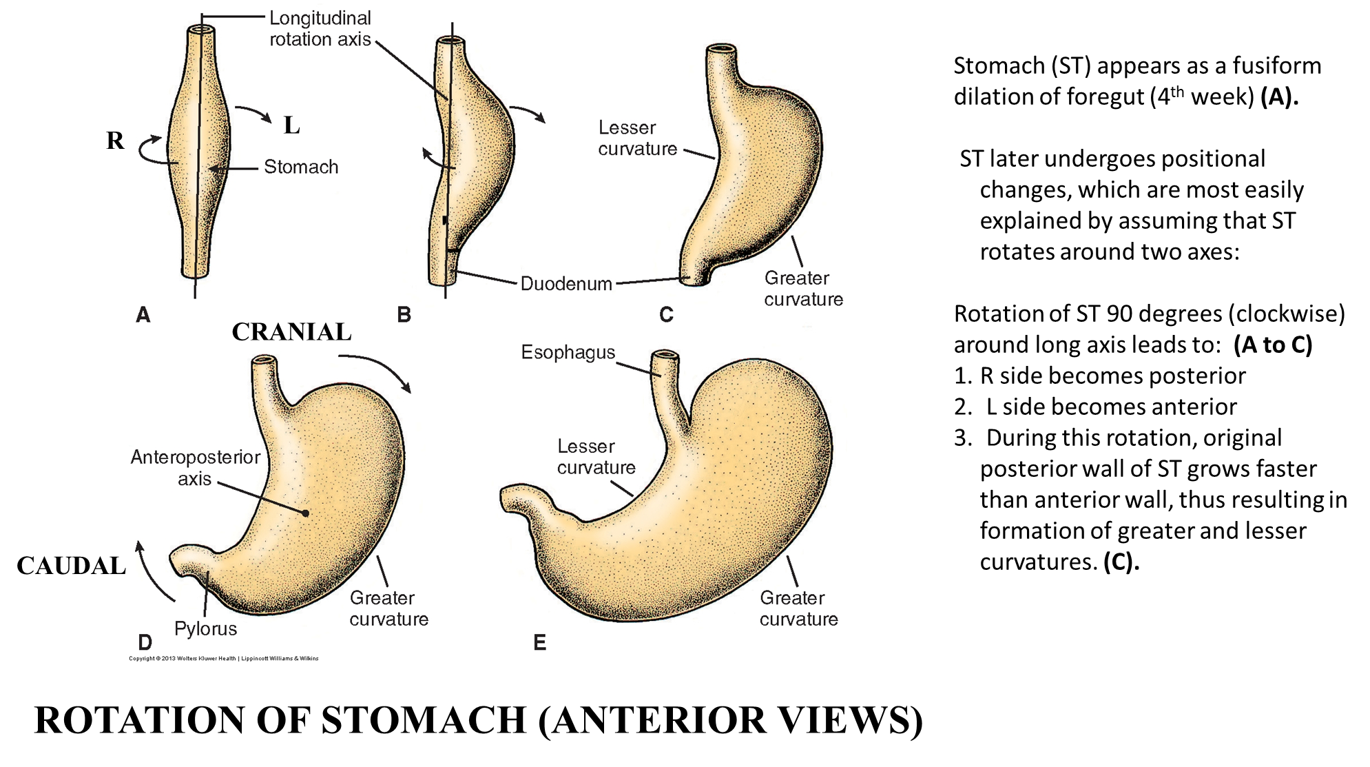
The Stomach - begins at the fifth week of development in four stages
1) The gut tube begins to expand, especially on its posterior side
2) The gut tube will begin to rotate longitudinally causing the anterior side of the stomach to point right and the posterior side of the stomach to point to the left
3) The gut tube will continue to expand, with the posterior portion (now facing left) continuing to bulge
4) The stomach will then rotate on its anteroposterior axis, essentially leaning backward with the superior portion rotating down to the left and the inferior portion rotating up to the right
→ establishes the final of the stomach
What is a pyloric stenosis
Pyloric stenosis occurs when the circular muscle of the pyloric region becomes hypertrophied (overgrowth of muscle)
→ this causes severe vomiting due to the narrow lumen of the pylorus, but the vomit will lack bile
What organs will also move following the formation of the lesser sac?
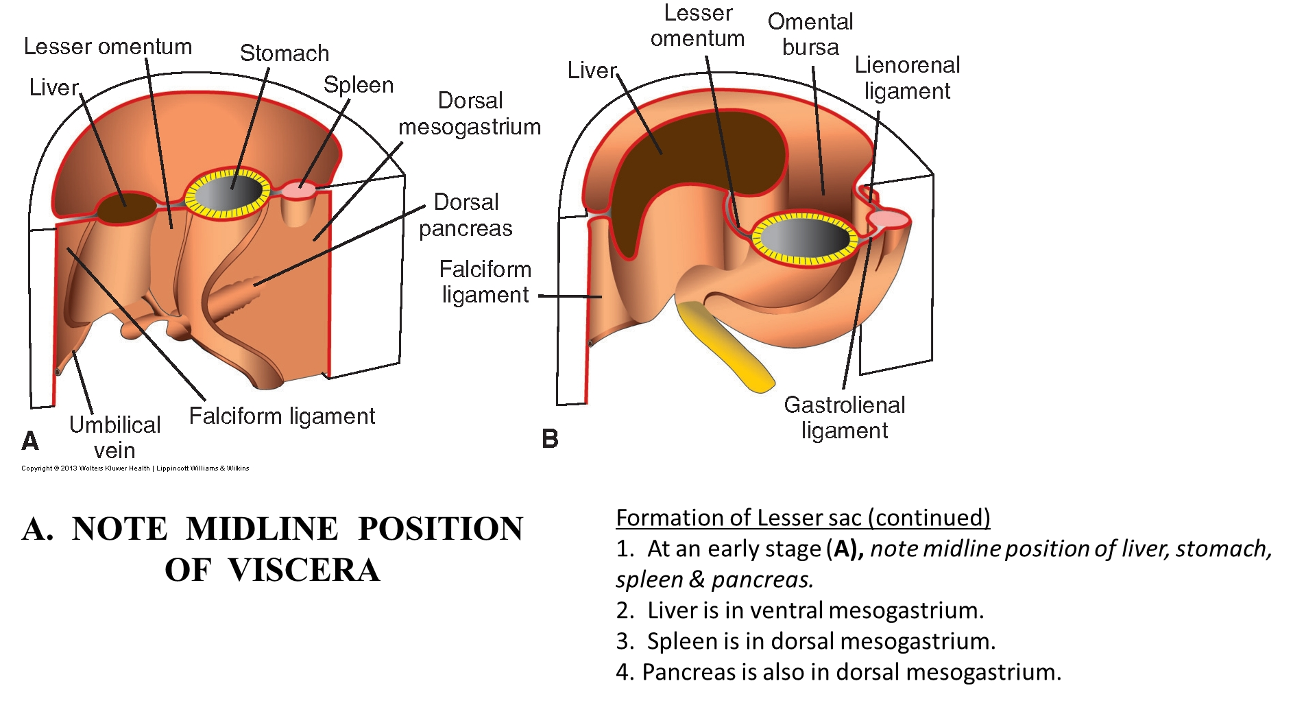
The lesser sac will form in response to rotate of the stomach, moving the mesenteries as it rotates. This rotation of the stomach and its mesentery will move the following organs
→ Liver is in the ventral mesogastrium and ends up on the right side
→ Spleen is in the dorsal mesogastrium and ends up on the left side
→ also causes the pancreas to become secondarily retroperitoneal
this rotation will also form the omental bursa also known as the lesser sac
How does the liver develop?
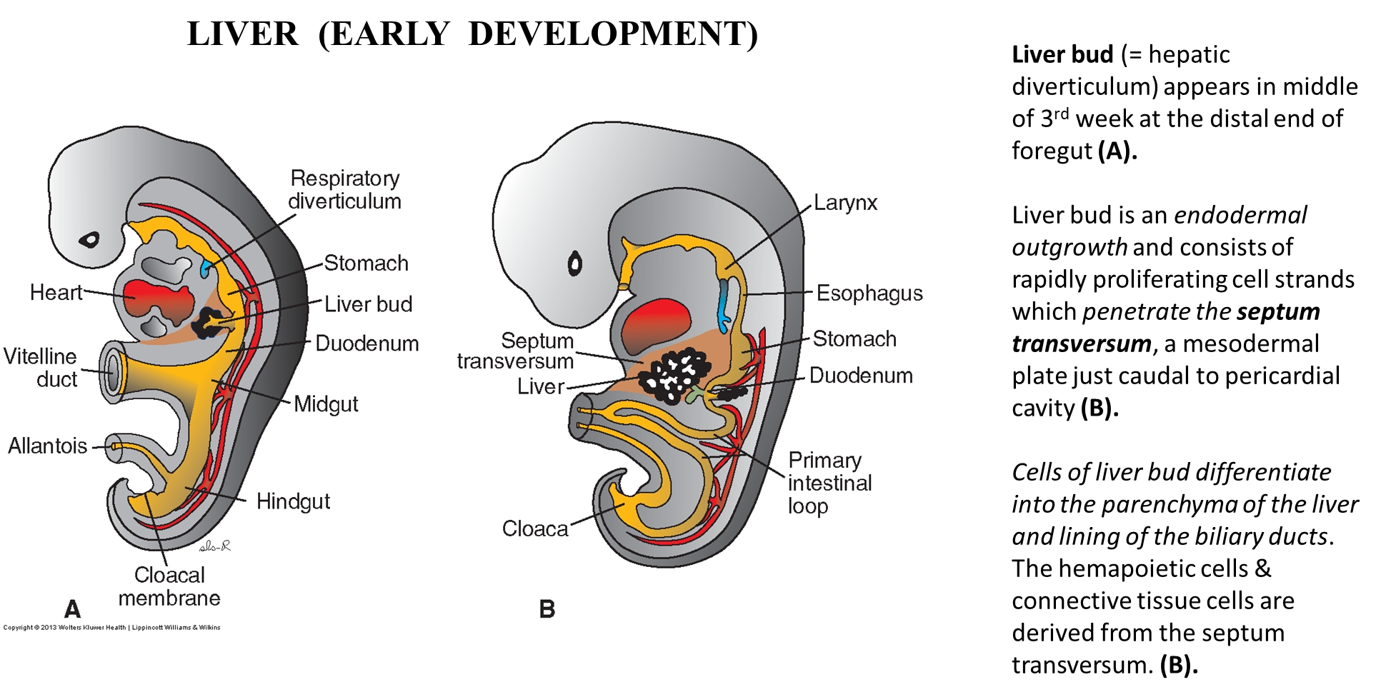
1) Liver will develop as a small pocketing of the foregut known as the liver bud or hepatic diverticulum
→ the liver bud is a endodermal outgrowth which proliferates outward and penetrates the septum transversum which is the developing diaphragm
→ the liver buds will differentiate into the different parts of the liver and forming the biliary tree
2) originally it is a hematopoietic organ that will eventually start making bile at 12 weeks of development where it will eventually lose its hematopoietic function
How does the gall bladder develop
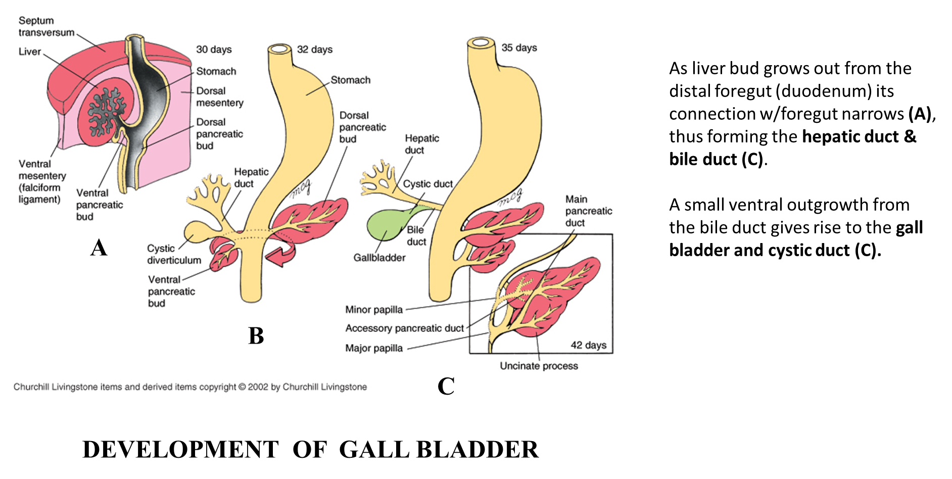
Occurs simultaneously with liver development
→ as the liver bud grows out from the distal foregut (duodenum) its connection with the foregut will narrow out
→ this narrowing will form the hepatic duct and bile duct
→ a small ventral outgrowth from the bile duct will give rise to the cystic duct and eventually the gall bladder
How does the pancreas develop?
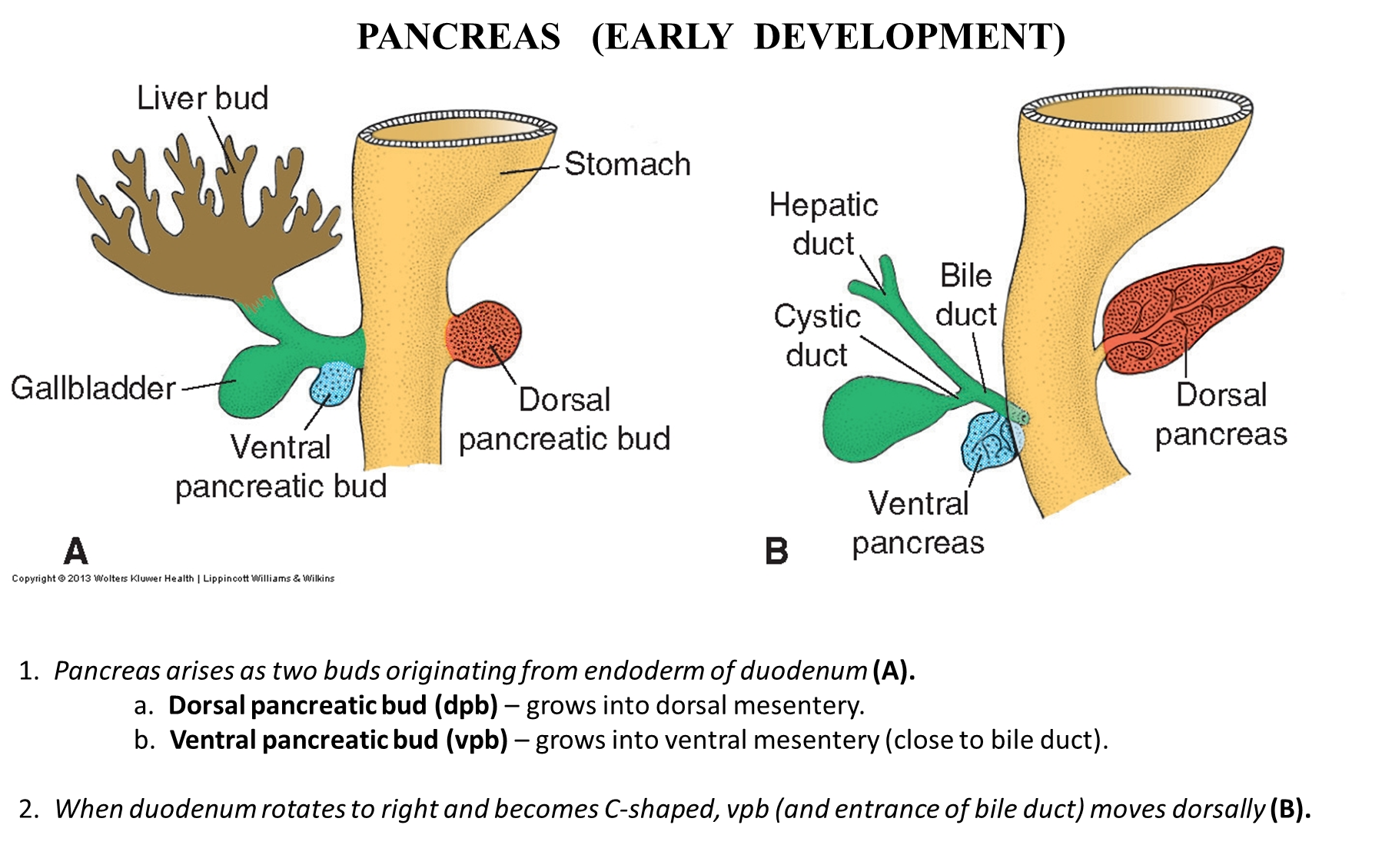
1) The pancreas will arise from two buds originating from the endoderm of the duodenum
→ dorsal pancreatic bud - grows into the dorsal mesentery
→ ventral pancreatic bud - grows into the ventral mesentery
2) these two buds grow on opposite sides of the duodenum
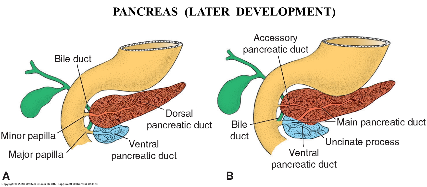
→ the ventral pancreatic bud will then grow around from the right side to the left posteriorally in order to join together with the dorsal pancreatic duct
→ the dorsal pancreatic duct will deviate downward to connect with the ventral pancreatic duct (which is the main one) and forming the main pancreatic duct
What is Annular Pancreas?
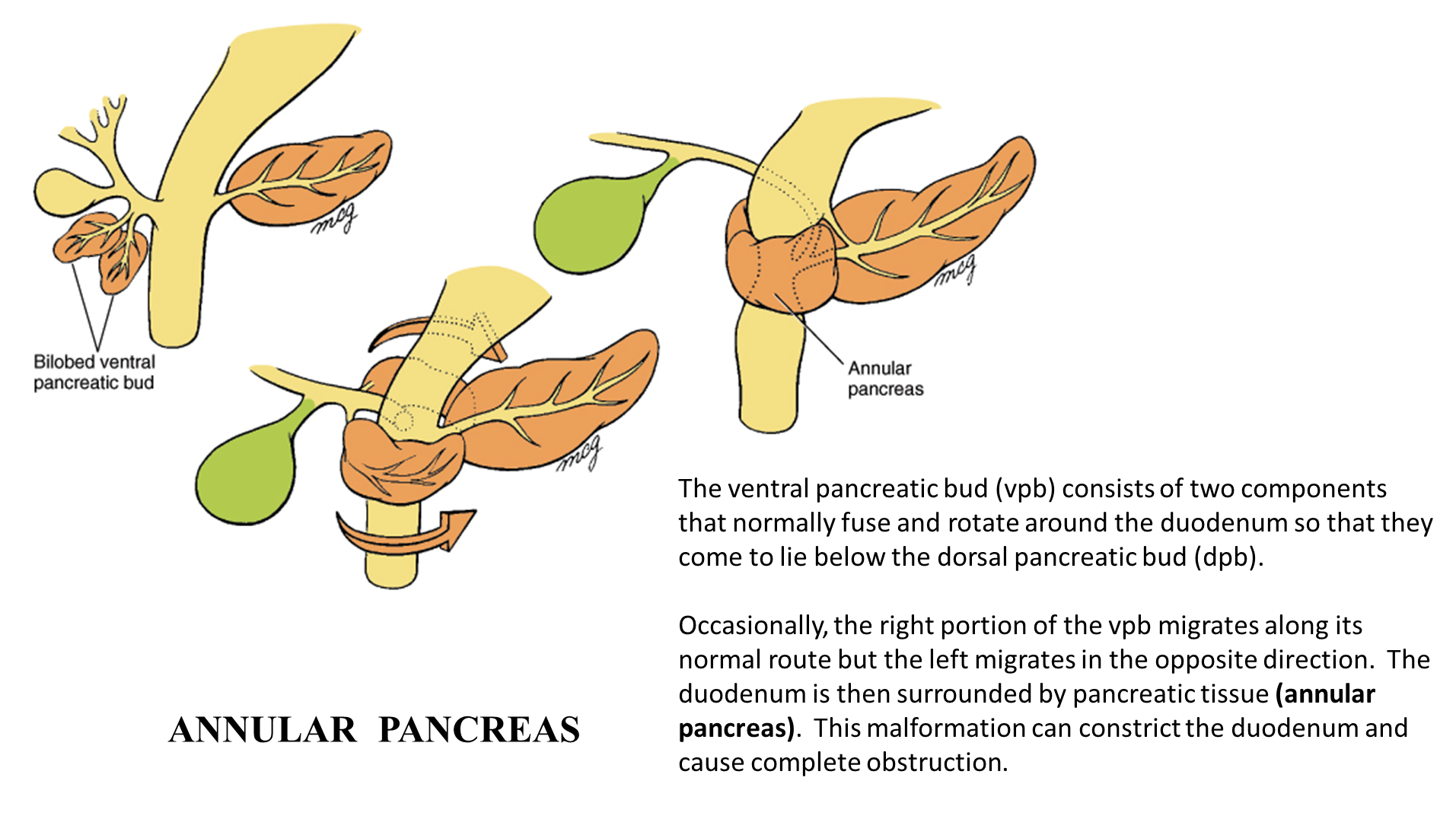
rare congenital condition where a ring of pancreatic tissue surrounds the duodenum
→ this can occur when we accidentally have two ventral pancreatic buds which can split off and loop around the duodenum in opposite directions forming a ring
→ can also occur if the ventral pancreatic bud grows around anteriorly
→ may cause duodenal stenosis which cause vomiting with bile present
What is herniation of the midgut loop
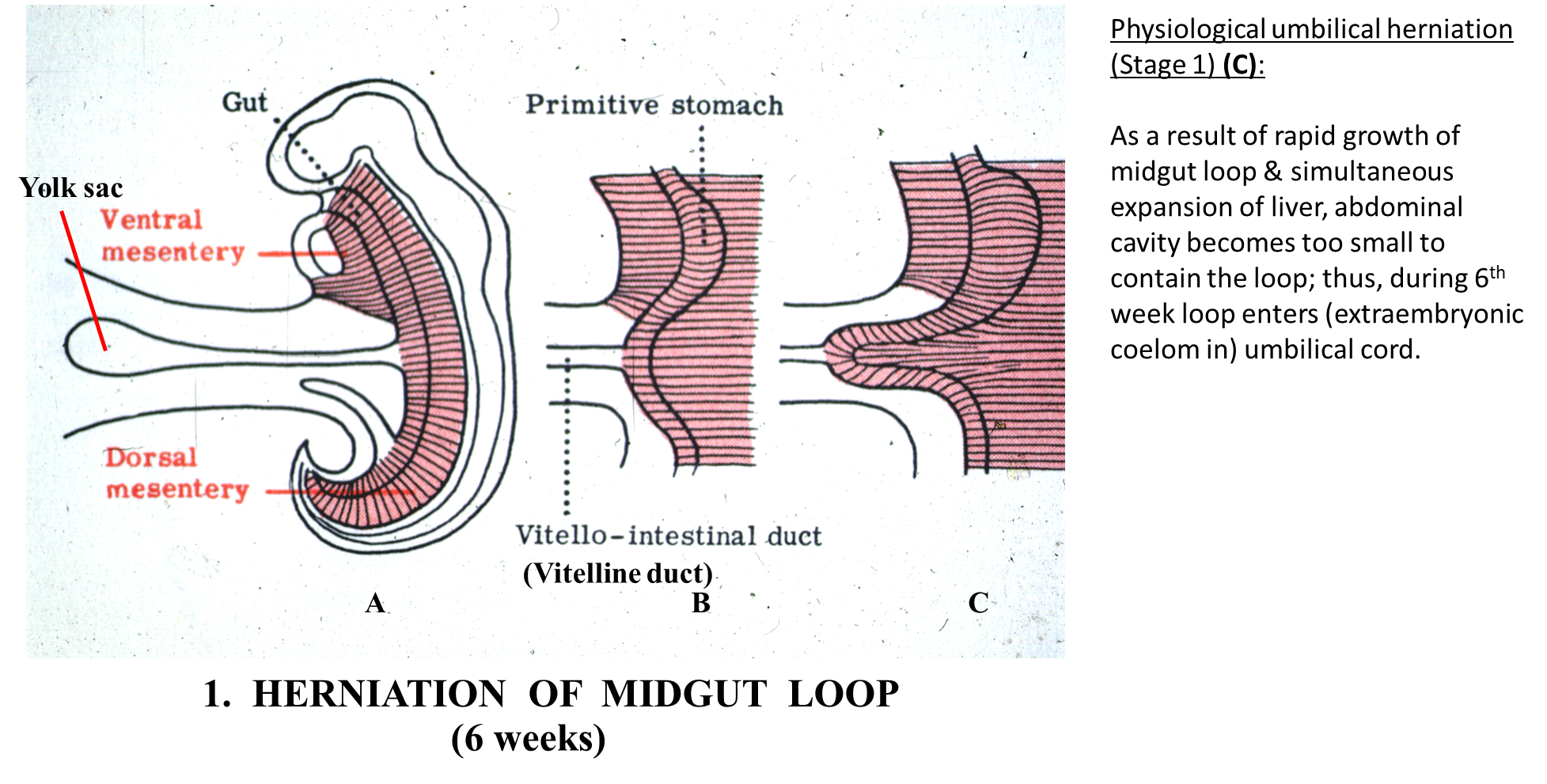
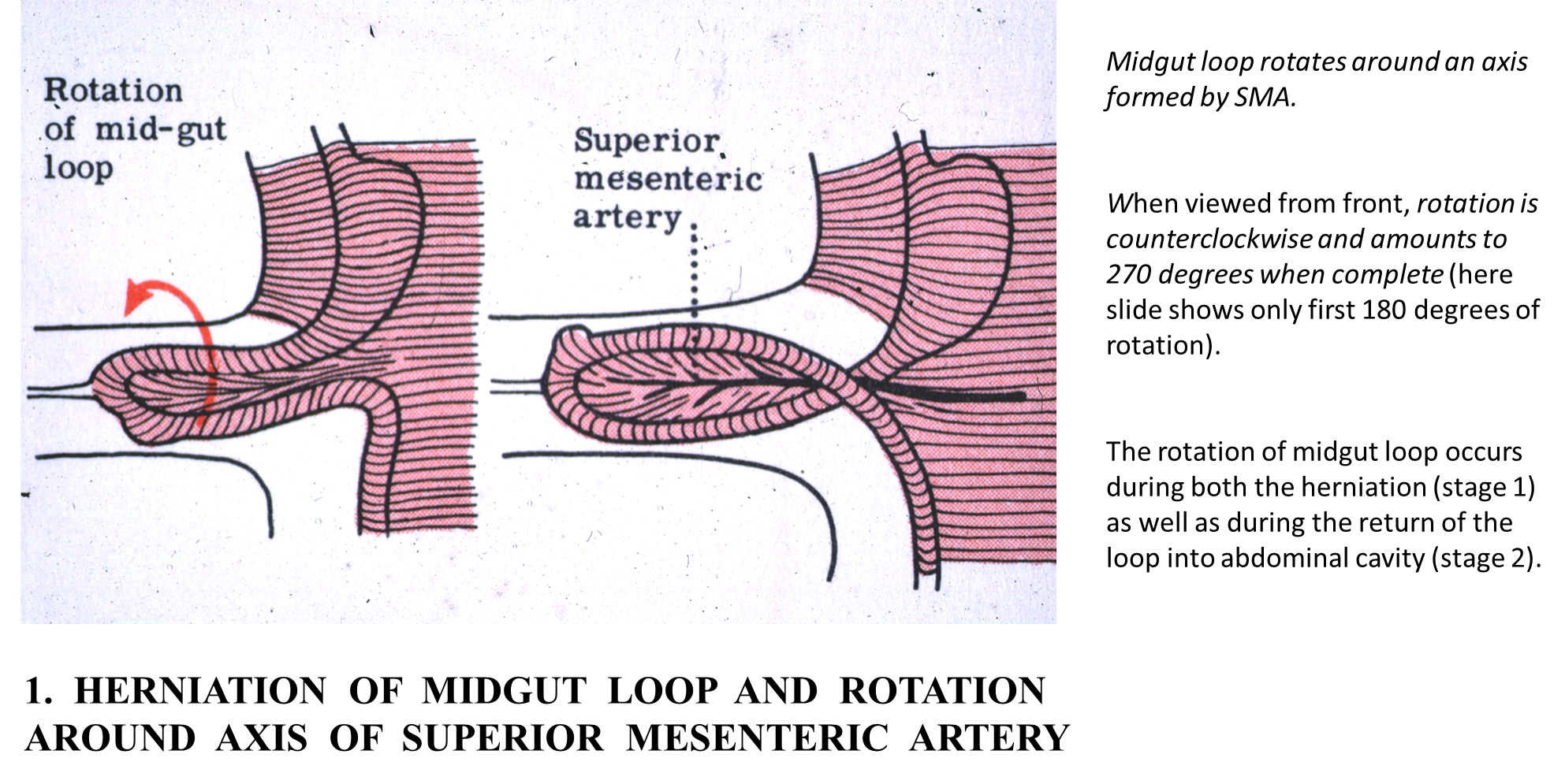
1) The midgut will begin to elongate causing the midgut to loop outward and protrudes outward into the umbilicus establishing several structures
→ the vitelline duct the connection between the loop and the yolk sac is at the apex of this loop
→ the upper part of the loop will be the cephalic limb giving rise to the majority of the small intestine
→ the lower part of the loop will be the caudal limb and forms the majority of the large intestine
2) This elongation will cause the loop to herniate outward (6th week)
→ after it has herniated outward it will rotate counter clockwise 180 degrees around the superior mesenteric artery
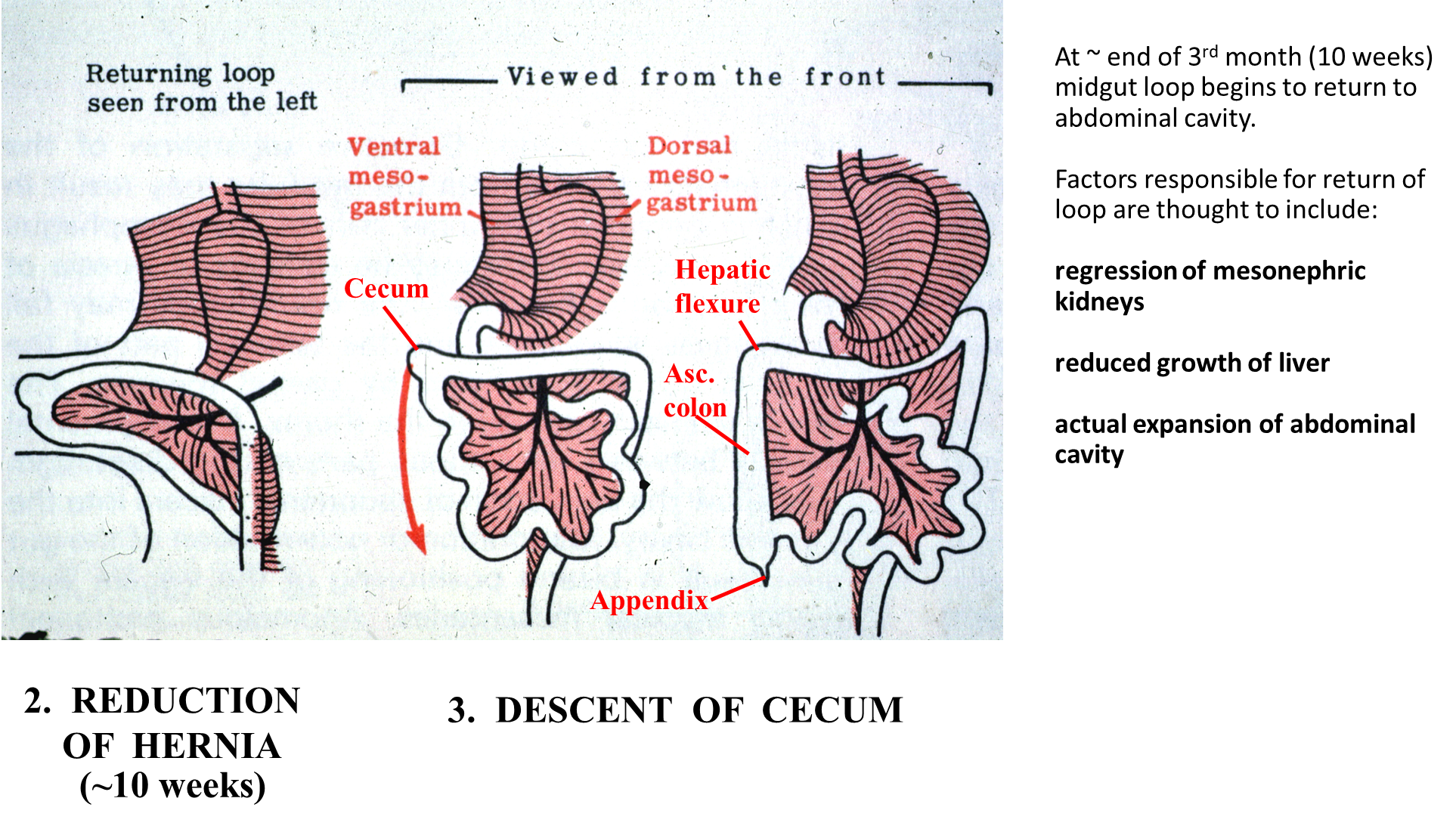
3) the herniated midgut loop is then brough back into the abdominal cavity (10th week)
→ the jejunum is pulled back in first and ends up on the upper left quadrant
→ the loop is then rotated downward 90 degrees, placing the cecum in the correct position
→ this causes the small intestine to be superficial
4) the intestines will then become fixated with some of them losing their mesenteries
What three factors are responsible for the return of the loop into the abdominal cavity?
1) Regression of mesonephric kidneys
2) reduced liver growth
3) actual expansion of the abdominal cavity
What is Fixation of the Intestines?
As the intestines return to the abdominal cavity, some of the mesenteries are pressed against the posterior abdominal wall where fusion occurs
1) ascending and descending colon lose their mesenteries becoming secondary retroperitoneal
2) transverse colon remains mobile but its mesentery fuses with the posterior layer of the greater omentum
3) sigmoid colon retains its mesentery
How does the greater omentum form?
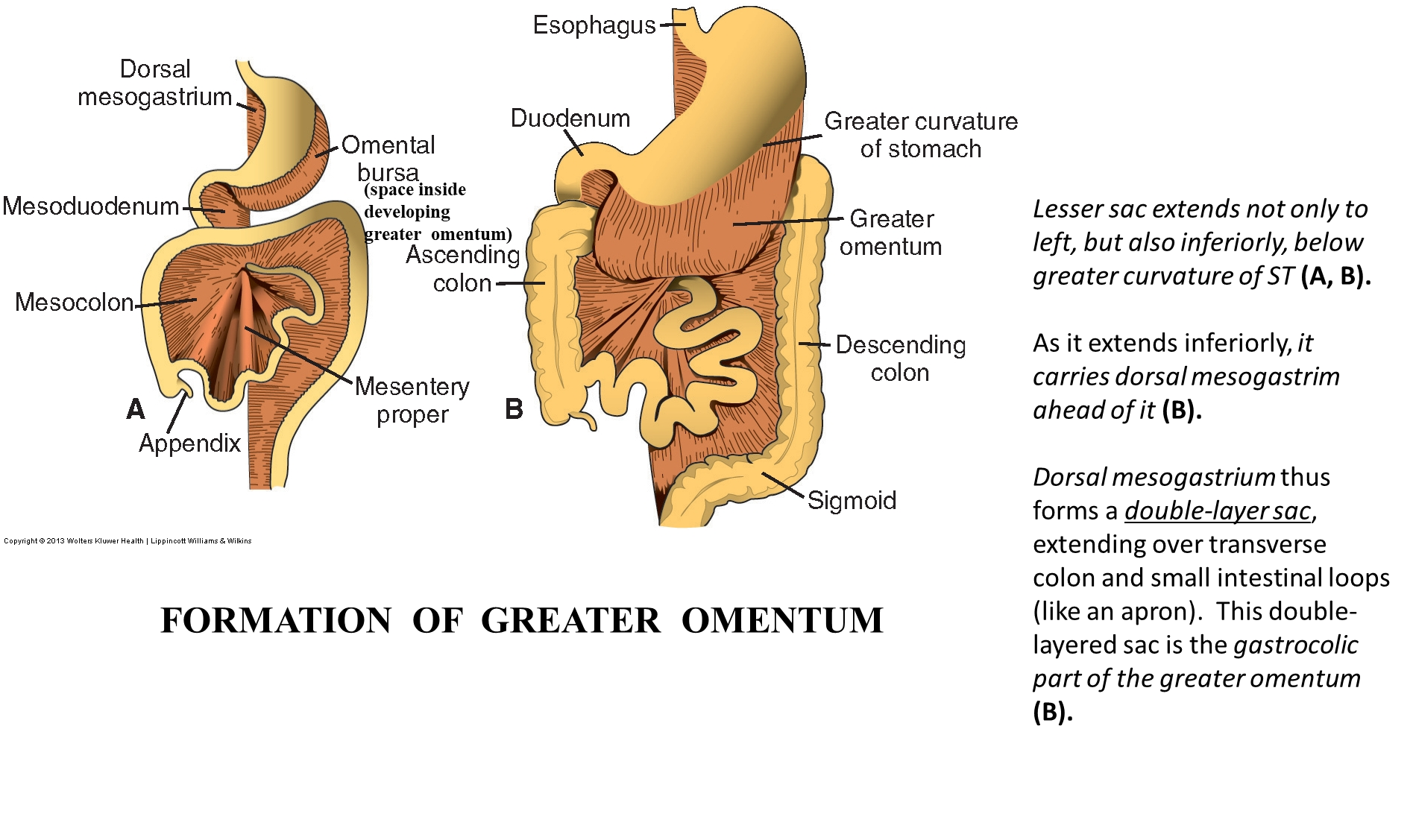
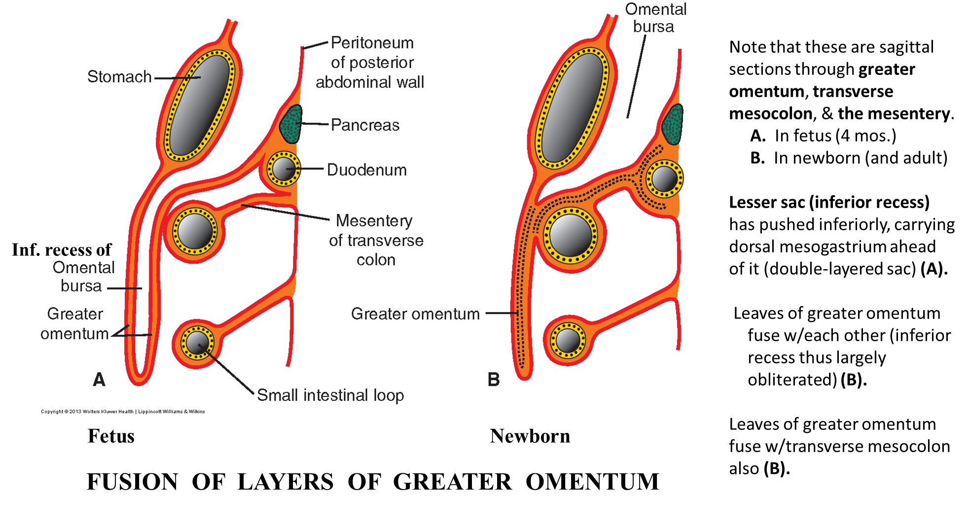
As the stomach rotates, the dorsal mesogastrium is carried and extends becoming a double-layered sac
→ the layers of the greater omentum will fuse with the mesentery of the transverse colon
→ the leaves of the greater omentum will also fuse together
What is an Omphalocele
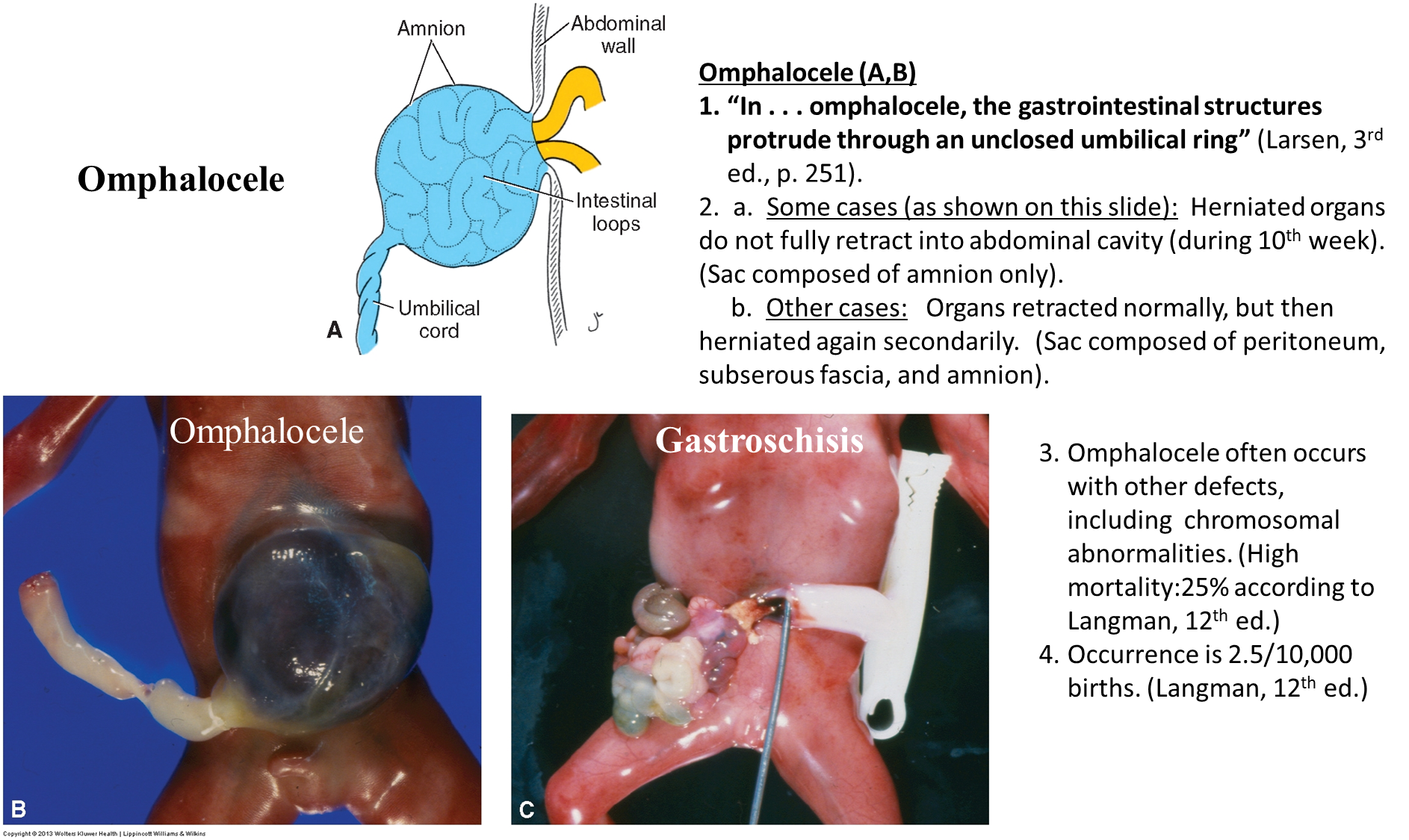
Omphalocele is when the midgut herniates outward into the umbilicus, but fails to go back into the body
→ primary - organs only covered with amnion
→ secondary - organs will herniate again covered with both serous membrane and amnion
Often associated with chromosomal abnormalities
What is Gastroschisis?
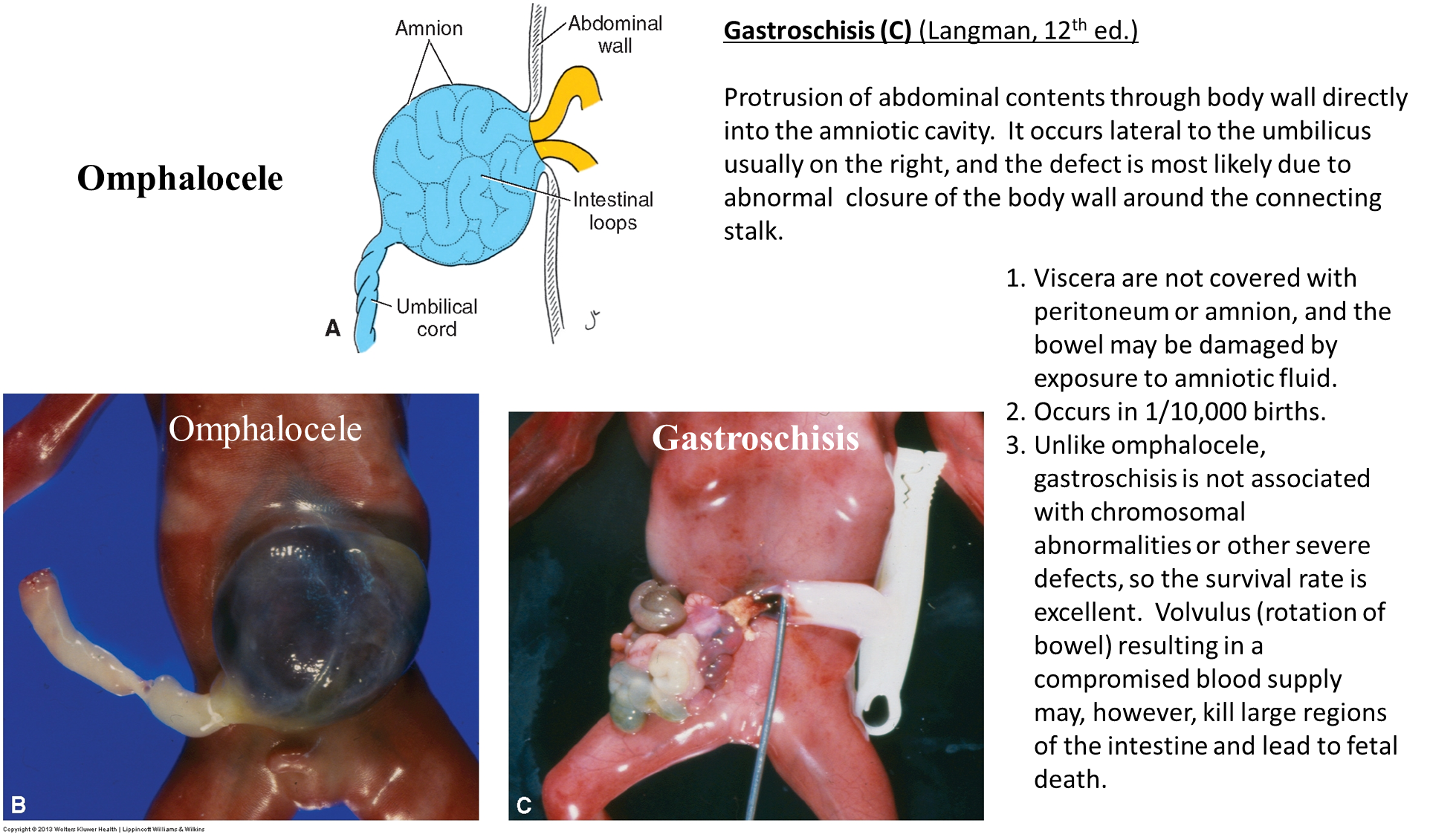
Gastroschisis is a hole in the abdominal wall, typically on the ventral right side
→ not associated with chromosomal abnormalities and is a issue of the body wall and not the GI tract
What are the three vitelline duct errors
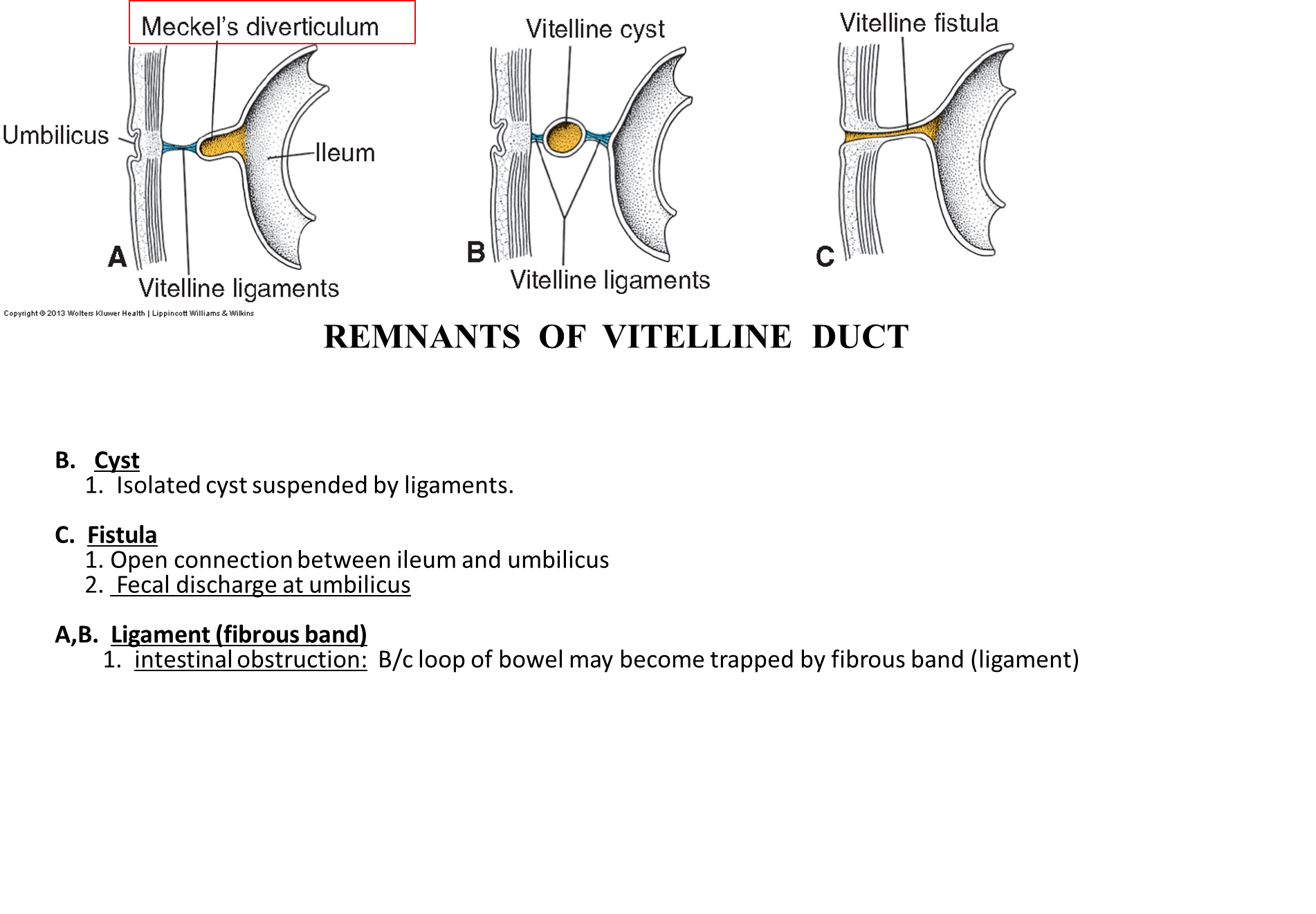
The Vitelline Duct will usually regress between the 5th and 8th weeks, but can often persist
1) Vitelline Fistula
→ the vitelline duct remains patent and connects the shit tube to the umbilicus
2) Vitelline Cyst
→ the vitelline duct will close on both sides but retain in the middle
→ forms a cyst in the middle
3) Meckel’s diverticulum
→ distal end of the duct closes while the proximal end stays open
What is Nonrotation of the midgut loop and reversed rotation of the midgut loop
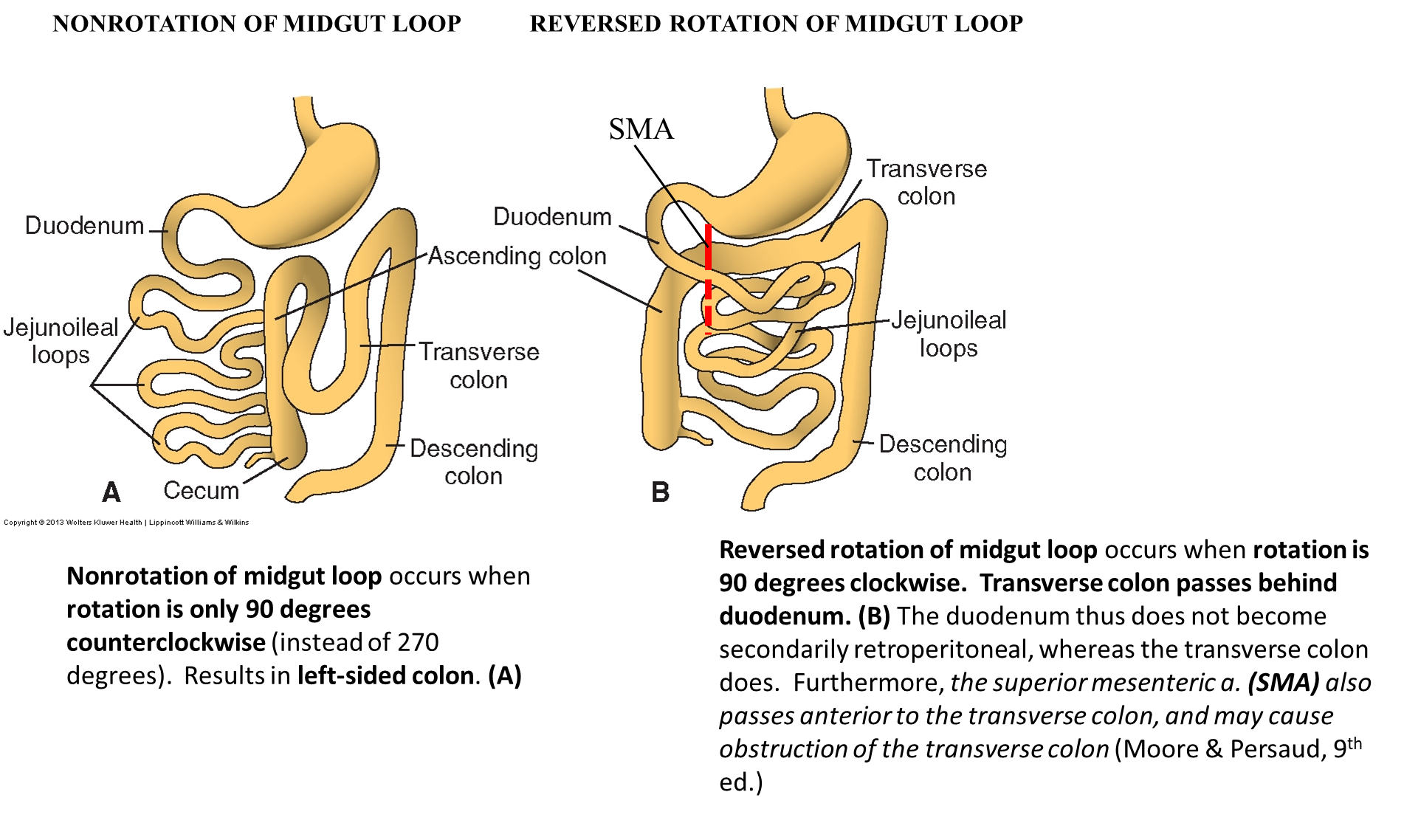
Nonrotation of the midgut loop
→ occurs when rotation of the gut tube is only 90 degrees
→ makes a left sided colon
Reversed rotation of the midgut loop
→ when rotation occurs 90 degrees clockwise instead of 180 degrees coutnerclockwise
→ causes the transverse colon to pass behind the duodenum and be retroperitoneal