Neuroscience: week 7: ADHD: a new theory
1/63
There's no tags or description
Looks like no tags are added yet.
Name | Mastery | Learn | Test | Matching | Spaced |
|---|
No study sessions yet.
64 Terms
Previous theories: in terms of ‘classical’ theories
Many psychiatric and neurological disorders are associated with frontal cortex dysfunction (from Sz to parkinson’s)
Another theory is that newer drugs, like atomoxetine and guanfacine, affect noradrenaline not dopamine, admittedly these drugs are not as effect as other stimulants, they are still effectively
Frontline treatments themselves have a lot of problems: DL-amphetamine or methylphenidate
both are Class B drugs according to the Misuse of Drugs Act (1971)
they have the potential for significant abuse of use, and yet we are giving them to children
this probably suggests that we need a new generation of therapy/ drug therapy
We need a new approach to ADHD
When people have thought about this, they often think incorrectly
there are several presentations of ADHD (inattentive, hyperactive/impulsive, combined)
so it may not be productive to look for 'single ‘causes’ or single therapeutically relevant actions of drugs
the lecturer thinks you should pick a specific ‘core’ symptom and go from there but idk
Since the work of A.A Staruss in the 1940’s and 50’s, distractibility has been considered key symptoms of ADHD
even now if you go to the DSM 5, it outlines a core aspect of the disorder as ‘is often easily distracted by extraneous stimuli’
another reason it is important or a useful focus is that: substrate mediating distractibility is well known
people often get distracted by trivial things and often divert attention from more important things, so actually focussing on this is also helpful in general
SC and distractibility
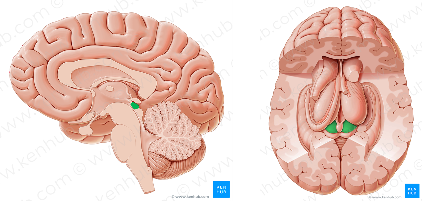
Distractibility is intimately linked with the superior colliculus
the SC sits in the dorsal/ back area of the midbrain and seems to be responsible for the act of pulling you away from what you are focussing on to something else
the SC receives a lot of information from the retina,
it is part of the most important subcortical visual system
SC seems to be at least partially involved in highly conserved eye movements/ saccades
if you lesion the colliculur area in rats, cats and monkeys, this has resulted in a decrease of distractibility
Goodale and Murison (1975)
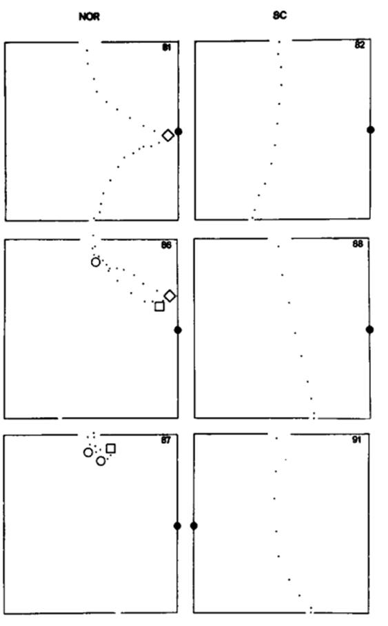
one rat had to put its head through the luminated door, open it and gets a reward
did this repeatedly
then one time when the rat ran, there was a flashing light not seen or displayed before to the side
each dot is the movement of the rats nose, so we can see in the example, the first animal sees the new flashing light, goes to it, investigates it and then completes the task
however, when you do this with a rat that had a removed colliculus they don’t even notice the flashing light and go straight past it
which demonstrates a reduction in distractibility
this isn’t just in rats though
SC and humans- Gaymard et al. (2003)
this still appears to be functional in humans
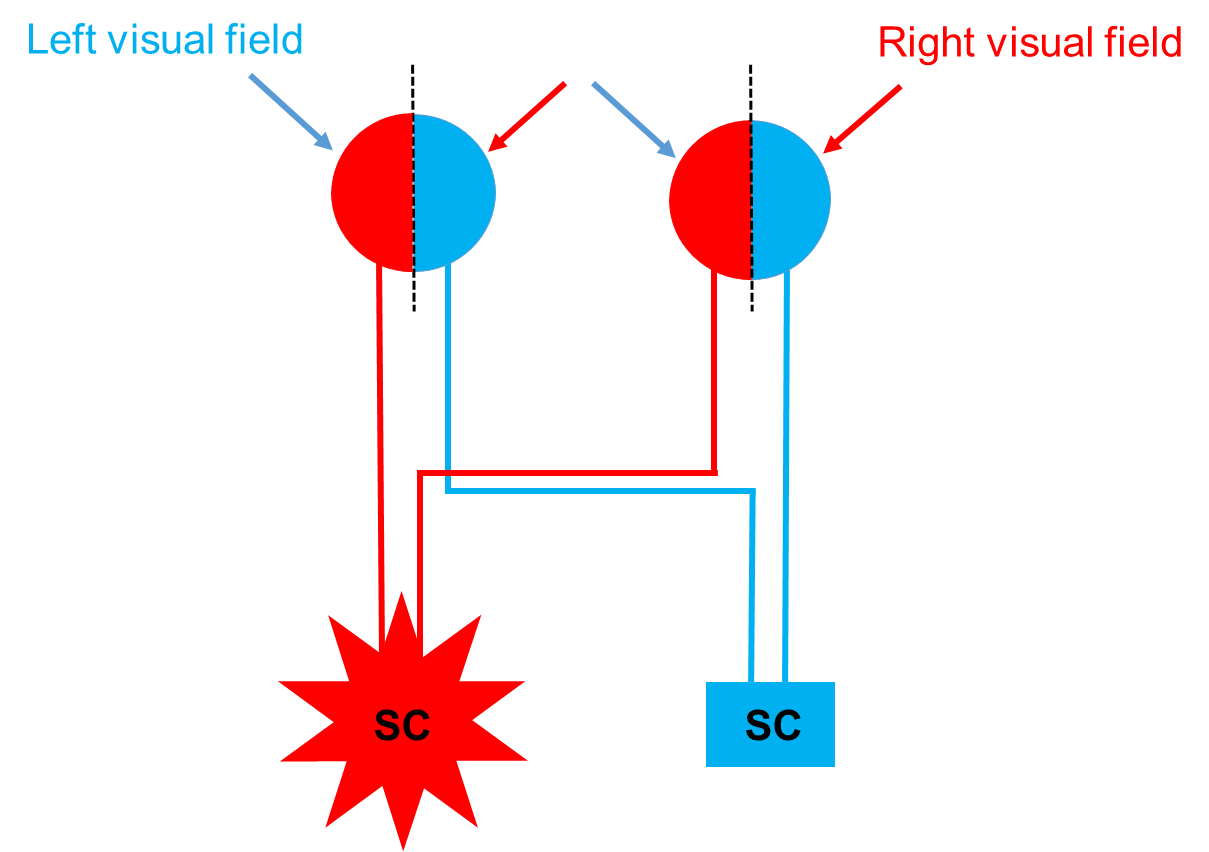
this 51 year old woman with a lesion affecting the projection from the cortex to the SC (prefronto-tectal tract) on the left hand side
lesion is in the inferior colliculus, not really relevant but still but just to show it till goes through the pathway that controls the SC
what did that do behaviourally?
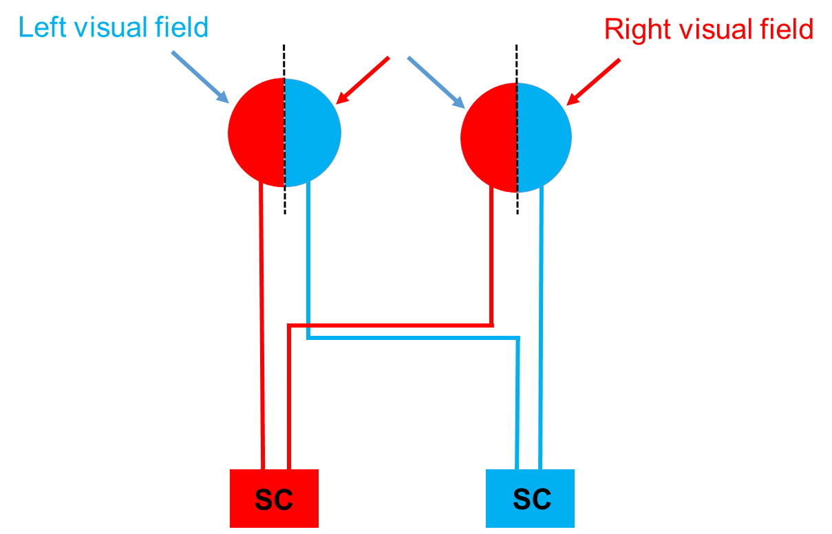
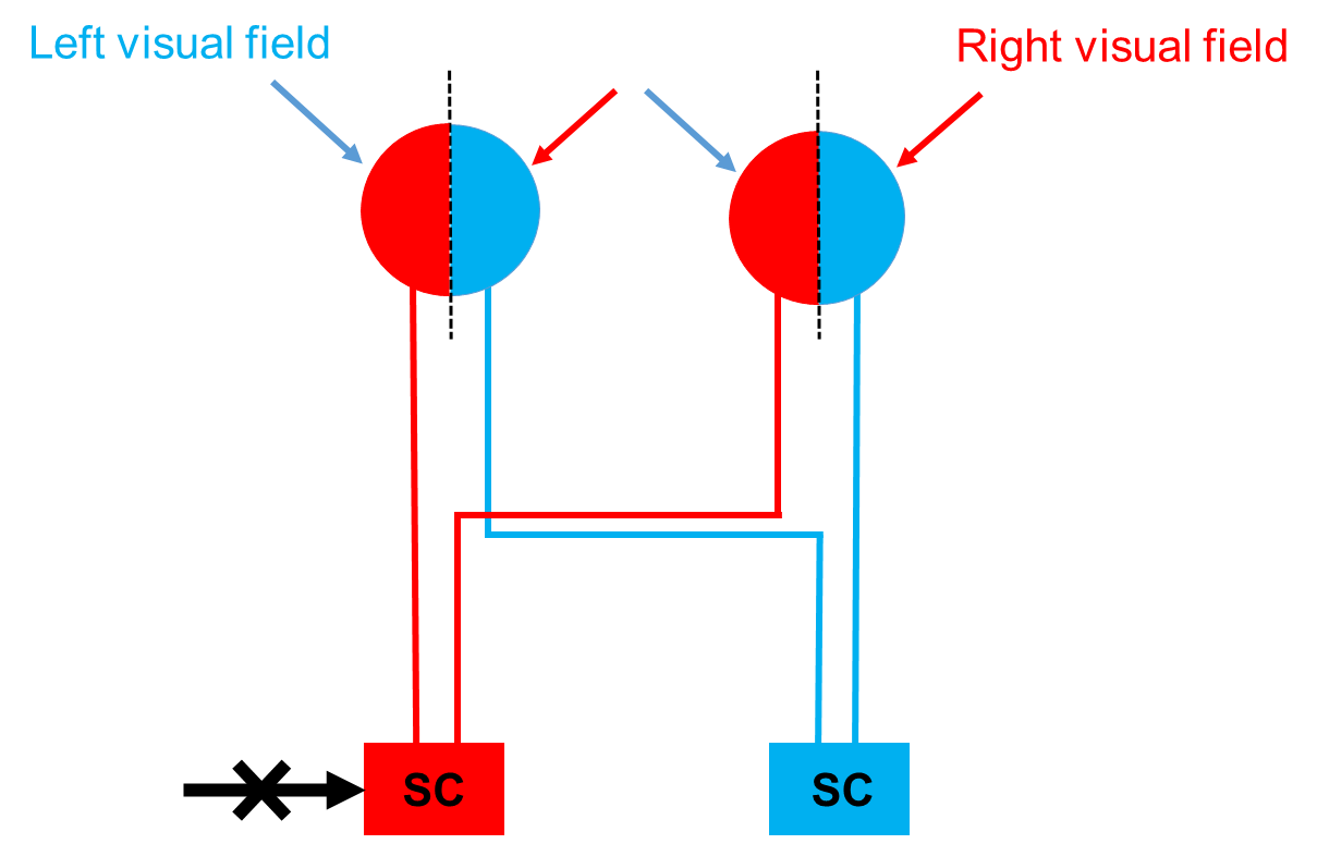
this is a representation of your brain’s hemispheres
the right hand side colliculas, they receive info from the temporal retina on the LEFT side of the head and you need to go back and hear what he said because i am confused
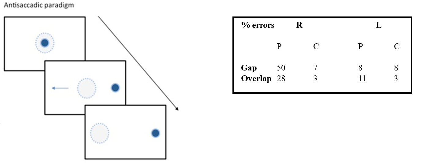
in the experiment look at the blob in the middle of the screen, then it will move in each trial and you have to move your eye in the opposite direction, you could have to pause before moving you eye or straight away
this is sometimes called an anti-saccade
the study tests whether the behaviour will be different for looking to one side than to the other for the 51 year old woman
SC and humans- Gaymard et al. (2003) results
the results show the number of errors
when the blob moved to the right, she made a huge number of errors, she basically couldn’t stop looking at the blob
whereas on the left side, her ability and results were similar to the control group
it seems as though the woman’s right side SC now failed to have the ability to disinhibit a saccade,
making it too responsive to sensory stimuli
What did Gaymard et al. (2003) bring forth?
This brings a new theory:
distractibility in ADHD reflects a hyper-responsive colliculus
background for the superior colliculus
Vertebrate brains have had a superior colliculus for 500 million years
If distractibility in ADHD reflects a hyper-responsive colliculus, what sort of evidence is needed to support this?
At least 4 types
hyper-responsiveness in an animal model?
is there a direct connection from the colliculus to the brains ‘off’ switch/ interrupt system?
any collicular impairments in ADHD?
do ADHD treatments affect the coliculus?
Hyper-responsiveness in an animal model
brain cells encode information by ‘firing’
which allows a message to get sent from one structure and delivered to another
cells in sensory structures fire when an appropriate sensory stimulus is encountered
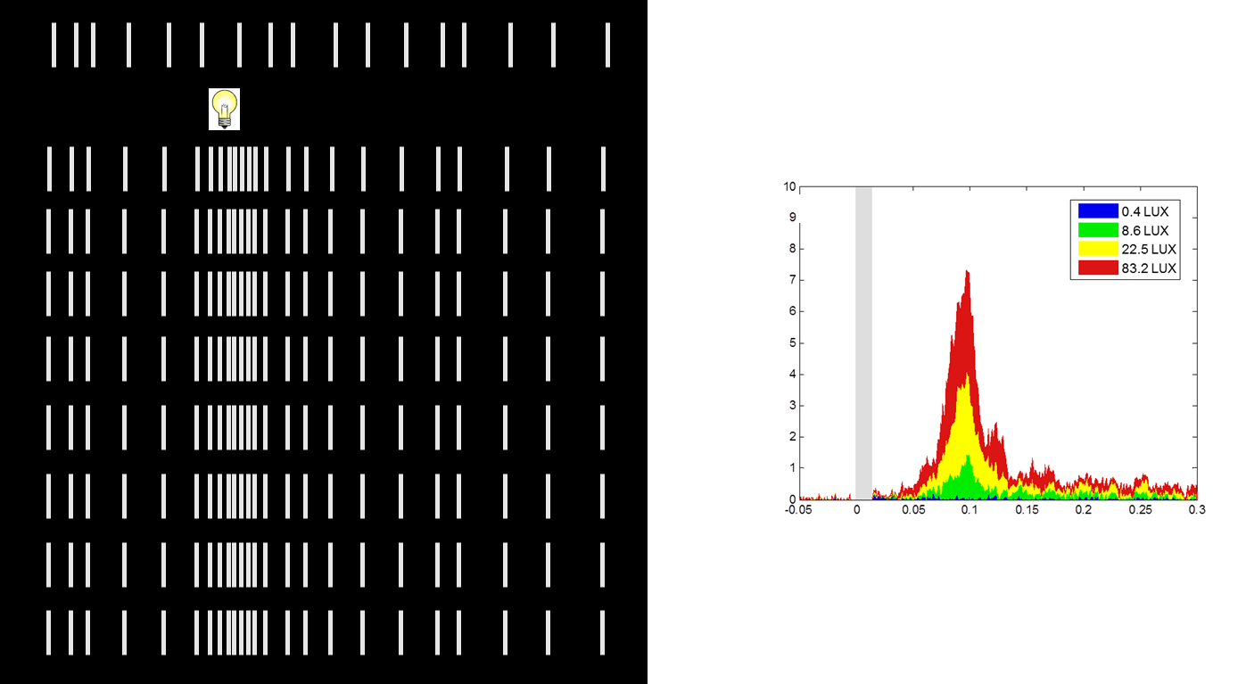
In the SC in the rat, the cells are spontaneously active (with no stimulus), what we are interested in is how the colliculus responds to the flashes of light, so what would happens if we flashed a light into the rats eye whilst measuring brains activity in the colliculus?
when flashing a light, there is an increase in the firing rate as seen in the black image
then it goes back down
the graph shows the response that this cell makes to flashes of various intensities
there is an intensity reliant variability in response
we use this paradigm to look at responses in the colliculus from flashing lights
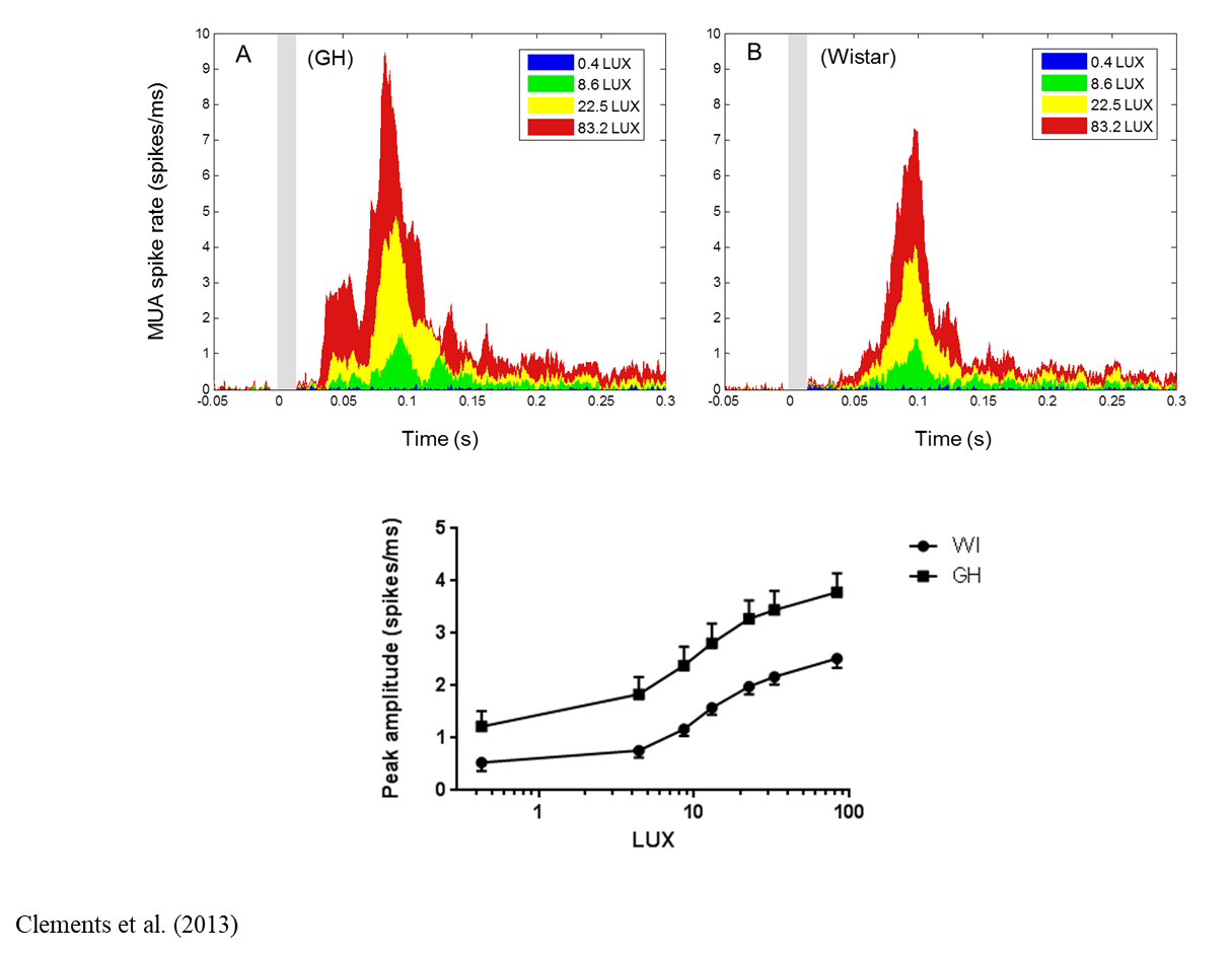
for each intensity of light, there is a bigger response in the colliculus (the higher line in the black graph) than in the control strain (the bottom line)
Hotline to the brain’s interrupt system?
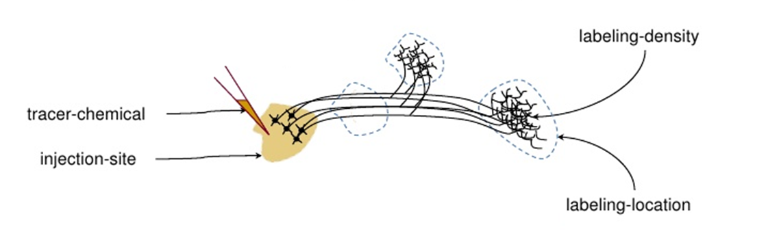
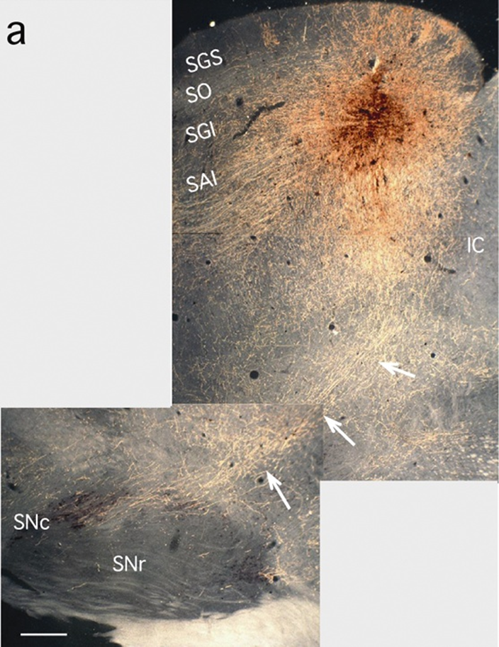
Using a neuronal tract tracer:
you inject the chemical locally in the area of cells bodies that we’re interested in studying
they transport it all the way down the axon to the terminal zone
we can use chemical reactions to demonstrate in the brain where these cells ended up
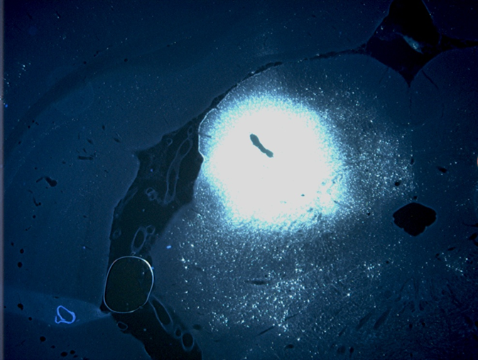
so we injected the SC
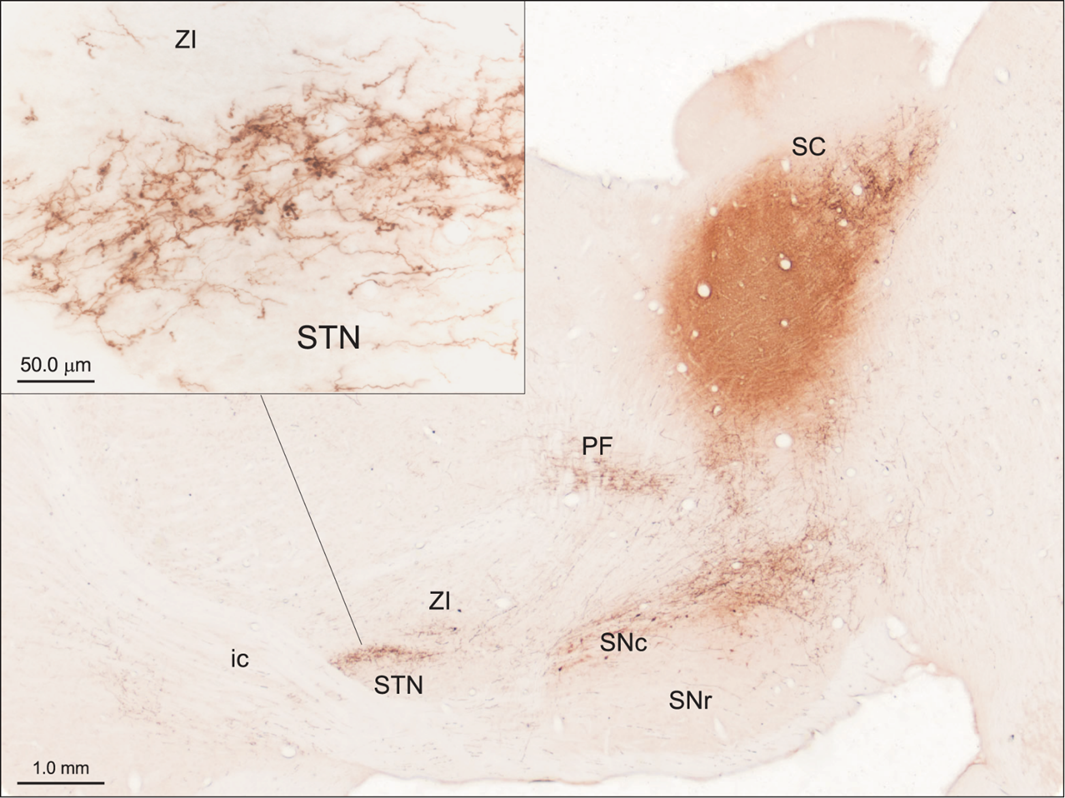
This is a rat brain that has been cut down the middle
the tract tracer is put in the dark pink area
the thin threads are the cells that contain the tracer
the lines demonstrate the movement of the cells to the synapse terminal
the SNc is the brain’s ‘off switch’
what this figure shows is that the SNc is the final destination in the transportation of cells from the SC
Any collicular impairments in ADHD?
Working on a clinical (children with ADHD) population in the UK is difficult
Fortunately:
ADHD is present in adults
ADHD is a continuum disorder (ASRS)
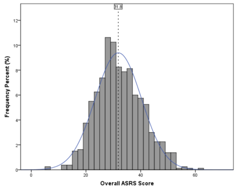
beyond the median, these pps are ‘highly likely to have ADHD’
Colliculus is involved in multi-sensory integration
visual, auditory and somatosensory inputs converge onto a common pool of neurons
some neurons respond to multiple senses, even sometimes olfactory
multisensory neurons show enhanced responses to multisensory stimuli, if the stimuli are close together in space and time’
Any collicular impairments in ADHD- Panagiotidi et al. (2017)
Simultaneity judgement task
presented two stimuli, auditory and visual
sometimes these stimuli occur together, but mostly occur slightly apart
the distance between the two changes, very close together or apart (the most is a 1/3 of a second apart)
all the person has to answer is if they were presented simultaneously or not
looked at people with high levels and low levels of ADHD based on the bell curve from before (ASRS)
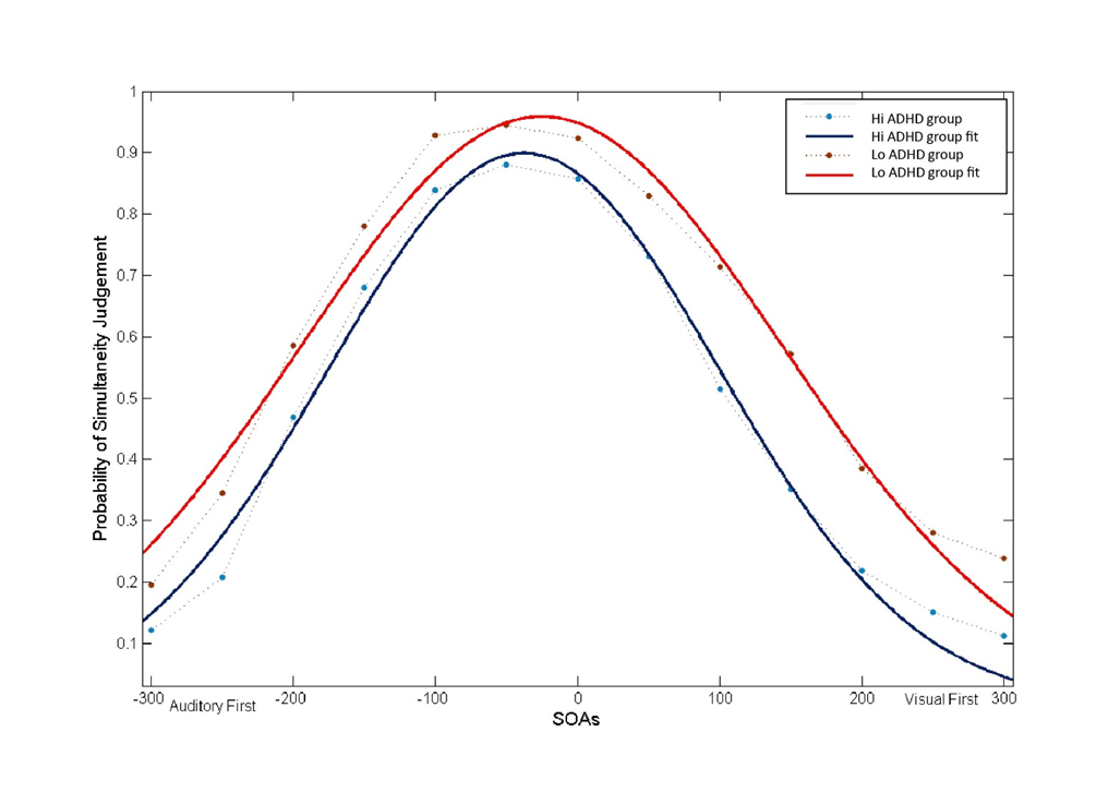
Gradually the difference between the two stimuli got bigger
the high ADHD group were less likely to say they were simultaneous than the low ADHD group across all separations between the two stimuli in each trial
referred to the temporal (something) of integration
this means people with high levels detect stimuli to be more individual and observant of each individual stimuli, whereas low levels sort of fuse them all together
this could perhaps speak more about there distractibility levels
Do ADHD treatments affect the colliculus?
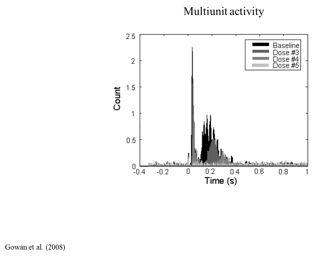
light flash makes a big increase in activity in their eye’s response
then gave them a dose of amphetamine, higher each time and recorded their responses to flashes still
as they received higher doses, the collicular activity goes from really big to virtually nothing at all
greater doses of amphetamine seemed to turn off the SC
Summary:
Superior colliculus is involved in distractibility in animals and humans
Animal models of ADHD exhibit visual hyper-responsiveness
The colliculus has a hot line to the brain’s interrupt system
Multisensory integration in people with high levels of ADHD-like traits is suggestive of collicular hyper-responsiveness
Amphetamine is able to ‘turn off’ or ‘turn down’ the colliculus
Would bias system so that distractions only occur to more salient stimuli
Integrating these ideas with classical theories of ADHD: making a global theory: Dopamine
colliculus not only mediates distractibility but it also regulates and has an anatomical projection to dopamine neurons
The tectonigral projection: A direct pathway from the deep layers of the colliculus to the ventral midbrain: it is one of the regulators of the dopamine system
Terminates on dopamine and non-dopamine neurons....
proving this connection uses the same methods as before

transported to terminal regions- are these terminal regions in any area of dopamine neurons in the brain
the chemical gets injected in the SC and transported by the blobs, which are the cells that pick up and transport the chemical
the SC produces fibres that directly influence the substantia nigra
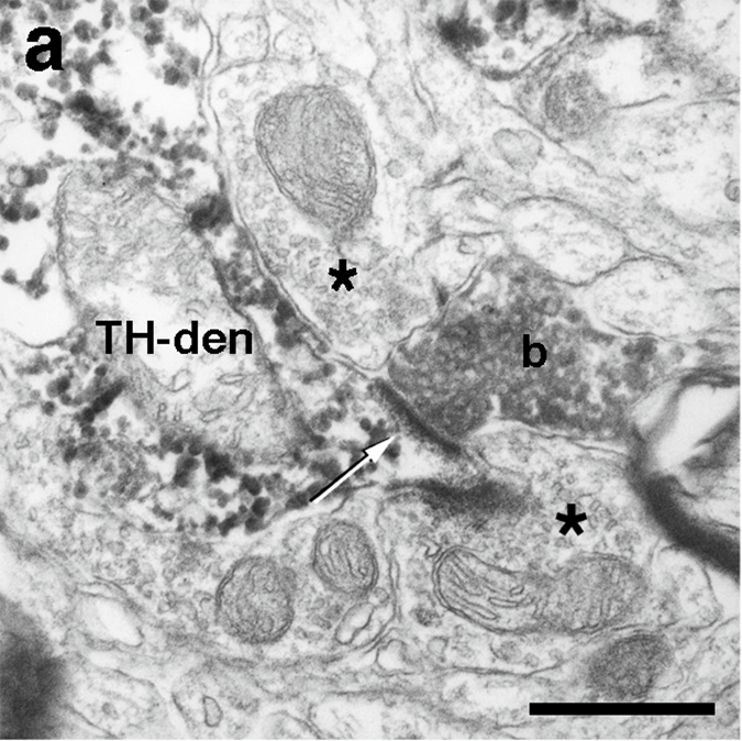
Integrating these ideas with classical theories of ADHD: making a global theory: Dopamine: Electron photo microgram
a- the blobs of the top left is stained is a dopaminergic dendrite
b is a terminal that is coming from the SC, stained from the chemical dye
this terminal is forming a synapse (where the arrows is)
whenever there is a thick dark line like this in a microgram means it is excitatory, when activated will make this cell more likely to produce and action potential
Colliculus transmits visual information to dopamine neurons
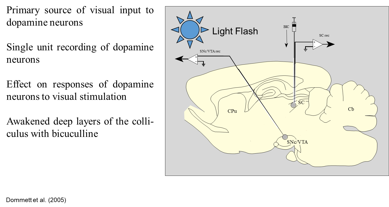
how do we prove that the colliculus is the part of the brain that is particularly important for sending visual information to the whatever he said
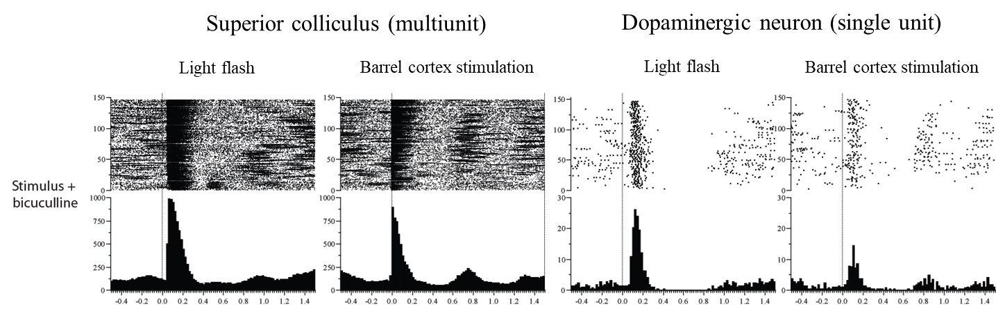
when you shine the light in the animals eye, the SNc is relatively inactive from anesthesia, unless you put a chemical in to wake the animal up
so flash the light in the animals eye before putting them to sleep and insert the chemical Bicuculline, and then you keep flashing it
there is two conditions: one with a chemical to wake them up, one without
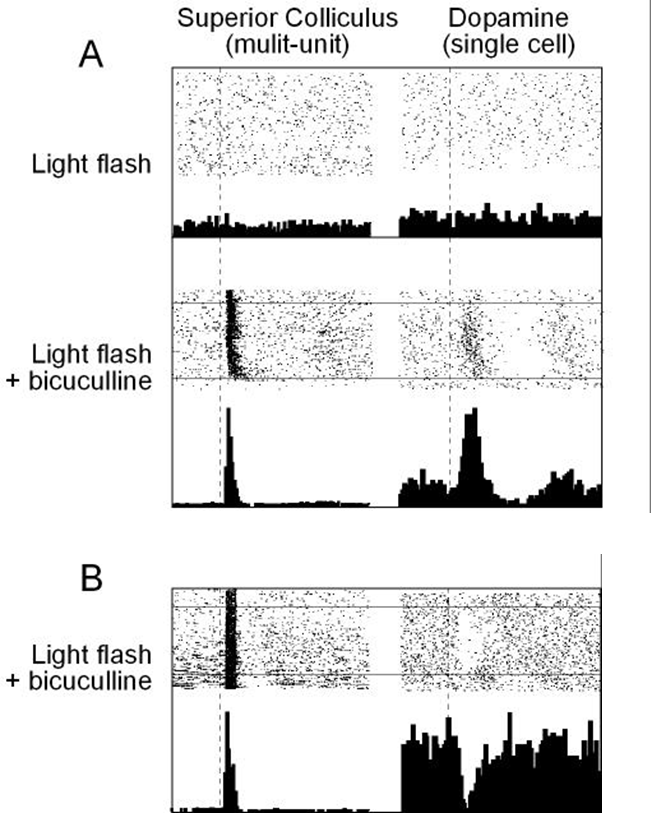
each line and dot is a trial, you flash the light trial after trial
at baseline, now we are shining the light deep into the colliculus, pre-drug there is no light response
after bicuculline: colliculus starts to ‘see’ the light
so do dopamine neurons
so the colliculus must be a primary source of information for dopamine
If we imagine we are activating the dopamine neurons via colliculus, will that activate the dopamine release in the forebrain?
the answer is yes but how do we find this out?
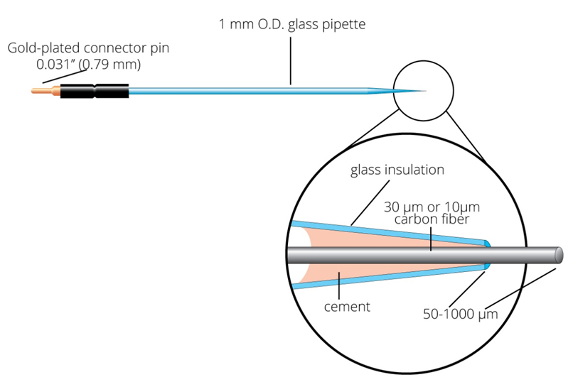
We can use microdialysis OR stick a small probe in the area you want to observe the dopamine levels of to chemically react the dopamine using amperometry, there is a voltage in the tip of the probe, the electrons form a little current that can be picked up and demonstrates that there is dopamine near where the tip of the probe is (electrolysis)
Visual activation of dopamine neurons via the input from the colliculus leads to dopamine release in the forebrain
Can measure dopamine release by amperometry
A constant potential (voltage) is applied to the working electrode
Will oxidise dopamine at the tip, which then creates a small current that can be measured
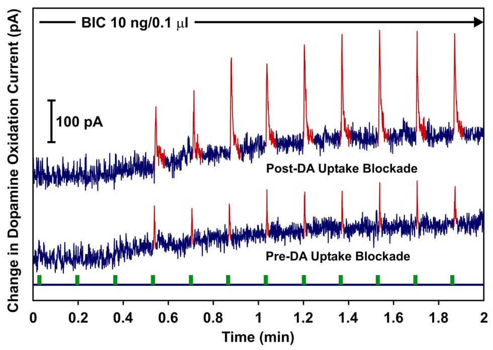
no electrochemical response to light without collicular bicuculline
bicuculline into colliculus induced light response
amplitude and duration of this response increased by nomifensine [selective dopamine reuptale inhibitor]
Each green bit is a flashing light, the reactions are with or without the colliculus, the dopamine is being squirted out of the forebrain, how do we know it is dopamine though?
the spikes of dopamine are much bigger as it hangs around the tissue area much longer
Where are the prescribing hot spots for methylphenidate in England?
Northwest and Southeast
Cortex
So, the colliculus is involved in the regulation of dopamine neurons, which may explain the dopamine dysfunction in ADHD
However, ADHD has also been thought to arise from a dysfunction of the cerebral cortex, so how does the collicular dysfunction idea fit in with that?
Colliculus receives from widespread areas of the cortex

the SC receives input significant input from the orbito-frontal cortex including the frontal cortex
retrograde traces can be injected to the SC and will be transported to the cell bodies, to show which cells project INTO the sc, found that pretty much all areas projected in the SC
this is a retrograde tracer injection site
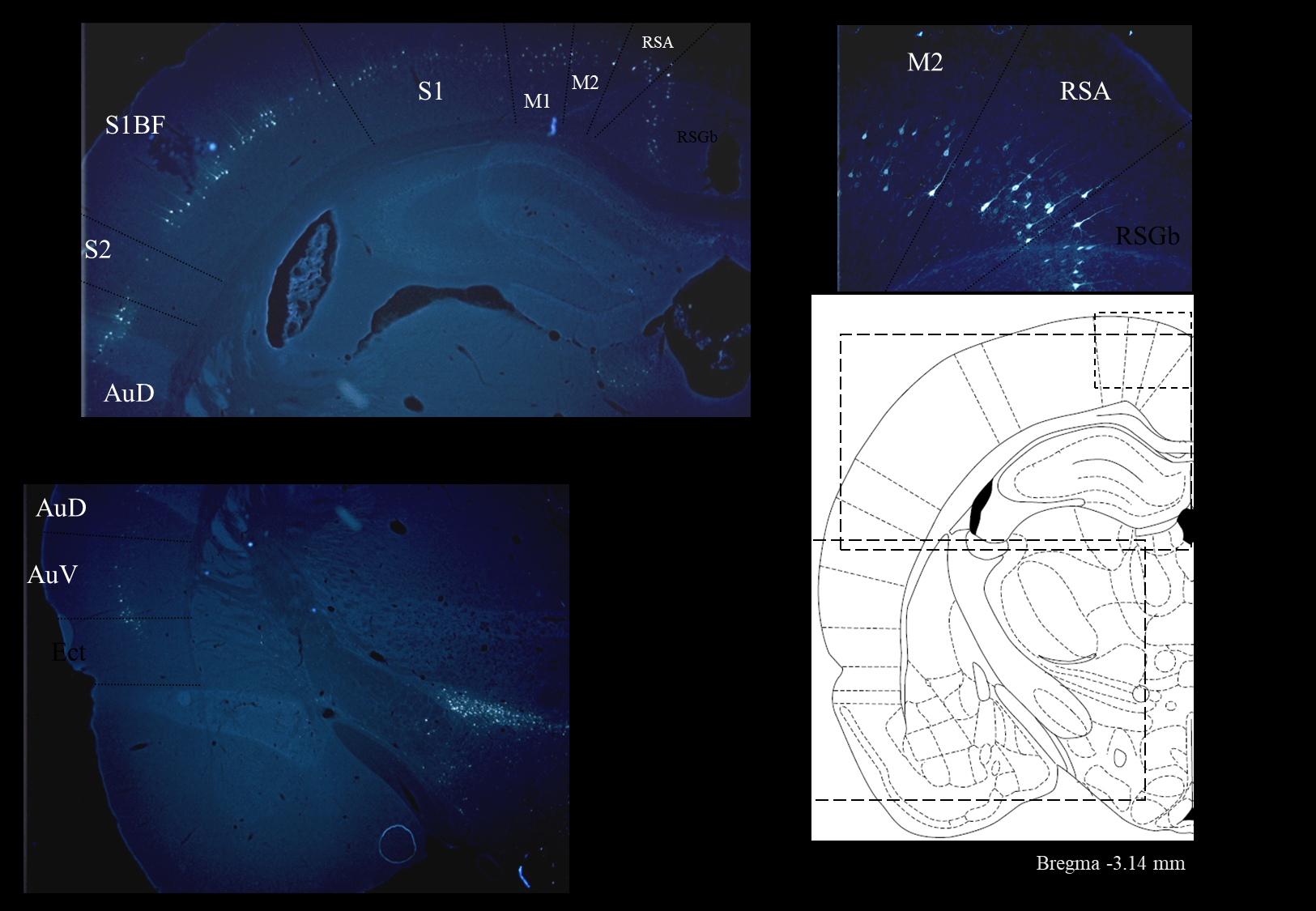
all the dots are cells that project into the SC, they contain the tracer that made its way to the cell bodies
auditory cortex, motor cortex, sensory cortex, retrosplenial area over the corpus callosum, frontal cortex etc
the frontal cortex projects really heavily into the SC
these are all paramital neurons with long axons
there is a strong relationship between the cortex and the SC
We’ve shown that the cortex transmits sensory information to dopamine neurons via the colliculus
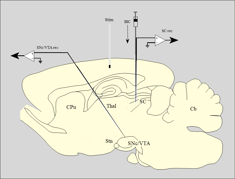
Effects of local chemical manipulation of colliculus on responses of dopamine neurons to barrel cortex stimulation
if the collicular was a route by which the cortex can reach and communicate with dopamine, we can record the activity of the SC with stimulating electrodes
if you stimulate the cortex with the colliculus asleep, are the dopamine neurons still responsive? when you wake the SC up does it alter the dopamine’s behaviour?
light flashes are used as control
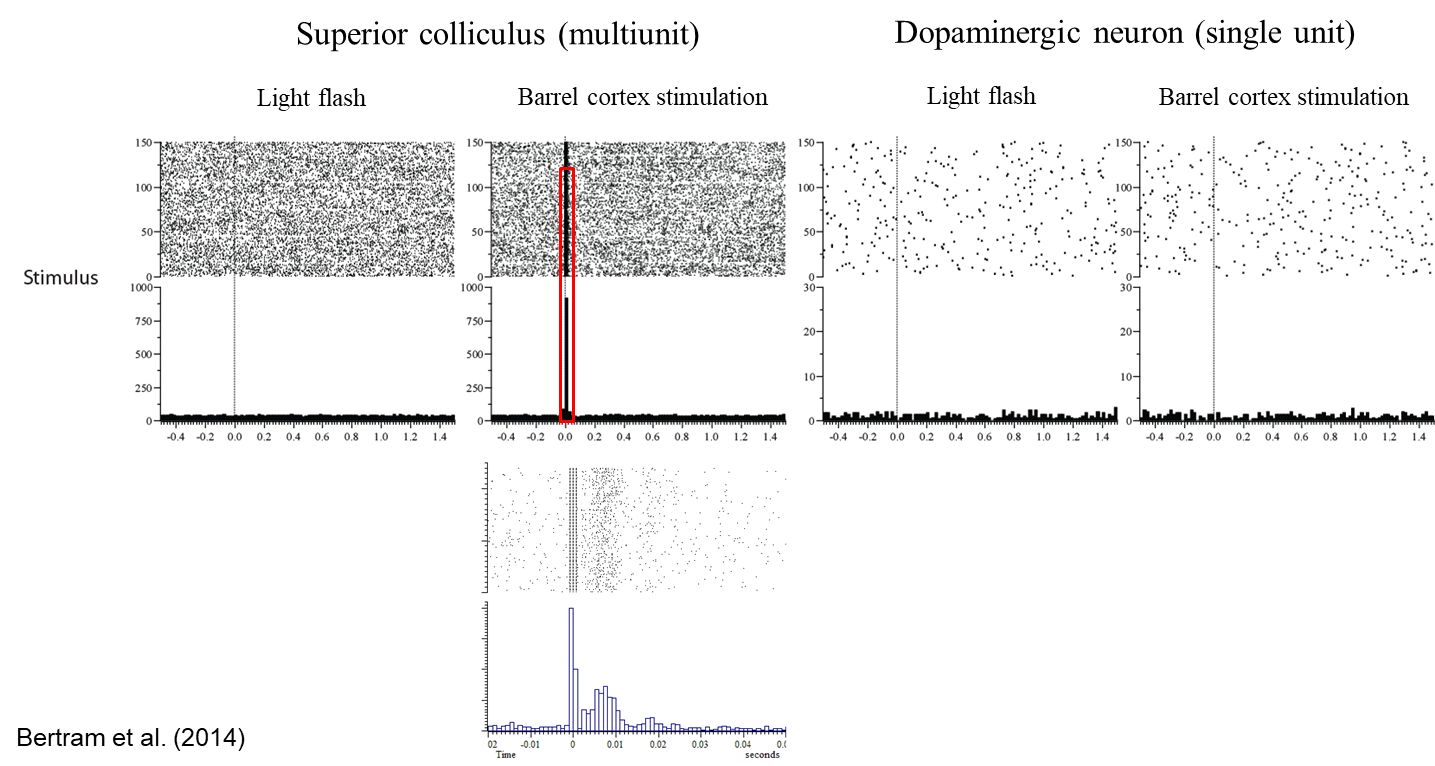
stimulate the barrel cortex, but with the dopamine neurons, no activation is really altered
however, everything changes when the colliculus is up
there is now a massive response to the flashing light
now the dopamine neurons are really starting to respond once the colliculus is up and about
the colliculus seems to pull together the two classical theories of ADHD like a third dimension
Summary-
How does the collicular theory of ADHD sit alongside the classical theories?
1. Frontal cortex
The colliculus receives inputs from widespread areas of the cerebral cortex, including the frontal cortex
Primary dysfunction?
2. Dopamine
The colliculus sends projection to the dopamine neurons in the midbrain
Colliculus involved in sensory regulation of dopamine neurons
Implications:
Step closer to understanding the underlying neural dysfunction in ADHD
Can now think of screening non-addictive drugs for their ability to depress collicular function
Opens up the possibility of drug companies developing new drugs that have a collicular target
Pharmacotherapies for ADHD
The current front-line pharmacotherapies for ADHD - the psycho-stimulants methylphenidate and amphetamines - have clear abuse potential, hence there is a strong need to develop new drug treatments for this disorder.
what is important to developing a new drug treatment?
Central to this process is the identification of the pathophysiological changes which underlie ADHD: however, these are still poorly understood. Here, we make the novel proposal that one locus of change is the superior colliculus (SC), a sensory structure intimately linked with distractibility and the production of eye and head movements.
Why suggest the superior colliculus in making new treatments for ADHD?
Here, we make the novel proposal that one locus of change is the superior colliculus (SC), a sensory structure intimately linked with distractibility and the production of eye and head movements. We suggest that in ADHD the colliculus may be hyper-responsive to sensory inputs, leading to the core symptom of increased distractibility.
Outline how the idea that the super colliculus plays a role in the development of ADHD is supported
This proposal is supported by:
1. ADHD patients show increased distractibility in tasks which are sensitive to collicular function;
2. ADHD patients have a general problem inhibiting eye movements (saccades), the generation of which involves the SC;
3. Saccadic deficits in ADHD include defects in the production of saccadic types which are particularly associated with the colliculus;
4. Covert shifts in attention, which have been argued to involve the SC, are also impaired in ADHD;
5. Reading disorders are frequently co-morbid with ADHD; dyslexia, which is associated with eye movement problems, has been linked to a specific visual perceptual deficit in the magnocellular (M) pathway, a major recipient of which is the colliculus;
6. Aberrant reward processing identified in ADHD could also reflect a collicular dysfunction, given the role played by the SC in the regulation of the dopamine systems.
Can the SC be involved in more?
In addition to the possibility that the colliculus may be dysfunctional in ADHD as the evidence above suggests, the therapeutic effects of current psychostimulant drugs may involve the SC, as the colliculus and structures immediately afferent to it contain the neurochemical machinery necessary to respond to these drugs, and D-amphetamine depresses visually evoked activity in the colliculus. We therefore suggest that not only does collicular dysfunction consitiute an important component of the pathophysiological changes that underlie ADHD, but also the SC may be a therapeutic target for current drugs. As a consequence, the colliculus may offer a novel direction for the development of new, non-addictive pharmacotherapies for ADHD.
Introduction
Attention Deficit Hyperactivity Disorder (ADHD) is the most common childhood behavioural disorder, the main clinical symptoms of which are overactivity, impulsiveness and inattentiveness.
ADHD prevalence
Estimates of the prevalence of the disorder in various countries range from 1.7% to 16.1%, depending on where and how the studies were conducted, and the diagnostic criteria employed.
Is ADHD a childhood disorder?
Although ADHD is primarily thought of as a childhood disorder, studies have found that ADHD symptoms persist into adulthood in between 8% and 43% of sufferers. As well as the adverse effects of ADHD on academic and social functioning in children, ADHD is a major risk factor for later delinquency, substance abuse and personality disorders, and hence the disorder represents a significant public health issue.
What is the most common treatment for ADHD?
At present, the most common treatment for ADHD is pharmacotherapy with psycho-stimulants – either methylphenidate (Ritalin) or amphetamines. Evidence suggests that psychostimulants are particularly effective at ameliorating attentional problems in ADHD. Therapeutic efficacy aside, methylphenidate, like amphetamines, has clear abuse potential, and hence these substances are far from ideal as long-term treatments. Indeed, there is a clear link between ADHD and substance abuse.
What makes the development of non-addictive pharmacotherapies difficult?
However, the development of non-addictive pharmacotherapies is hampered by two inter-related obstacles.
Firstly, despite the proposal of several models of the pathophysiological changes underlying ADHD, the neurobiology of ADHD is far from completely understood.
Secondly, possibly partly because of this pathophysiological uncertainty, the therapeutic mechanism of action of current psychostimulant medications in ADHD is also far from clear.
How should treatments be developed for ADHD?
Attention Deficit Hyperactivity Disorder is a heterogeneous disorder, and sub-types lie at the heart of the ADHD criteria in the fourth edition of the Diagnostic and Statistical Manual. As a consequence, it is possible that several pathological loci exist in this population. Arguably a better strategy therefore for the development of treatments would be to focus on a specific aspect of symptomatology and attempt to trace its underlying cause, rather than trying to develop more global and integrative theories. In terms of ADHD symptomatology, distractibility has been considered by many to be one of most common symptoms (along with overactivity) of the disorder in children, a position that goes back to the neuropsychiatrist A Strauss in the 1940s and 50s. Clinical accounts often describe ADHD children as distractible. For example, Thorley states that hyperkinetic children show significantly more ‘distractibility on examination’ than controls, and ‘Is often easily distracted by extraneous stimuli’ is one of the core symptoms for ‘Predominantly Inattentive Type’ (and ‘Combined Type’) ADHD children according to the DSM IV.
Superior Colliculus and Distractibility
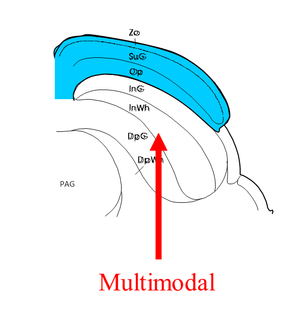
A structure which is intimately linked with distractibility is the midbrain superior colliculus (SC). The SC constitutes the main subcortical visual system, and is highly conserved across species. It appears to play a particular role in detecting and organising responses to unexpected phasic stimuli in a range of modalities, and considerable research emphasis has been placed on the involvement of the SC in orienting the head and eyes toward unexpected objects in the visual field. This facility is enabled by the fact that the superficial layers, which are exclusively visual (receiving inputs from the retina, primary and secondary visual cortices, contain a reintotopic map of the visual field. The deeper layers, which are multimodal, also contain sensory maps which are in register with each other and the superficial layer visual map.
Work in a range of species has shown that collicular lesions lead to a decease in distractibility (rat; cat; monkey). In humans, disconnecting the colliculus from the controlling influence of the prefrontal cortex leads to an increase in distractibility, suggesting that the structure’s function of detecting and responding to unexpected (distracting) stimuli is preserved in humans.
How do eye movements demonstrate that the SC and ADHD could be linked?
Given the presence of distractibility in ADHD and the link between the SC and distractibility, we make the novel proposal here that ADHD may be associated with a collicular dysfunction. The SC is an important part of the circuitry which generates eye movements (saccades). Research in the monkey suggests that superficial layer neurons respond more vigorously to a stimulus if the stimulus is the target for a saccadic eye movement. In terms of recent conceptualisations of the manner in which bids for motor expression, by systems (like the colliculus) capable of specifying actions, are processed by the brain, enhanced activity can be seen as putting a stronger ‘bid’ into the central selection device (hypothesised to be the basal ganglia), which makes the bid more likely to win and generate a motor output, hence the eye movement. As a consequence, it might be suggested that in ADHD, sensory responsiveness in the colliculus is abnormally high, leading to an increased tendency to respond - make eye and head movements - to stimuli activating the structure (this is referred to below as ‘collicular hyper-responsivity’ for short). Such an increased tendency to respond would be manifested outwardly as an increase in distractibility. In this regard it is interesting that decreasing inhibitory GABAergic tone in the SC with bicuculline (a GABAA receptor antagonist), which increases sensory responsiveness in the SC, produces irrepressible saccades in the monkey to the area of visual space subserved by the disinhibited part of the colliculus.
Although distractibility links ADHD and the SC, making the case for the involvement of the SC in ADHD plausible, further evidence is required if the case is to be truly convincing.
what may be the reason behind the increase in sensory responsiveness in the SC?
This hypothesised increase in sensory responsiveness in the SC might arise as a consequence of a primary collicular deficit, for example due to changes in the regulation of the structure by monoamines (monoaminergic innervation of the SC is discussed below). Alternatively, it might arise as a secondary consequence of the dysfunction of a system afferent to the colliculus. The most likely contender here is probably the frontal cortex. The symptoms of ADHD have often been likened to those which follow frontal lobe lesions in humans and animals, and ADHD children show deficits on tests of frontal lobe function. Functional neuroimaging studies frequently report hypo-frontality in ADHD, and structural imaging has revealed reduced prefrontal cortex volume in the disorder. Frontal cortical hypofunction in ADHD might be expected to lead to an increase in distractibility which is qualitatively similar to that following disconnection of the SC from the prefrontal cortex.
more direct evidence for a collicular dysfunction in ADHD comes from three major sources:
1. Studies of distractibility in ADHD patients,
2. Studies of eye movements (and covert shifts in attention) in ADHD patients
3. Studies of reward processing in ADHD patients.
Why is it important to examine studies of distractibility specifically in ADHD patients?
Although collicular lesions in animals lead to a decrease in distractibility, the conditions used to test distractibility are dissimilar to those used in the majority of laboratory tests of distractibility in ADHD. The majority of studies of distractibility in ADHD to date have used distractors of the intra-task type. Although deficits (greater distractibility) have often been reported for these distractor types (Stroop interference; Erikson flanker task, incongruent condition; Test Of Auditory Discrimination, noise subtest; speeded classification task with high salience irrelevant stimulus dimensions; distracting visual borders around mathematics problems), current models of attention suggest that tasks like Stroop and Flanker tap into the function of Posner and Petersen’s ‘anterior attentional system’, of which the SC does not form part.
intra vs extra- task distractions:
In the latter, a distinction has been made between distractors which occur within a task that is being undertaken (intra-task distractors) and those that occur outside the task that is being undertaken (extra-task distractors). The former are usually temporally predictable and intrinsic to the task, whereas the latter are usually unpredictable (unexpected) and extrinsic to the task.
Distractors
It is also quite difficult to see how the increases in distractibility revealed by tests that have used intra-task distractors map onto the distractibility observed in a clinical setting (e.g. ‘distractibility on examination’). Distractors occuring in these contexts seem to accord more closely with the concept of extra-task distractors, i.e. temporally unpredicted (unexpected) stimuli extrinsic to the task. Such stimuli are much more closely related to those used in animal studies where collicular lesions have been found to lead to a decrease in distractibility. For example, in the work of Goodale and colleagues, distractibility was measured as a change in responsiveness of experimental subjects (rats) to novel, unexpected light and sound stimuli presented as the animals performed a brightness discrimination task.
increases in distractibility have been reported in ADHD patients, for example…
Dykman et al. presented a very loud hooter - 90 dB SPL – unpredictably during a reaction time task (telegraph key pressing in response to a target). Although the data in this paper were not clearly reported, from Figure 1 it is apparent that for distractors given immediately before target presentation, press reaction times in hyperactive children increased more than in controls, and for distractors given at the same time as the target, release reaction times in hyperactive children increased more than in controls. Similarly, Gumenyuk et al. presented novel sounds unpredictably prior to presentation of visual stimuli in a categorisation task. Novel sounds enhanced the number of response omissions significantly more in ADHD children than controls.
Issues with Dykman and Gumenyuk
Although supportive of the basic hypothesis of collicular hyper-responsivity in ADHD, Dykman et al. and Gumenyuk et al. both used an indirect measure of distraction, namely changes in general task performance. When responses to the distractors themselves have been examined, again deficits have been reported in ADHD.
Bremer and Stern
Bremer and Stern measured eye movements whilst hyperactive and non-hyperactive children read stories. During the reading of the stories, subjects were presented unpredictably with distracting visual and auditory stimuli. More hyperactive children responded (moved their eyes) to the stimuli than controls and more stimuli elicited a response in hyperactive children than in controls.
Sykes et al
Sykes et al. found significantly more glances off task (‘non-observing responses’) among hyper-active children than among controls during the performance of a range of tasks, including reaction time and vigilance tasks. These studies are particularly significant in the current context because, as stated above, in primates the SC plays a key role in the generation of saccades.
Generally how does eye movement emphasise the link between SC and ADHD?
Indeed, given the intimate link between the SC and saccades, the literature on eye movements in ADHD is a fruitful source of evidence relevant to the collicular hyper-responsivity hypothesis. There are numerous reports of eye movement abnormalities in ADHD: in particular, ADHD sufferers seem to have problems with inhibiting saccades to visual targets. Impaired saccadic inhibition fits well with the idea that the SC might be hyper-responsive in ADHD. The inability to inhibit saccades to targets could arise because of the increased ‘pull’ of the target location as a result of collicular hyper-responsivity
Saccades and collicular dysfunction in ADHD
However, a more compelling case for collicular dysfunction in ADHD could be made if sufferers had difficulties with aspects of saccades in which the SC was particularly implicated. Evidence at present suggests that this does appear to be the case for two specific saccadic types: anti-saccades and express saccades. Anti-saccades are saccades performed (in an experimental setting) in the direction opposite to a suddenly presented visual target. The SC appears to be involved in the generation of this saccadic type, and ADHD sufferers have problems making these saccades. Presumably, making a saccade in the direction opposite to a target is more difficult for ADHD sufferers because of the increased pull of the target. Interestingly, disconnecting the colliculus from the inhibitory controlling influence of the prefrontal cortex in humans leads to an increased error rate in anti-saccade tasks, and methylphenidate, the most widely used pharmacotherapy for ADHD, decreases both the error rate and anti-saccade latencies in anti-saccade tasks.
SC and express saccades
As well as playing a role in the production of anti-saccades, the SC also plays a role in express saccades: short latency saccades elicited under experimental conditions in which a temporal gap exists between the extinguishing of a fixation point and the appearance of a saccadic target. Children suffering from ADHD make fewer of these express saccades. Although, as with the data on anti-saccades, this is suggestive of a collicular dysfunction in ADHD, it is unclear why ADHD patients (with putative collicular hyper-responsivity) might make fewer express saccades. However, ADHD patients in this study were found to make more saccades with latencies shorter than traditional express saccades. Hence, one intriguing possibility is that, under conditions of collicular hyper-responsivity, express saccades could be speeded so that some of them now fall outside the accepted range of latencies for express saccades.
Reading difficulties and ADHD
Finally on the subject of eye movements in ADHD, reading difficulties and ADHD are frequently co-morbid, and poor saccadic control has been reported in dyslexics. It is now widely acknowledged that developmental dyslexia is associated with visual perceptual deficits, in particular with a deficit in the magnocellular (M) pathway that carries visual information centrally from the Y-type ganglion cells in the retina. Although the M pathway is normally considered in relation to the geniculostriate visual system, in the present context it is potentially significant that SC receives inputs from Y-type retinal ganglion cells (which give rise to the M pathway), but not from X-type cells (which give rise to the parvocellular or P pathway). The possibility therefore exists that ADHD and dyslexia may both arise in part due to a collicular dysfunction.
attention and saccades
As well as overt shifts of attention using eye movements, attention can also be shifted covertly in the absence of eye movements. According to the ‘premotor theory’ of attention, saccades and spatial shifts of attention (covert orienting) share a common functional module with a distinct neuronal basis, and hence the colliculus is considered to be involved in such shifts. Covert shifts in attention are usually examined using the Posner paradigm. In this, a target is presented after a cue. The target may either agree in position, when it appears, with direction implied by the cue, or it may not - so the cue is valid or invalid. Responses (button presses) to targets preceded by valid cues are faster than those preceded by invalid cues (or no cue), arguably because of covert shifts in attention to the cue’s location. Thus far, three studies have found slower responses in the invalidly cued condition in ADHD patients than in controls, when attention was explicitly drawn to a target location by a cue (‘exogenous cuing’). The increased reaction times to invalidly cued targets in ADHD may arise as a result of the increased pull of the cued location as a result of collicular hyper-responsivity.
SC and dopamine systems
In addition to the collicular efferent systems which underlie the control of saccades and covert orienting, recent evidence suggests that the SC also plays a critical role in regulating the ascending dopamine systems, central pathways involved in the processing of rewards and other salient stimuli. Given this, collicular hyper-responsivity may underlie the alterations in reward processing (e.g. increased reward sensitivity; increased preference for immediately available rewards) reported in ADHD patients, which have been argued to account for all the major symptoms of the disorder. A corollary of that is that dysregulation of the dopamine systems following collicular dysfunction may underlie some of the abnormalities in the dopamine systems found in ADHD, leading directly to changes in dopamine levels and indirectly to compensatory changes in dopamine transporters, dopamine receptors and dopamine synthesis.
Psychostimulants have been found to be effective against distractibility in ADHD patients- how would this work with the SC hypothesis
Our suggestion that the SC may be hyper-responsive in ADHD raises the possibility that these drugs may act therapeutically at the level of the SC (or afferent regulatory structures) in the disorder, presumably to dampen collicular sensory responsivity. Actually, this possibility is to some extent independent of the collicular hyper-responsivity hypothesis. If distractibility in ADHD did not involve the SC, arising instead from some other parallel dysfunction, targetting the colliculus may still be one of the therapeutically relevant actions of psychostimulants in ADHD. Psychostimulants reduce distraction in normal humans and rats, and hence a collicular dysfunction (as postulated in ADHD) is not required to benefit from the decrease in distractibility.
Is there any evidence that psychostimulants effect distractibility improvements in ADHD by modulating sensory processing in the SC?
Evidence for such a possibility comes from two major sources;
1. Neuropharmacological studies of the SC;
2. Recent studies from our laboratory on the effects of d-amphetamine on visually evoked activity in the SC of the rat.
amphetamines and SC
The proximal effects of amphetamines are mediated by increasing synaptic levels of the monoamines dopamine (DA) and noradrenaline (NA) and, at higher doses, serotonin (5-HT]). Methylphenidate has a similar profile of action. Evidence suggests that in a wide range of species, including rats and humans, structures immediately afferent to the SC receive monoaminergic innervation (e.g. visual cortex; rat; human). Evidence also suggests that the SC itself receives extensive noradrenergic and serotonergic innervation, as well as a limited dopaminergic input. Noradrenergic and serotonergic afferents appear to preferentially (but not exclusively) target the superficial visual layers. Since the visual response properties of deep layer neurons are critically dependent on superficial layer inputs, disrupted superficial layer function will affect all visually-related efferent activity from the colliculus.
The neurochemistry of SC
So, structures immediately afferent to the SC, and the colliculus itself (especially the superficial layers) contain the neurochemical substrate necessary for psychostimulant action. As a consequence, we recently performed an investigation into the effect of d-amphetamine on visual responses within the superficial layers of the SC in the rat. To this end, two studies were performed. In the first, d-amphetamine was administered systemically to rats under urethane anaesthesia whilst local field potential (LFP) and multi-unit (MUA) responses were recorded from the superficial layers of the SC in response to whole-field light flashes. In the second, d-amphetamine was administered directly into the colliculus itself whilst animals underwent the same stimulation and recording protocol. Systemic d-amphetamine dose-dependently decreased the responsiveness of cells in the superficial layers of the SC to visual stimuli, measured both by LFPs and MUA. Similar results were obtained with intra-collicular administration.
The results of intra-collicular administration
The results suggest that the effects of systemic d-amphetamine on visual responses in the superficial layers of the SC appear to be produced, at least in part, by a local action in the colliculus. Hence, a natural question arises as to which monoamine systems are most likely to mediate the effects. Since there is an extensive noradrenergic and serotonergic innervation of the rat SC, whilst the dopaminergic input is small, the involvement of DA is unlikely. Instead, the effects are more likely to be produced by an alteration of NA and/or 5-HT-mediated transmission. Iontophoretic NA has been reported to both depress and facilitate superficial layer visual responses, although depressions are far more prevalent. In contrast, the effects of 5-HT are uniformly depressant. Given that d-amphetamine tends to affect 5-HT-mediated transmission at higher doses, it is likely that lower dose effects are mediated primarily by NA, with the action of 5-HT super-imposed at higher doses.
The main effect of d-amphetamine in our study was to reduce visual responsiveness in the superficial layers of the SC. By depressing visual responses in the SC, it might be anticipated that the expression likelihood of eye and head movements to unexpected visual stimuli would be reduced by d-amphetamine, leading to a reduction in distractibility and a correlative improvement in sustained attention. In support of this, intra-collicular micro-injection of S-CM-GTNH2, a 5-HT 1B-1D receptor agonist has been found to reduce distractibility in behaving rats. Similarly, intra-collicular injections of the GABA agonist muscimol in the monkey, which lead to localised inhibition of the SC, decrease the production of saccades to the area of visual space subserved by the inhibited region.
Were the results consistent with the hypothesis?
Consistent with the hypothesis that the colliculus is a locus of therapeutic action in ADHD, noradrenaline selective (atomoxetine) and 5-HT selective (fluoxetine) drugs, i.e drug that work on the systems which are probably involved in d-amphetamine-induced suppression of visual responsiveness in the SC, appear to be therapeutically efficacious in ADHD, whereas dopamine-selective agents are not currently in clinical use for this disorder. Although attentional improvements in rats can be found at doses of d-amphetamine (0.1–0.4 mg/kg ) below those which lead to depression of visual responses in the superficial layers of the SC (>2 mg/kg i.v.), it is difficult compare doses in anaesthetised and unanaesthetised preparations. Similarly, although effective doses in ADHD patients (0.3-1.0 mg/kg p.o.) are much lower than the inhibitory doses used here, anaesthetic and species-specific factors make it difficult to compare doses used in human treatment with those used in our studies.
Conclusion
The current frontline pharmacotherapies for the treatment of ADHD, namely methylphenidate and amphetamines, have clear abuse potential, hence there is a strong need to develop new drug treatments for this disorder. Central to this process is the identification of the pathophysiological changes which underlie ADHD. Given the heterogeneity of the disorder, multiple loci are probably involved, providing multiple potential therapeutic targets. Here, we propose that one such locus is the SC.
Whether or not the SC is indeed hyper-responsive in ADHD, the SC may well represent an important therapeutic target for drugs. Fortunately, again, even if psychostimulants do not achieve their therapeutic effects on distractibility in ADHD by an action at the level of the SC, then the development of drugs which dampen stimulus-related activity in the colliculus could still represent an important novel path for drug development to take. Pharmacological manipulations of this structure are able to decrease distractibility. As a consequence, sensory responsiveness in the SC may represent a new model system for use in the development of non-addictive pharmacotherapies for ADHD, and as such may be of interest to the pharmaceutical industry.
Clearly, the evidence for collicular hyper-responsivity in ADHD, and for psycho-stimulant-induced changes in responsivity, are at present indirect. To move these ideas forward it will be necessary to obtain more direct evidence. To achieve this, two approaches seem potentially fruitful. Firstly neuroimaging: to date, little relevant data concerning collicular function in ADHD has been gathered. Although early imaging studies occasionally had a remit broad enough to encompass sensory systems (Zametkin et al. reported decreased somatosensory cortical responses), most imaging studies in humans have focused on frontal cortical areas normally involved in attention/ cognition, and the neostriatum. Since activation of the colliculus has now been successfully imaged in humans using functional magnetic resonance imaging (fMRI), the possibility that the SC is hyper-responsive to phasic sensory stimuli in ADHD could now be investigated, as could the possibility that psychostimulant pharmaco-therapies decrease stimulus-related activity in the colliculus.
The second useful approach to try to obtain more direct evidence for the hypotheses under discussion is to pursue the investigation of eye movement production in ADHD further. Collicular lesions in monkeys have been shown to lead to characteristic deficits in eye movement production, both to distractor stimuli and others. It should be possible to test ADHD patients in similar or identical tasks and compare outcomes. The effects of psychostimulant medication should qualitatively resemble those of collicular lesions, although this may appear against a background (drug-free) increase in responsivity (relative to controls) to the same kind of sensory stimuli negatively affected by the drugs
current psychostimulant therapies may already work at that level, a contention which is supported by
1. The colliculus and structures immediately afferent to it contain the neurochemical machinery necessary to respond to these drugs;
2. D-amphetamine depresses visually evoked activity in the rat colliculus in a dose-dependent manner.
It is suggested that in ADHD, the colliculus is hyper-responsive, leading to the core symptom of increased distractibility. This proposal is supported by:
1. ADHD patients show increased distractibility in tasks which are sensitive to collicular function;
2. ADHD patients have a general problem inhibiting saccades, the generation of which involves the SC;
3. Saccadic deficits in ADHD include defects in the production of saccadic types (anti-saccades and express saccades) which are particularly associated with the colliculus;
4. Covert shifts in attention (which also have been argued to involve the SC) are also impaired in ADHD;
5. Reading disorders are frequently co-morbid with ADHD; dyslexia (which is associated with eye movement problems) is linked to a specific visual perceptual deficit in the M pathway, a major recipient of which is the colliculus.
6. Aberrant reward processing identified in ADHD could also reflect a collicular dysfunction, given the role played by the SC in the regulation of the dopamine systems.