NEURAL TISSUE AND BRAIN- JONES
1/66
There's no tags or description
Looks like no tags are added yet.
Name | Mastery | Learn | Test | Matching | Spaced | Call with Kai |
|---|
No analytics yet
Send a link to your students to track their progress
67 Terms
The nervous system is divided into what 2 systems/categories?
CNS and PNS
What 2 components made up the CNS?
brain and spinal cord
What 2 types of neurons made up the PNS?
Which sends afferent signals and where?
Which receives efferent signals from the CNS?
motor neurons —> receives efferent signals from CNS
sensory neurons —> sends afferent signals to CNS
What are the 2 categories of motor neurons?
Which controls voluntary movement?
Which controls involuntary movement?
somatic and autonomic nervous system
voluntary movements—> somatic
involuntary responses —> autonomic
What division of the autonomic nervous system deals with “fight or flight”?
sympathetic division
What division of the autonomic nervous system deals with “rest or digest”?
parasympathetic
What is the functional unit of the nervous system?
neuron
What part of the neuron receives signals?
dendrites
What part of the neuron is also known as the cell body and where the organelles are?
soma
What part of the neuron is a long projection that conducts signals from the dendrites to terminal buttons?
axon
What part of the neuron releases NTs?
terminal buttons
What acts as an insulator around axons and allows signals to be sent quickly?
myelin
Most neurons are _________________.
a. unipolar
b. bipolar
c. pseudounipolar
d. multipolar
d.
Bipolar and pseudounipolar cells deal with what process?
a. motor
b. transport
c. senses
d. structure
c.
What’s the function of the glial cells in the PNS?
satellite cells
Schwann cells
satellite cells—> support cell bodies
Schwann cells—> form the myelin sheath
What’s the function of the glial cells in the CNS?
oligodendrocytes
astrocytes
microglia
ependymal cells
oligodendrocytes- form the myelin sheath
astrocytes- support CNS, FORM BBB, secrete neurotropic factors, take up K+ and NTs
microglia- act as scavengers, immune cells that destroy invading pathogens
ependymal cells- create barriers between compartments… lines the SPACES in the brain w/CSF not blood
Gray matter in the CNS is the _______________.
a. axon
b. cell body
b.
White matter in the CNS is what? why is it white?
is the axon
white bc it doesn’t stain bc axon is fatty
PRACTICE: Which region of a neuron contains the highest concentrations of ligand-gated channels which are activated by neurotransmitters?
a. axon
b. axon hillock
c. myelin
d. terminal button
e. dendrites
e.
What are some things that protect the brain and spinal cord?
skull/ vertebrae
meninges
CSF
BBB
What are the meninges?
What are the names of the layers? innermost? outermost?
tissue layers that surround CNS
always 3 layers
pia mater (innermost)
arachnoid mater
dura mater (outermost)
Arachnoid mater is made of what components?
subarachnoid space—> filled with CSF, contains major blood vessels
arachnoid villus—> cushion, DRAINAGE point
A lumbar puncture samples CSF from which space?
subarachnoid space
What is the function of ventricles (in the brain)?
Which ventricles are the largest?
What does each ventricle contain? fxn?
function: store CSF
lateral ventricles the largest
each ventricle contains a CHOROID plexus (mass of capillaries)—> what MAKES the CSF
Explain how CSF is made, flows through CNS, and is reabsorbed.
choroid plexus secretes CSF in lateral ventricles
flow of CSF:
lateral ventricle—> interventricular foramina—> 3rd ventricle—> cerebral aqueduct—> 4th ventricle—> median and lateral apertures —> subarachnoid space
reabsorbed at arachnoid villi
What happens if there is a blockage in the apertures of the brain?
hydrocephaly—> swelling in brain
What is the difference between communicating and noncommunicating hydrocephalus?
Communicating: CSF flow blocked after ventricles
Noncommunicating: Blockage within ventricles.
What is called the barrier between the brain circulation and tissue fluid of the brain?
blood-brain barrier
What forms tight junctions to limit movement out of capillaries aka allows endothelial cells to be VERY VERY tight?
astrocytes
The brainstem is composed of what 3 main structures?
midbrain
pons
medulla oblongata
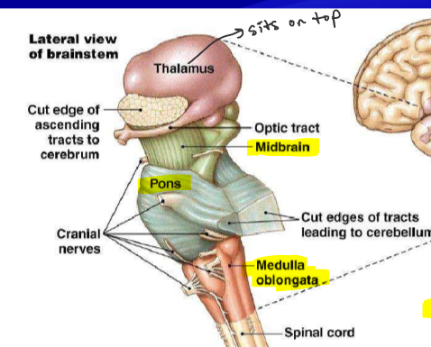
What is the function of the medulla oblongata in general?
CONNECTS spinal cord to brain
involuntary/autonomic controls
vomiting
respiration
speech
sweating
CV, GI
Name 2 important structures within the anterior and posterior medulla:
anterior
pyramids/ white matter
olives (olivary nucleus)
posterior
nucleus cuneatus and gracilis
What is the function of each of the following:
olives
pyramids/white matter
nucleus cuneatus and gracilis
olives- relaying SOUND/auditory
pyramids/white matter- axons run up medulla
nucleus cuneatus and gracilis- sensory perception
What is the primary function of the pons?
Relay station between cerebrum/cerebellum
coordinates breathing and urination
What connects the cerebellum to the pons?
cerebellar peduncles (bundles of nerves)
What are pontine fibers?
Transverse white matter tracts connecting the cerebrum to the cerebellum
(part of the pons)
Describe EACH of the following components of the MIDBRAIN:
tegmentum
substantia nigra
cerebral crus
superior colliculi
inferior colliculi
tegmentum: fine motor movement
substantia nigra: relays inhibitory signals to thalamus and basal nuclei to control unwanted movement
cerebral crus: white matter of corticospinal tract
superior colliculi: visual movements and tracking of head (visual reflexes)
inferior colliculi: receives info from inner ear (auditory relay)
What is the reticular formation?
nerve cell bodies forming gray matter which runs vertically throughout the brainstem
What does the reticular formation of the brainstem regulate?
arousal
sleep
somatic muscle tone
breathing
blood pressure
pain modulation
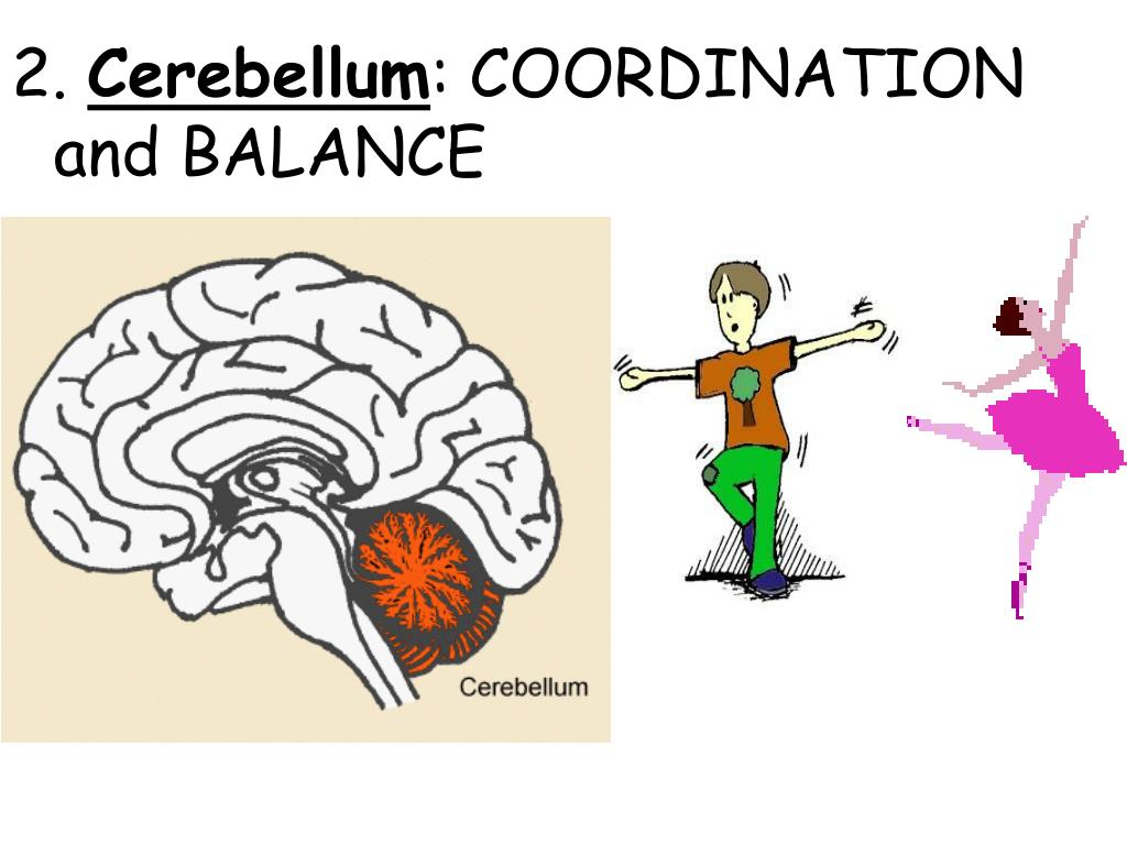
What is the function of the cerebellum?
controls coordinated movements, posture, balance
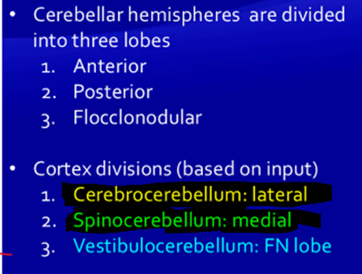
There are 2 ways to classify the cerebellum…by lobe or based on input.
what are the 3 lobes?
what are the 3 cortex divisions (based on input)?
(idk how important)
Function of each of the following in the cerebellum:
peduncles
arbor vitae
deep nuclei
Peduncles (connect cerebellum to other regions)
Arbor vitae (white matter within each hemisphere)
Deep nuclei (gray matter within each arbor vitae)
What is the function of granule neurons in the cerebellum?
What is the role of Purkinje neurons in the cerebellum?
They are located in the cortex layer and modify Purkinje cells.
They project from the cortex to the deep nuclei.
The Thalamus and Hypothalamus make up the ____________________.
Diencephalon
Function of the thalamus?
integration center/relay for sensory and motor info
(chat: Acts as the brain’s "relay station," processing and transmitting sensory and motor signals to the cerebral cortex)
Function of the hypothalamus?
nuclei which regulate homeostasis and behavioral drive
endocrine/hormone
autonomic nervous system
pituitary gland
Function of the cerebrum? (overview)
cerebral cortex
basal ganglia
limbic system
cerebral cortex
sensory fields
motor fields
association area
basal ganglia
movement control
limbic system
emotions
learning
memories
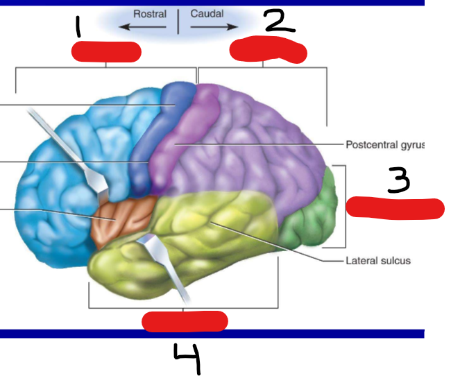
Be able to identify visually the lobes of the brain:
frontal lobe
parietal lobe
occipital lobe
temporal lobe
The cerebrum is divided into white matter and grey matter.
Describe the WHITE matter of the cerebrum:
what does it includes?
what are projection tracts?
what are commissural tracts?
what are association tracts?
includes myelinated AXONS and glial cells
projection tracts- vertical connections between cerebrum and lower areas (ex: spinal cord, brainstem, etc.)
commissural tracts- connections from one hemisphere to another
association tracts- connections within same hemisphere
The cerebrum is divided into white matter and grey matter.
Describe the GRAY matter of the cerebrum:
grey matter also called what?
what cells?
layered structure is called what?
all inputs go into what layer?
grey matter of cerebrum: cerebral cortex
cells: stellate cells and pyramidal cells
layered structure called neocortex
all inputs go into the IV layer of the neocortex
We know the basal nuclei play a role in movement… but more specifically its function is what 3 things?
integrated motor movement
motivation
addiction
What are the 3 main basal nuclei and what do they combine to form?
3 main nuclei:
caudate
putamen
globus pallidus
combine to form the CORPUS STRIATUM
Which two major structures send inputs to the basal nuclei?
Where do the basal nuclei send their final output signals?
inputs: cerebral cortex, substania nigra
outputs: internal, thalamus to cortex
How does Huntington’s disease affect the basal nuclei? Results?
corpus striatum degenerates/missing
results in uncontrollable jerky movements
What are the 3 core structures of the limbic system?
amygdala
cingulate gyrus
hippocampus
What part of the limbic system serves as nerve bundles from the hippocampus to the hypothalamus?
fornix
What part of the limbic system is responsible for MEMORY?
hippocampus
What part of the limbic system is responsible for pleasure, reward, FEAR, aggression and motivation?
AMYGDALA
Which of the following is a function of the limbic system:
a. eyesight
b. olfaction
c. hearing
d. taste
b.
The cerebral cortex (grey matter of the cerebrum) can be split into 4 lobes.
What’s the function of each:
occipital
temporal
parietal
frontal
occipital- VISION
temporal- auditory, olfaction, language
parietal- taste, somatosensory area
frontal- olfactory, motor skills, language
IDK HOW IMP:
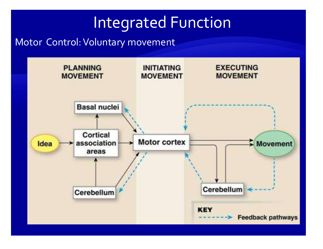
Explain the step-by-step process of voluntary movement:
Which brain region initiates voluntary movement planning, and where does it send signals?
What is the role of the corticospinal tract in motor control?
How do the basal nuclei contribute to movement?
What two roles does the cerebellum play in motor control?
Where are lower motor neurons located, and what do they activate?
premotor cortex sends signal to motor cortex
through the corticospinal tract carries signals from motor cortex to spinal cord/lower brain regions (basal nuclei/cerebellum)
basal nuclei regulate movement and tell when to start/stop
cerebellum deals with coordination/posture and ADDITIONALLY involved in error correction
ventral horn of spinal cord → directly stimulate muscle groups.
Language is an integrated function and deals with reading, writing, speaking, and speech comprehension.
What are the 2 main areas associated with language and where are they located?
Wernicke area- left hemisphere, temporal lobe
Broca area- left hemisphere, frontal cortex
Function of Wernicke’s area?
recognition of spoken/written language
How does Broca’s area interact with Wernicke’s area and the motor cortex?
Receives interpreted language from Wernicke’s → sends speech/writing commands to motor cortex.
What is a language deficit due to lesion in the left hemisphere called?
aphasia
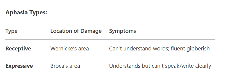
What are the 2 types of aphasia?
Where is the location of damage?
What are the symptoms?
2 types:
Receptive
Damage to Wernicke’s area
cannot understand spoken or visual information
fluent gibberish
Expressive
Damage to Broca’s area
understands, but cannot chose words properly, slow speech, or write clearly
PRACTICE:
A patient speaks fluently but uses incorrect words (e.g., "fork" for "pen"). Where’s the lesion?
Wernicke’s area