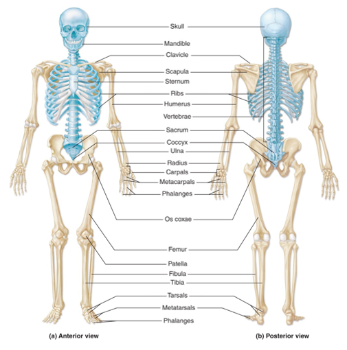
Axial Skeleton
-forms the long axis of the body
-includes: skull, vertebral column, rib cage
-bones involved with protecting, supporting, and carrying other body parts
Appendicular Skeleton
-bones of the upper and lower limbs & girdles
-includes hip and shoulder
-bones involved in locomotion
The Skull
-most complex bony structure in the body
-composed of 22 bones *Cranial bones (8 bones) *Facial bones (14 bones)
-most are flat bones
-bones are united together by sutures *interlocking joints with saw-toothed or serrated appearances *exception is mandible which is freely moving
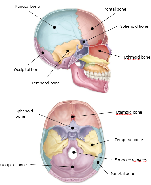
The Cranium
-encloses the brain and furnishes the attachment sites for the head & neck muscles
-single and paired bones
-divided into 2 parts (Cranial vault & Cranial base)
Single bones of the cranium
-frontal
-occipital
-ethmoid
-sphenoid
Paired bones of the cranium
-temporal
-parietal
Cranial vault
-round portion that makes up the superior, lateral, and posterior portions of the skull
Cranial base
-makes up the floor or interior portion of the skull
-Foramen magnus: where spinal cord exits the cranium
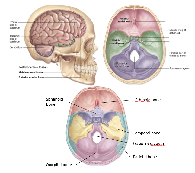
Cranial Fossae
-contoured depressions that make up the floor of the cranium
-Anterior cranial fossa
-Middle cranial fossa
-Posterior cranial fossa
Anterior cranial fossa
-frontal bone, ethmoid bone, and sphenoid bone
-supports the frontal lobe
Middle cranial fossa
-sphenoid bone and temporal bone
-supports the temporal lobe and the pituitary gland
Posterior cranial fossa
-temporal bone and occipital bone
-supports the cerebellum and part of the brainstem
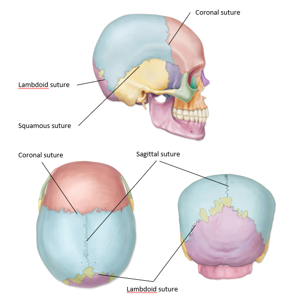
Cranial Sutures
-immovable joints that form boundaries between the cranial bones
-4 major sutures: *Coronal suture *Lambdoid suture *Sagittal suture *Squamous suture
Coronal suture
-extends laterally across the superior surface along the coronal plane
-articulation between frontal and parietal bones
Lambdoid suture
-arc across the posterior surface of the skull
-looks like a lambda (l)
-articulation between parietal and occipital bones
Sagittal suture
-extends between coronal and lambdoid sutures along the midsagittal plane
-articulates between the paired parietal bones
Squamous suture
-one on each side of the skull
-articulates between the temporal and parietal bone (on a side)
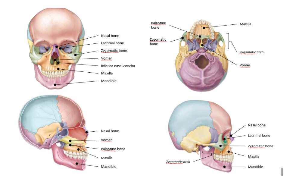
Facial Skeleton
-bones that form the framework for the face
-single bones
-paired bones
Single bones of facial skeleton
-mandible
-vomer
Paired bones of facial skeleton
-nasal bones
-lacrimal bones
-zygomatic bone
-palatine bones
-maxilla
-inferior nasal concha
Function of facial skeleton
-contain or form cavities for the special sense organs (sight, smell, taste)
-turbinate air
-provides openings for the passage of air and food
-securing teeth
-anchor facial muscle
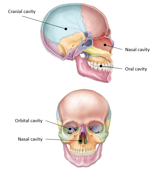
Cavities of skull
-cranial
-orbital
-nasal
-oral
Cranial cavity
-largest cavity
-surrounds the brain
Orbital cavities
-contain eyeballs, blood vessels, muscles, nerves, and lacrimal glands that secrete tears
-formed by frontal, sphenoid, ethmoid, zygomatic, palantine, maxilla, and lacrimal bones
Nasal cavity
-contains passages for air and special sensory neurons for smell; it is considered the first part of the respiratory tract
-divided by the septum (formed by vomer & ethmoid bone)
-formed by sphenoid, ethmoid, palantine, nasal bones, maxilla, & inferior nasal concha
Oral cavity
-contains teeth, tongue, a passage for both food and air, and most of the salivary glands
-formed by mandible and maxilla
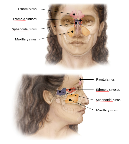
Nasal and paranasal sinuses
-air filled chambers within the bones of the skull
-located around the nasal cavity *4 paranasal sinuses *named for which bone they are located
-possess small openings between sinuses and nasal cavity
-mucus-lined and air-filled *air moves in from nasal cavity *mucus drains out to nasal cavity
Function of nasal and paranasal sinuses
-help to warm & humidify air
-lighten the skull
-enhance resonance of the voice
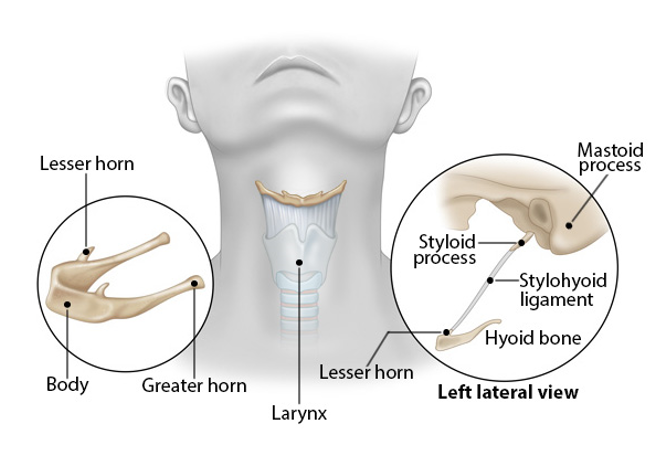
Hyoid bone
-found in the anterior neck region
-no direct articulations with any other bones *position maintained by a combination of ligaments and muscles
Lesser and greater horns
-2 pairs of projections in hyoid bone
-important sites of attachment for muscles involved in swallowing and speech production
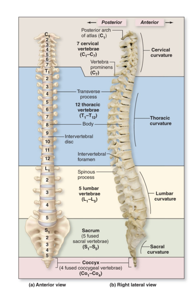
Vertebral column
-4 divisions & 4 curvatures *Cervical *Thoracic *Lumbar *Sacral *Coccygeal
(letter designates the type of vertebrae and a subscripted number its position)
Cervical vertebrae
-7
-concave curvature
Thoracic vertebrae
-12
-convex curvature
Lumbar vertebrae
-5
-concave curvature
Sacral vertebrae
-5
-vertebrae are fused, called the sacrum
-convex curvature
Coccygeal vertebrae
-4
-vertebrae are fused, called the coccyx
Why does the spine curve?
-provides flexibility & increased resilience
-better supports the weight of body
-functions more like a spring
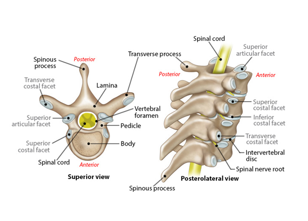
Body (structure of vertebrae)
-weight-bearing portion
-located anteriorly
Vertebral foramen (structure of vertebrae)
-space between the body & arch
-make up the vertebral canal
Vertebral arch (structure of the vertebrae)
-composite structure
-located posteriorly
-composed of 2 pedicles and 2 laminae
Spinal process
-single
-extends anteriorly
Articular processes
-2 pairs
-extend superiorly & inferiorly
Transverse processes
-one pair
-extend laterally
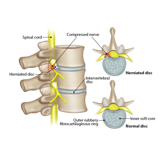
Intervertebral discs
-reside between the vertebrae to provide cushioning
-2 parts: *Anulus fibrosis *Nucleus pulposus
Anulus fibrosis
-outer portion
-composed of collagen fibers and fibrocartilage
-limits the expansion of the nucleus pulposus
Nucleus pulposus
-inner portion of the disc
-elastic and compressible
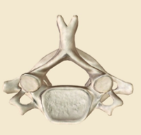
Cervical vertebrae
-Body shape & size: *small and oval *C1 lacks a body *C2 has the dens on the superior surface of its body
Vertebral foramen shape: triangular
Transverse processes: contain transverse foramina
Spinous processes: *most are fork-shaped *C1 lacks a spinous process
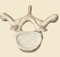
Thoracic vertebrae
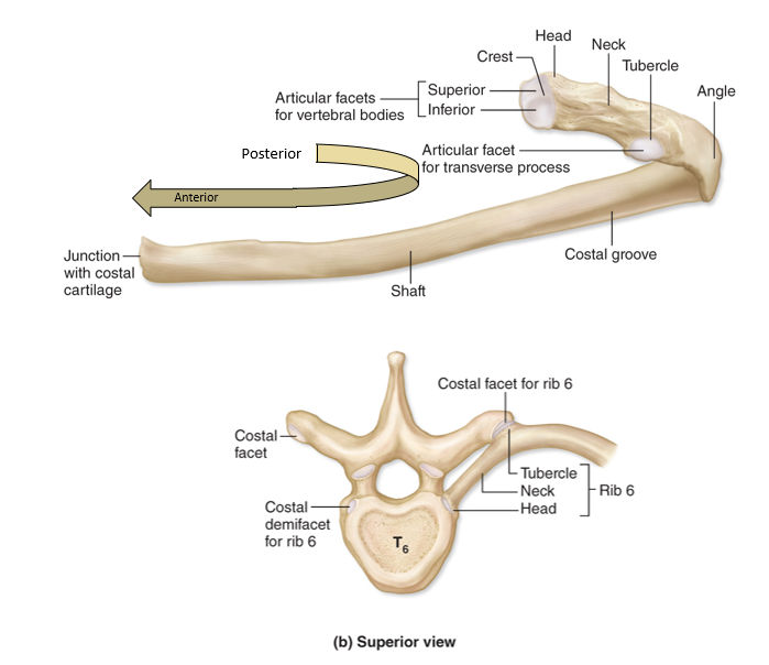
-Body shape & size: *larger and heart-shaped *contain costal facets
Vertebral foramen shape: circular
Transverse processes: *long *contain articular facets for ribs
Spinous processes: *long *point inferiorly
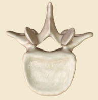
Lumbar vertebrae
-Body shape & size: *largest *kidney-shaped
Vertebral foramen shape: -flattened triangular
Transverse processes: *short *no facets or foramina
Spinous processes: *thick *point posteriorly
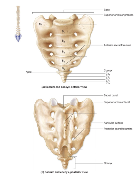
Sacrum
-formed by 5 fused vertebrae
-superior articular process connects to hip
-possesses foramen for blood vessels & spinal nerves to pass through
Coccyx
-formed by 4 fused vertebrae *(occurs around age 25)
-attachment site for several ligaments
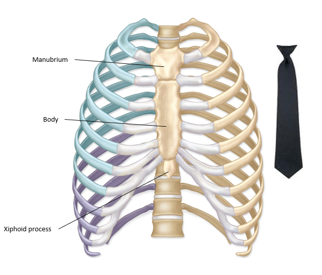
Thoracic cage: Sternum
-breastbone
-stabilizes the thoracic cage
-protects the heart, vena cava, and thymus
-made of 3 bones: *Manubrium *Body *Xiphoid process
Manubrium
-articulates with clavicle and ribs
-part of the sternum
Body
-articulates with ribs
-part of sternum
Xiphoid process
-initially composed of hyaline cartilage, ossifies by age 40
-attachment point for the abdominal muscles
-part of sternum
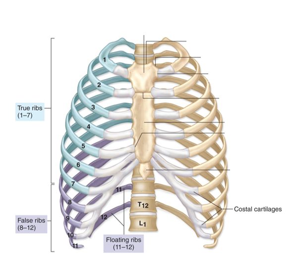
Thoracic cage: Ribs
-all attach posteriorly to the thoracic vertebrae
-true & false ribs
True ribs
-ribs 1-7
-attach directly to sternum
False ribs
-ribs 8-12
-ribs 8-10 attach to rib 7
-ribs 11 & 12 are floating ribs
Rib structure
-bowed flat bone
*Shaft *Head *Neck *Angle *Tubercle
Shaft
-comprises the bulk of rib
Head
-articulates with the thoracic vertebrae at the costal groove
-divided into the superior and inferior articular facets
Neck
-area between the head and tubercle
Angle
-the point where the ribs curves toward the sternum
Tubercle
-articulates with the transverse process of the vertebrae
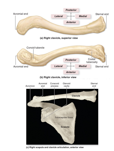
The pectoral girdle: Clavicle
-composed of 2 bones: Clavicle and Scapula
(aka collar bone)
-located anteriorly
-attachment point for many muscles
-acts as a brace to hold the arms and scapula away from the body
-Sternal end *articulates with the manubrium of the sternum
-Acromial end *articulates with the acromion of the scapula
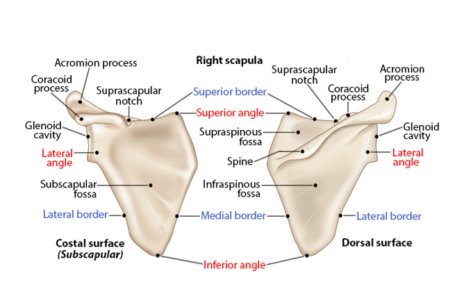
The pectoral girdle: Scapula
(aka shoulder blade)
-located posteriorly *attached to the axial skeleton via articulation with the clavicle and various muscles
-Dorsal surface possesses ridge called the spine *ends at the acromion which articulates with the clavicle
-Lateral border *Glenoid cavity serves as the site of articulation with humerus
-3 fossae for muscle attachment *(Ventral surface) - subscapular *(Dorsal surface) - supraspinous & infraspinous
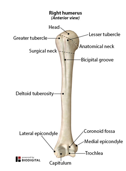
Humerus
-articulates with scapula to form the shoulder
*head *tubercules (greater & lesser) *deltoid tuberosity
articulates with ulna and radius to form the elbow *distal end *capitulum *trochlea *epicondyles (lateral & medial)
Head of humerus
-proximal end
-fits into the Glenoid cavity
Tubercules (Greater & Lesser)
attachment sites for the rotator cuff
Deltoid tuberosity
attachment for the deltoid muscle
Capitulum
-lateral
-articulates with the head of the radius
Trochlea
-medial
-articulates with the trochlear notch of the ulna
Epicondyles (lateral & medial)
-attachment sites for muscles
-ulnar nerve travels posterior to the medial epicondyle
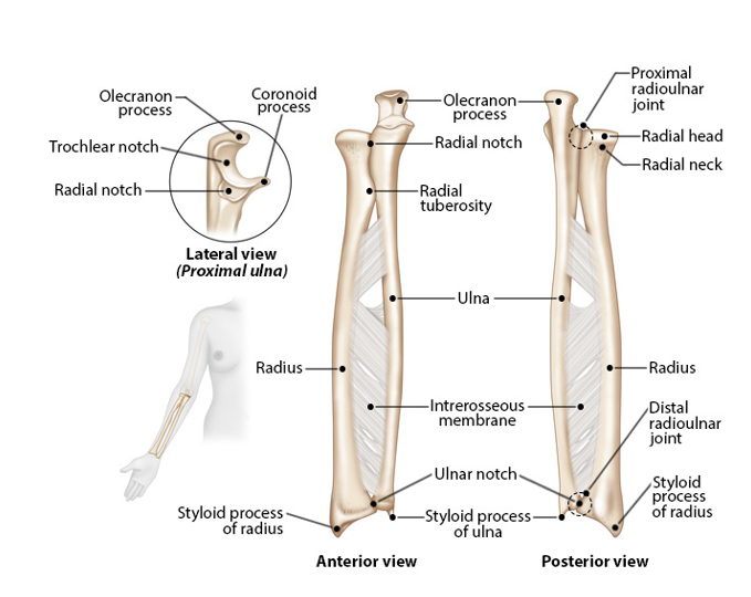
Forearm
-includes the ulna and radius *connected via interosseous membrane
-ulna
-radius
Ulna
-medial bone
-slightly longer than the radius
-trochlear notch
-olecranon
-styloidprocess: connects to the wrist
Trochlear notch
forms the elbow joint with humerus
Olecranon
bony end of the elbow
Radius
-lateral bone
-styloid process: connects to the wrist
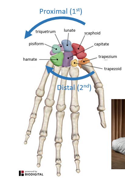
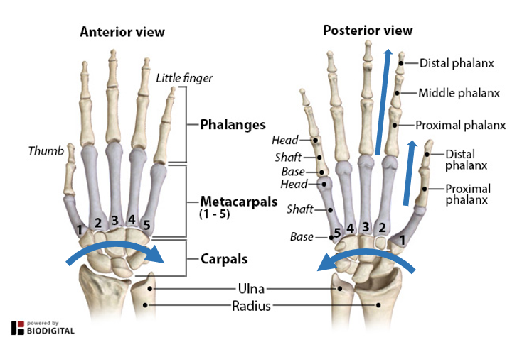
Carpal bones
-wrist bones
-8 per wrist
-closely united by ligaments
-2 rows, lateral to medial *proximal row *distal row
Proximal row of carpal bones
-Scaphoid, Lunate, Triquetrum, Pisiform
(Some Lunatics Try Positions)
Distal row of carpal bones
-Trapezium, Trapezoid, Capitate, Hamate
(That They Can't Handle)
Hands
-palms
-fingers
Palms
-5 metacarpal bones
-thumb or pollux (l) to Pinky (V) *lateral to medial
-heads of metacarpals make up the knuckles
Fingers
-14 phalanges (s. phalanx)
-Thumb (I) has 2 phalanges (proximal & distal)
-Index finger (II) to Pinky (V) each have 3 phalanges each (proximal, middle, and distal)
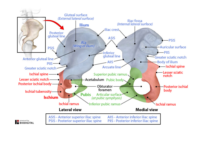
Pelvic Girdle
-attaches lower limbs to the axial skeleton
-refers to the pairs of os coxae (hip) bones *start out as 3 pairs of bones, but fuse age 13-15
3 bones -ilium (superior) -ischium (inferior) -pubis (anterior)
-Obturator foramen
-articulates with the femur at the acetabulum
Ilium
-superior
Iliac crest: protrudes, regarded as hip -serves as an attachment point for muscles of the trunk, hip, and thigh -articulates with the sacrum at the sacroiliac joint
Ischium
-inferior
-bear weight of the body when sitting via the paired ischial tuberosities
Pubis
-anterior
-jointed together by the pubic symphysis (fibrocartilage disc)
Obturator foramen
-formed by the pubis and ischium
-allows blood vessels and nerves to pass to lower limbs
Acetabulum
-the point of fusion for all 3 bones
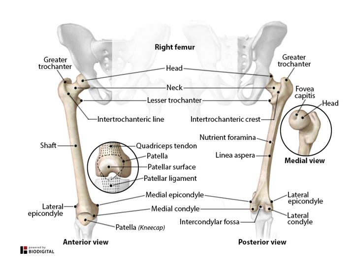
The thigh: femur
-largest, strongest, and longest bone in body (~1/4th size of height)
-covered with bulky muscles
-head articulates with hip
-shaft moves medially allowing knees to be closer together towards the body's center of gravity *gluteal tuberosity is the attachment site for the gluteus maximus muscle
-condyles (lateral & medial) articulate with the tibia to form the knee joint
-trochanters *greater projects laterally *lesser projects posteromedially *processes that serve as insertion sites for the gluteal and thigh muscles
-patella (kneecap) *enclosed in the quadriceps femoris tendon *protects the knee joint *improves the leverage of the thigh muscle
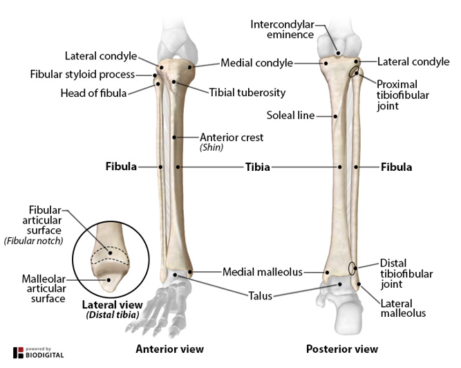
Lower leg
-tibia
-fibula
Tibia
-larger of the 2 bones
-head articulates with the femur
-located medially
-bears the weight of the body & transfers it to the foot
-articulates with the fibula & talus to form ankle *medial malleolus: bony process that forms the inside ankle bone
Fibula
-smaller of the two bones
-head articulates with the tibia
-located laterally
-articulates with the tibia & talus to form ankle *lateral malleolus: bony process that forms the outside ankle bone & provides lateral stability
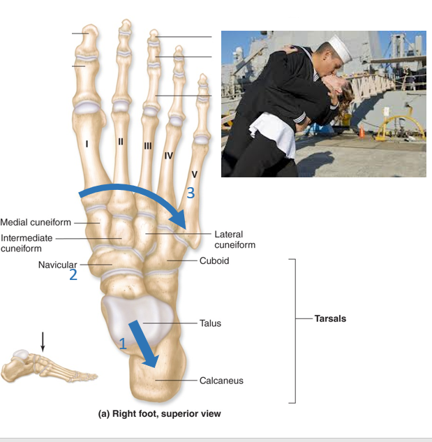
Tarsal bones
-ankle bones
-seven bones located distal to the lower leg *1st group (superior to inferior): Talus, Calcaneus *Middle medial loner: Navicular *Distal row (medial to lateral): Medial cuneiform, Intermediate cuneiform, Lateral cuneiform, Cuboid
-2 bones of interest *Talus (ankle) - articulates tibia and fibula *Calcaneus (heel) - under the talus, attached to the Achilles tendon
(Proximal group): Tall Californian (Middle loner): Navy (Distal row): Medical Interns Love Cuties
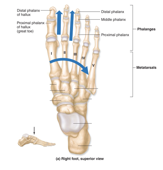
Metatarsals
-5 long bones located distal to the tarsal bones and proximal to the phalanges
-enumerated (I to V) *medial to lateral
Phalanges
-Hallux: big toe *has only a proximal and distal phalanx
-each of the other toes has 3 phalanx (proximal, middle, and distal) just as fingers do
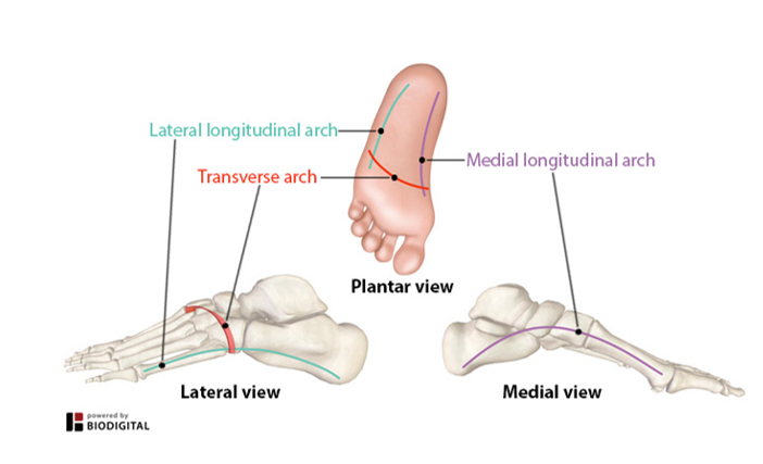
Arches of the foot
-provide strength to the foot
-allows for there to be some give in the foot
-2 longitudinal arches (medial & lateral)
-1 transverse arch
-maintained by the attachment between the bones, ligaments, and tendons
Medial longitudinal arch
-highest of the 3 arches
-prevents the medial side of the foot from touching the ground
-gives our footprint the characteristic shape
-extends from ball of foot (great toe) to heel
Lateral longitudinal arch
-not as high as the medial arch
-contributes to our footprint
-extends from little toe to the heel