neuroanatomy
1/80
There's no tags or description
Looks like no tags are added yet.
Name | Mastery | Learn | Test | Matching | Spaced |
|---|
No study sessions yet.
81 Terms
dorsoventral
dorsum (back) → entrum (belly)
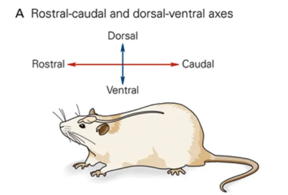
rostrocaudal/anterioposterior
longitudinal axis that runs from rostrum (beak) to caudal (tail)

mediolateral
horizontal axis that runs from the midline (medial) to the lateral margin of the animal

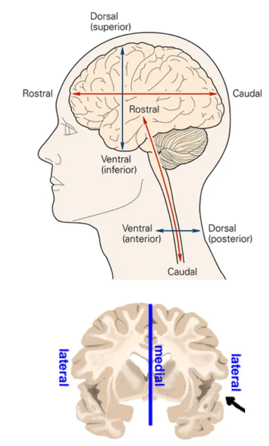
dorsoventral in human brain
top (head) to bottom (chin)

rostrocaudal/anteroposterior in human brain
front (eyes) to back

mediolateral in human brain
horizontal axis that runs from midline (medial) to the lateral margin of the animal
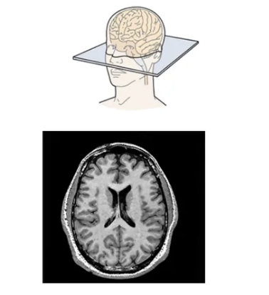
horizontal plane
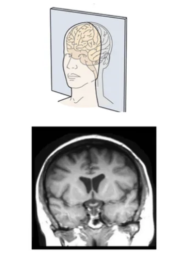
coronal plane
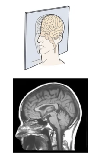
sagittal plane
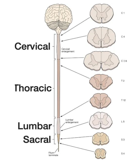
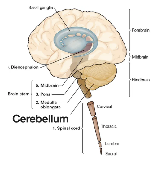
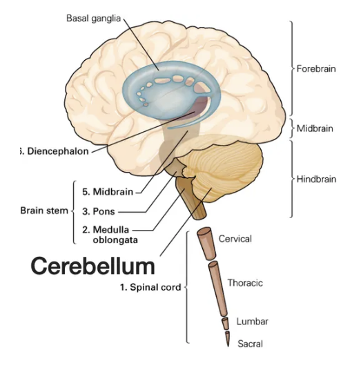
spinal cord has which regions
cervical, thoracic, lumbar, sacral
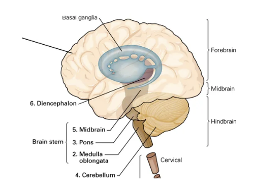
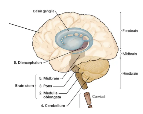
brain stem
necessary passage for all ascending and descending tracts between forebrain and spinal cord
modulates sensory, motor, and reflex behaviors (feeding, drinking, and respiration)
modulation of arousal; CNS activity
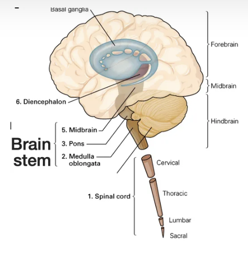
medulla
regulates autonomic functions like respiration, heart rate, and blood pressure
PreBotzinger complex: generates respiratory rhythm
pons
cranial nerve nuclei
mediates sensory and motor control of the head and neck
midbrain
voluntary motor control (substantia nigra)
processing of auditory and visual information (superior and inferior colliculi)
brainstem is the site of
cranial nerve nuclei
nucleus refers to
collection of neurons within a brain region
cells within a given nucleus share
the same projections, or inputs
cerebellum & motor control
modifies motor commands to adapt to desired output, accuracy of movement
cerebellum maintains
posture

cerebellum receives
sensory information from the spinal cord, balance from vestibular organs in the inner ear, and sensory information from the cortex
cerebellum is involved in certain forms of
learning

diencephalon is comprised of
thalamus, hypothalamus
thalamus
sensory relay center
information from visual, auditory, and somatosensory pathways relay here on their way to sensory cortices
hypothalamus
integrates physiology and behavior
regulates homeostatic behaviors including feeding, thirst, regulation of sleep-wake cycle, reproduction
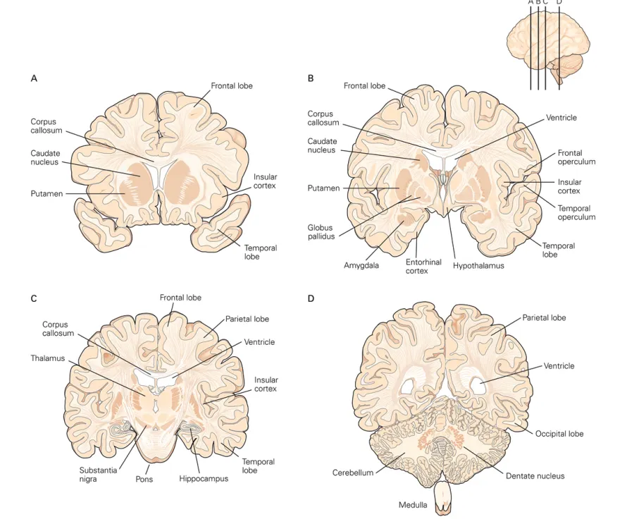
cerebrum is comprised of
cerebral cortex, hippocampus, amygdala
cerebral cortex
higher order cognitive processing
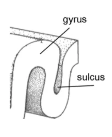
sulci and gyri increase the SA of the cortex
hippocampus
learning and memory
amygdala
formation and storage of memories associated with emotion
sulci and gyri properties
defined by anatomical features
not random outgrowths
consistent across individuals
serve as anatomical landmarks demarcating specific brain regions
sulci and gyri
sulci = in
gyri = out
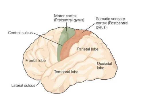
central sulcus and pre/post-central gyrus importance
precentral gyrus has motor cortex (control voluntary movement on the opposite side of the body)
postcentral gyrus has primary somatosensory cortex (processes sensations like touch, temperature, and pain from the opposite side of the body)
frontal lobe
short-term memory, planning, control of movement
temporal lobe
auditory information, learning and memory, emotion
parietal lobe
sensory processing, language processing
occipital lobe
visual processing
white matter has ___, but gray matter has ___
axon tracts (myelin)
cell bodies
what happens at the corpus callosum?
axons cross over so the sides can communicate with each other
true/false: in the human cortex, gray matter takes more more space than the white matter
FALSE
gray matter take up significantly less space than the white matter
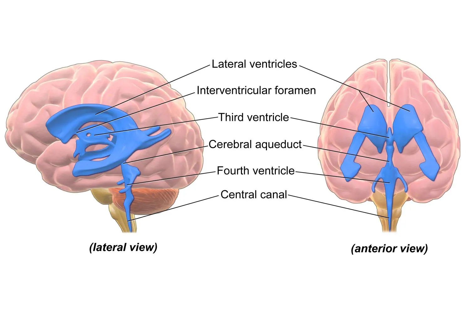
ventricles contain
CSF
how does the ventricles having CSF help?
provides protection by buffering the brain
“floats” the CNS, reduces weight of the brain and pressure at the base of the brain (brain is about 1300g, but in CSF is 25g)
important because bones are very stiff and thick, so floating the tissue helps to protect it from damage during movement
ventricles remove
waste from CNS to bloodstream
waste produced by
choroid plexus - epithelial cells found inside lateral ventricles
which ventricles are the most prominent?
lateral ventricles
meninges
protect the CNS
cells are fibroblasts
has 3 layers
3 layers of the meninges
dura → pia → arachnoid
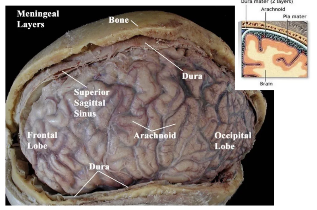
bacterial meningitis
results from bacterial infection and inflammation of meninges
cervical spine
sends/receives information to/from arms/upper body
lumbar spinal cord
sends/receives information to/from legs/lower body
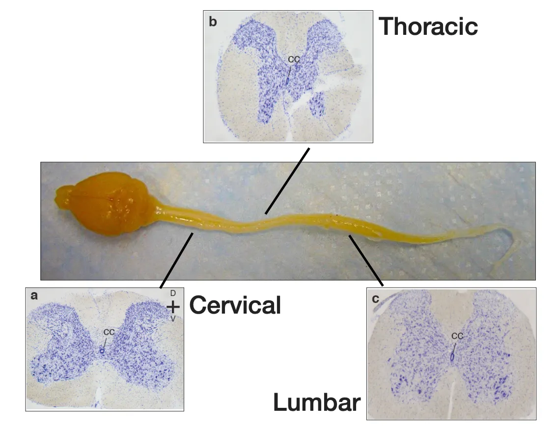
purple is gray matter (cell bodies)
white is axonal tracts
cervical innervates
arms
lumbar innervates
legs
thoracic innervates
core of our body
number of cells required to innervate arms/legs (and processes) is ____ than cells innervating our trunk
greater
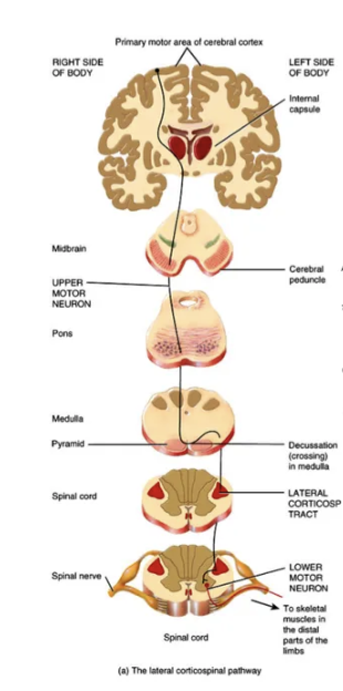
what does it mean that the brain and spinal cord are bilaterally symmetrical?
whatever is on one side is mirrored on the other side
decussation
many projections cross to the opposite hemisphere in the brain stem
retrograde tracers
travel from axon to cell body
axon terminal takes up the dye, and travels back to the soma
anterograde tracers
travel from cell body to axon terminal
cell body takes up due, travels down axon, crosses synapse and the connected cells will take up the due
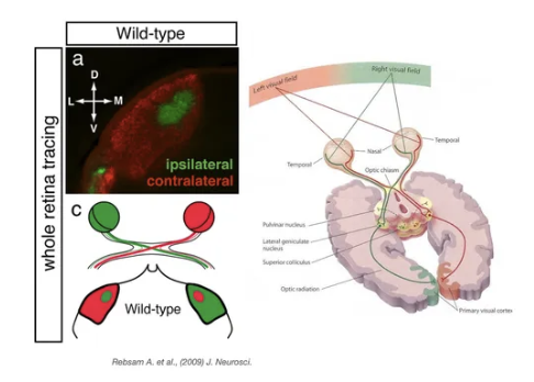
retina tracing
goes from cell body (retina), which sends projections into thalamus → send into visual cortex (occipital lobe)
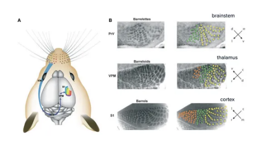
rodent whisker-barrel pathway
barrel processes whisker information
each whisker is mapped onto the somatosensory cortex (info from each whisker goes to corresponding barrel in cortex)
each dark structure = input from a specific whisker
there is a topographic organization to sensory input AND motor output
true/false: neurons projecting to specific muscle groups are organized and localized to specific regions
TRUE
inject tracers into muscle groups →
the cells there are more lateral compared to other group
can guess that the location of the muscle is more lateral relative to other one with more medial labelling
also look at shape of SC
motor neurons and the muscles that they innervate are ___ organized
topographically
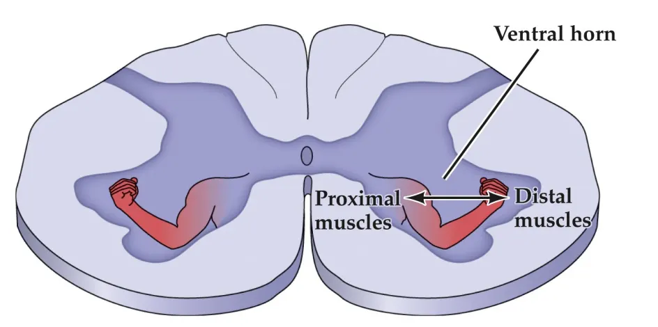
distal muscles have cell bodies in which region?
lateral

proximal muscles have cell bodies in which segment?
medial
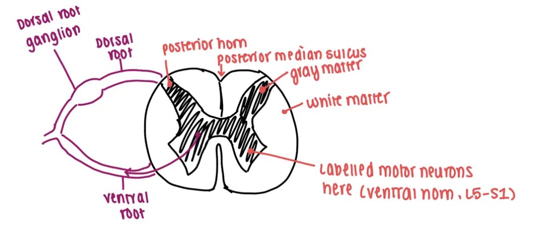
dorsal horn
receives and processes sensory information
ventral horn
contains motor neuron cell bodies; receive input from higher brain centers and from sensory neurons (via interneurons) to coordinate muscle contractions
lateral horn
contains neurons that control autonomic functions like heart rate, digestion, and blood vessel constriction
motor neurons innervating a single muscle distributed in
columns
medial motor column
innervates axial musculature; mediates posture
spinal accessory column
innervates neck muscles
phrenic motor column
innervates diaphragm, mediates respiration; rhythmic activity
pre-ganglionic motor column
spinal visceral motor neurons; innervate smooth muscle
hypaxial motor column
innervates musculature of the body wall (intercostal and abdominal muscles)
lateral motor column
innervates limb muscles
spatial arrangement and density of sensory receptors is mapped onto ___
the primary sensory cortex
spatial arrangement and density of sensory receptors reflects
degree of sensory sensitivity
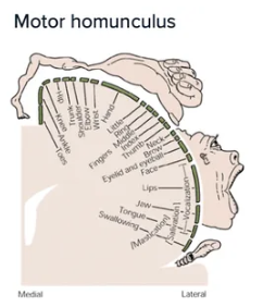
primary motor cortex
motor maps reflect density of neuronal innervation, correlated with degree of fine motor control
primary somatosensory cortex
abundance of neurons receiving input from specific region is associated with degree of sensory sensitivity
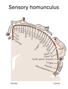
more sensitive the area → (more/less) sensory receptors/neurons needed to innervate it
MORE
Patient presents with difficulty controlling movement of their left arm and hand. You suspect a stroke and order an MRI. In which region of the brain would you expect to observe the lesion?
You would expect a lesion in the right motor cortex (since the right side of the brain controls the left side of the body, and the motor cortex governs movement)
More specifically, you could see this in the pre-central gyrus of right frontal lobe
It is the pre-central gyrus because this houses the primary motor cortex
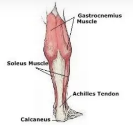
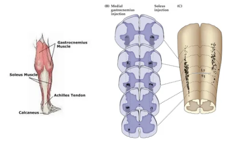
Consider an experiment in which you inject retrograde tracers into the gastrocnemius and soleus muscles. Where in the spinal cord would you expect to observe labeled cells? Draw your answer.
Retrograde tracer: travels from axon to cell body (the axon terminal takes up the dye and travels back to the soma)
You would expect to observe the labelled cells (motor neurons) in the ventral horn of the spinal cord’s gray matter, since this houses cell bodies
Specifically, they would be in the lumbar segments, since this is what innervates lower leg muscles
Labelled cells would be on the same side as the injected muscles