Histology - Gastrointestinal Tract
1/394
There's no tags or description
Looks like no tags are added yet.
Name | Mastery | Learn | Test | Matching | Spaced |
|---|
No study sessions yet.
395 Terms
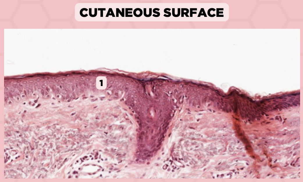
a. Epidermis
Identify the structure labelled in the given image?
a. Epidermis
b. Dermis
c. Hypodermis
d. Mucosa/Mucous Membrane

a. Keratinized stratified squamous epithelium
Identify the structure’s lining epithelium given in the image?
a. Keratinized stratified squamous epithelium
b. Nonkeratinized stratified squamous epithelium

Lip
What specimen is being showed in this picture?

Keratinized Stratified Squamous Epithelium (Epidermis)
Identify the structure labeled as 1.
d. Mucosa/Mucous Membrane
Identify the structure labelled in the given image?
a. Epidermis
b. Dermis
c. Hypodermis
d. Mucosa/Mucous Membrane

b. Nonkeratinized stratified squamous epithelium
Identify the structure’s lining epithelium given in the image?
a. Keratinized stratified squamous epithelium
b. Nonkeratinized stratified squamous epithelium
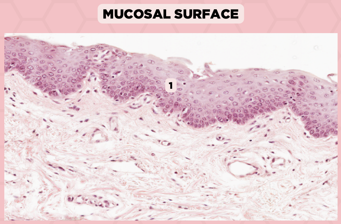
Lip
What specimen is being showed in the picture?
Non-Keratinized Stratified Squamous Epithelium (Mucosa/Mucous Membrane)
Identify the structure labeled as 1.

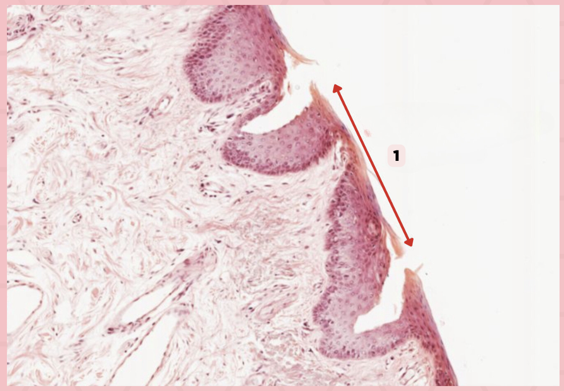
a. Abrupt Transition from keratinized to nonkeratinized epithelium
Identify the structure’s lining epithelium given in the image?
a. Abrupt Transition from keratinized to nonkeratinized epithelium
b. Abrupt Transition from nonkeratinized to keratinized epithelium
c. AOTA
d. NOTA

Lip
What specimen is being showed in the picture?

Mucocutaneous Junction
Identify the structure labeled as 1.
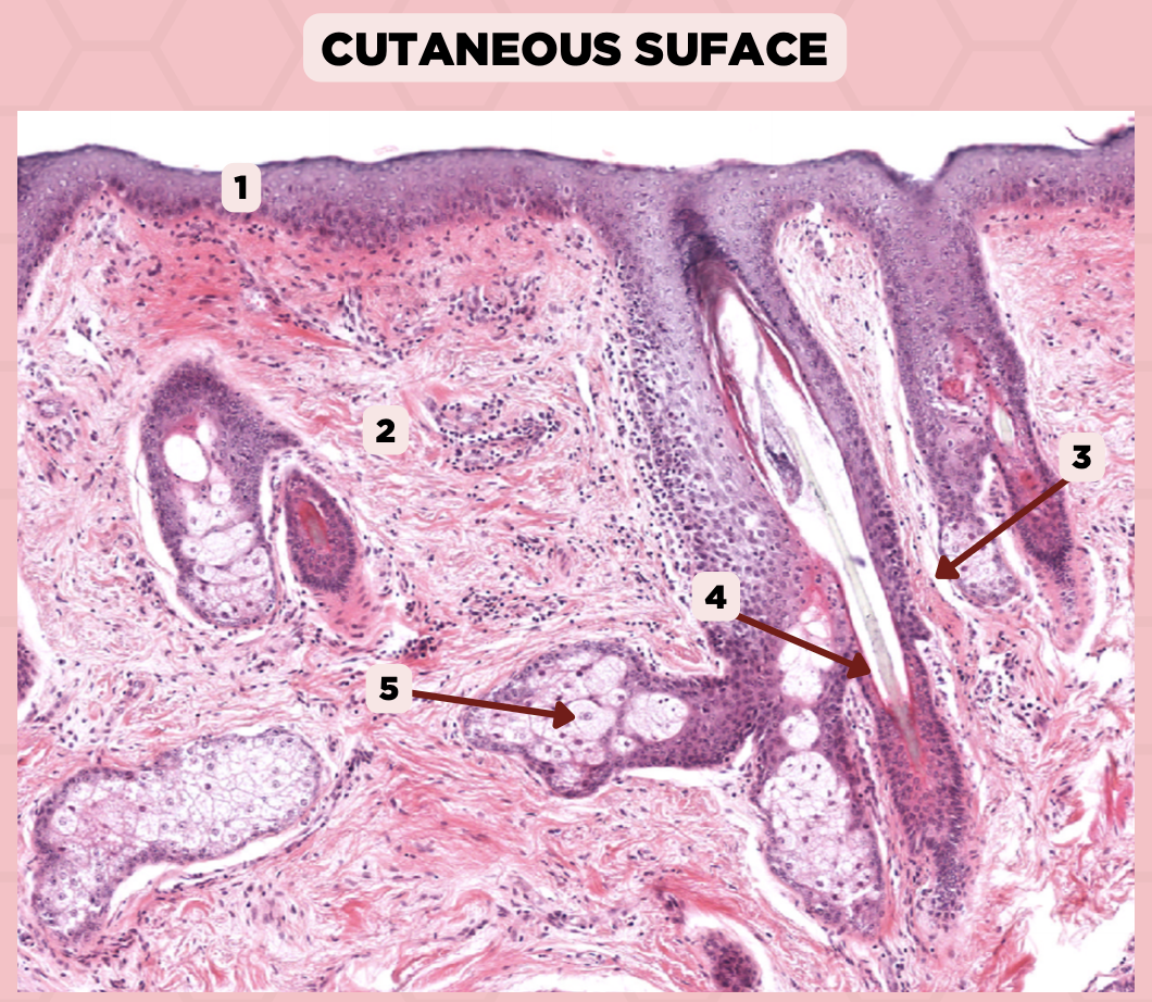
Lip
What specimen is being showed in the picture?
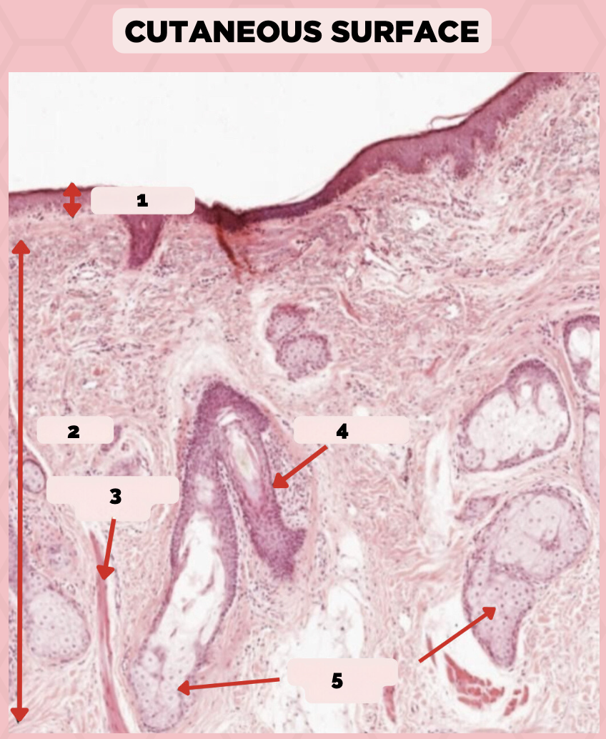
Epidermis
Identify the structure labeled as 1.

Dermis
Identify the structure labeled as 2.

Arrector Pilli Muscle
Identify the structure labeled as 3.

Hair Follicle
Identify the structure labeled as 4.

Sebaceous Glands
Identify the structure labeled as 5.

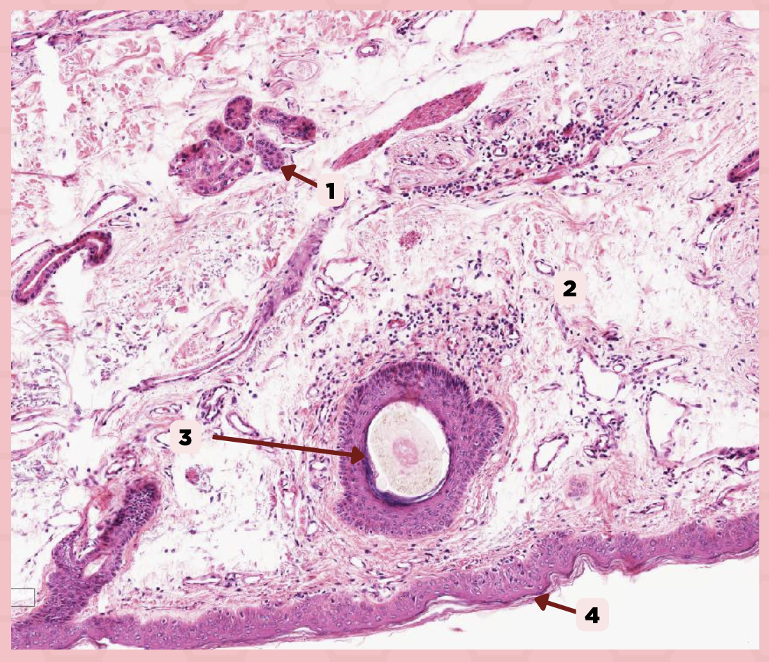
Lip
What is the specimen showed in the picture?
Epidermis
Identify the structure labeled as 1.

Dermis
Identify the structure labeled as 2.

Arrector Pilli Muscle
Identify the structure labeled as 3.

Hair Follicle
Identify the structure labeled as 4.

Sebaceous Glands
Identify the structure labeled as 5.

Lip
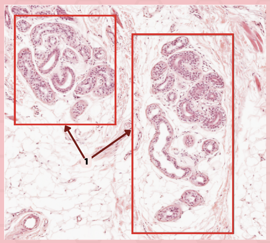
What specimen is showed in the picture?

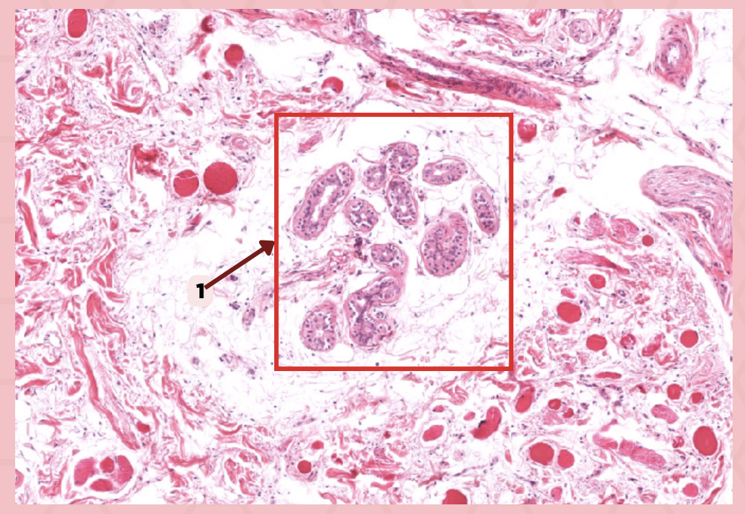
Labial Glands (in the Mucosa)
Identify the structure labeled as 1.

Skeletal Muscle Cells
Identify the structure labeled as 2.
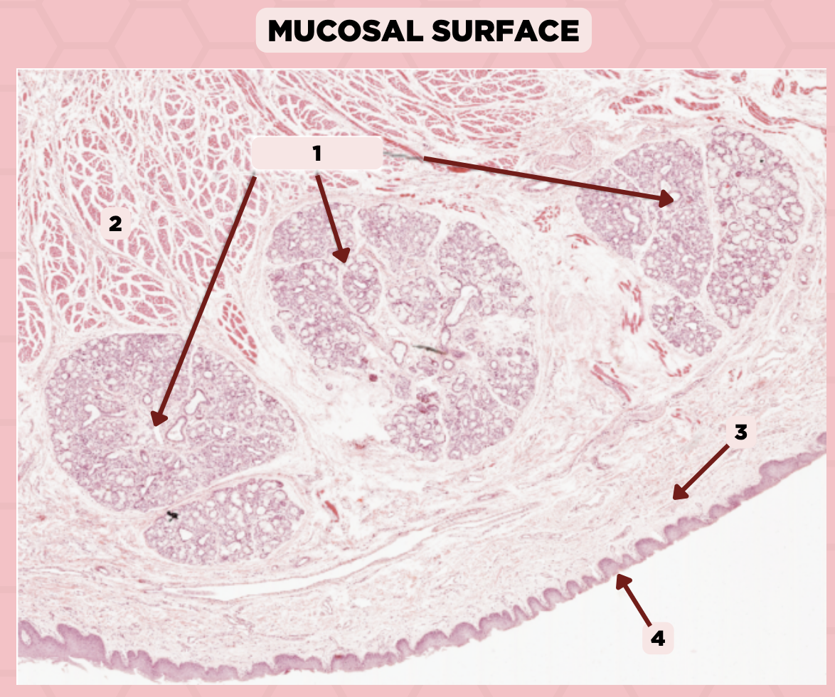
Lamina Propria
Identify the structure labeled as 3.

Mucosa
Identify the structure labeled as 4.

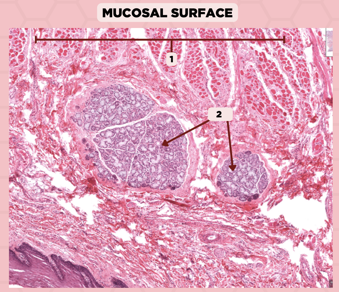
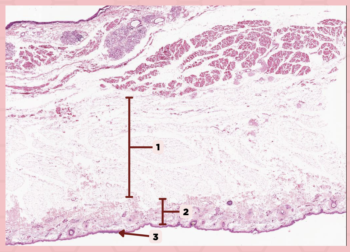
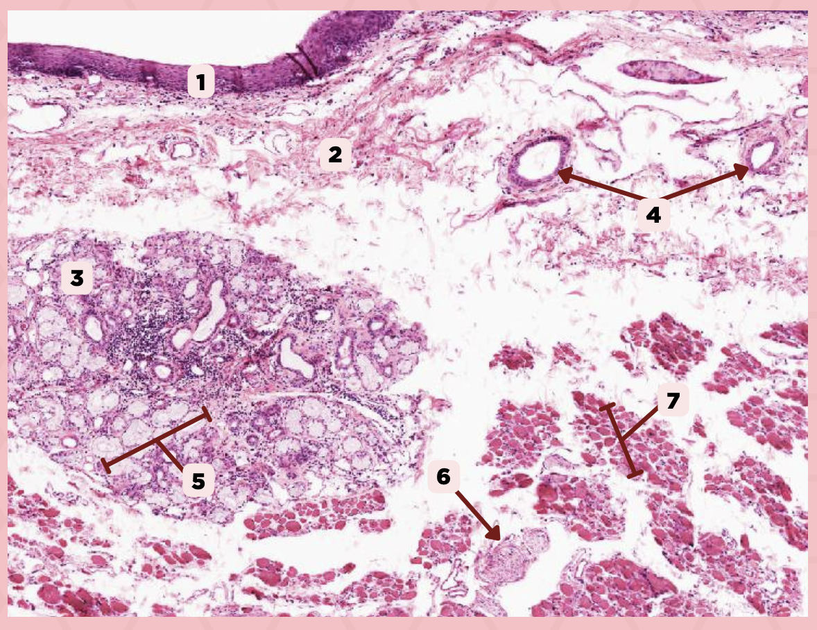
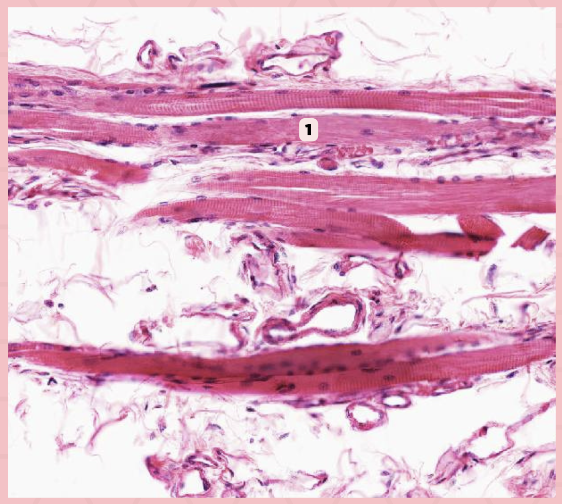
Lip
What specimen is showed in the picture?

Skeletal Muscle Fibers
Identify the structure labeled as 1.

Labial Glands
Identify the structure labeled as 2.
b. Dermis of thin skin
Identify where the structure is located?
a. Epidermis of thin skin
b. Dermis of thin skin
c. Epidermis of thick skin
d. Dermis of thick skin
Sweat Glands
Identify the structure labeled as 1.

b. Eccrine glands
What type of glands labelled as #1 as in the given image?
a. Apocrine glands
b. Eccrine glands
c. Merocrine glands
d. NOTA
Sweat Glands
Identify the structure labeled as 1.

Cheek (Coronal Section)
Identify the specimen.

Hypodermis
Identify the structure labeled as 1.

Dermis
Identify the structure labeled as 2.

Epidermis
Identify the structure labeled as 3.
a. Keratinized stratified squamous epithelium
What is the lining of the structure labeled as #1?
a. Keratinized stratified squamous epithelium
b. Nonkeratinized pseudostratified squamous epithelium
c. Stratified squamous epithelium

Cheek
Identify the specimen.

Sweat Glands
Identify the structure labeled as 1.
Dermis
Identify the structure labeled as 2.

Hair Follicle
Identify the structure labeled as 3.

Epidermis (Keratinzied Stratified Squamous Epithelium)
Identify the structure labeled as 4.

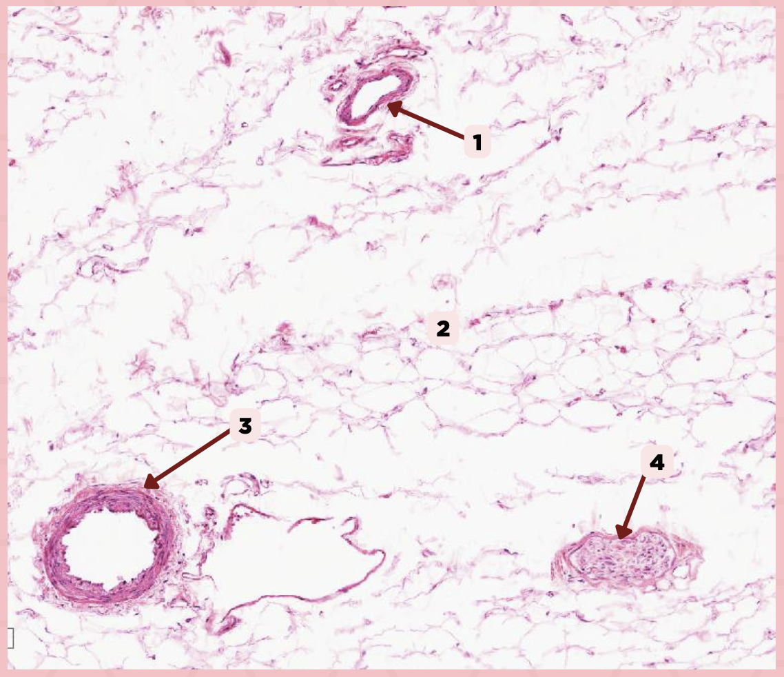
Cheek
Identify the specimen.

Vein
Identify the structure labeled as 1.

Hypodermis
Identify the structure labeled as 2.

Artery
Identify the structure labeled as 3.

Nerve
Identify the structure labeled as 4.
Cheek
Identify the specimen.

Mucosa
Identify the structure labeled as 1.

Lamina Propria
Identify the structure labeled as 2.

MALT
Identify the structure labeled as 3.

Blood Vessels
Identify the structure labeled as 4.

Buccal Glands
Identify the structure labeled as 5.

Nerve
Identify the structure labeled as 6.

Muscle Fascicles
Identify the structure labeled as 7.
c. Deep Submucosa Layer
Identify the layer of the tissue labelled as #1 in the given image?
a. Dermis Layer
b. Hypodermis Layer
c. Deep Submucosa Layer
d. Epidermis Layer
Cheek
Identify the specimen.

Skeletal Muscle Fibers
Identify the structure labeled as 1.

C) Septa
Which of the following structures shown delineate the lobes and lobules of the parotid gland?
A) Capsule
B) Lobule
C) Septa
D) Lymph Nodes

A) Capsule
Which of the following structures encapsulates the parotid gland?
A) Capsule
B) Lobule
C) Septa
D) Lymph Nodes

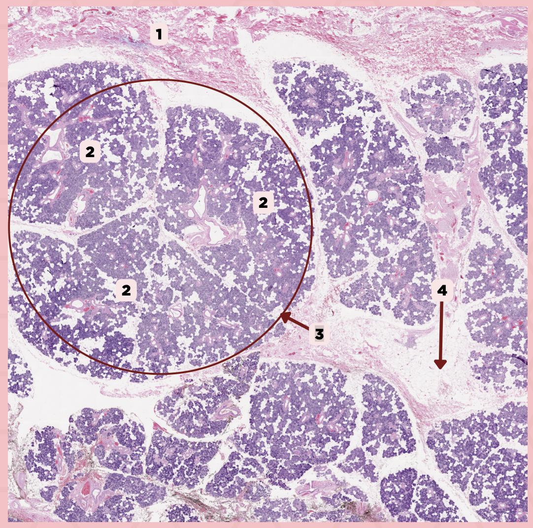
Parotid
Identify the specimen.

Capsule
Identify the structure labeled as 1.

Septa
Identify the structure labeled as 2.

Lobule
Identify the structure labeled as 3.
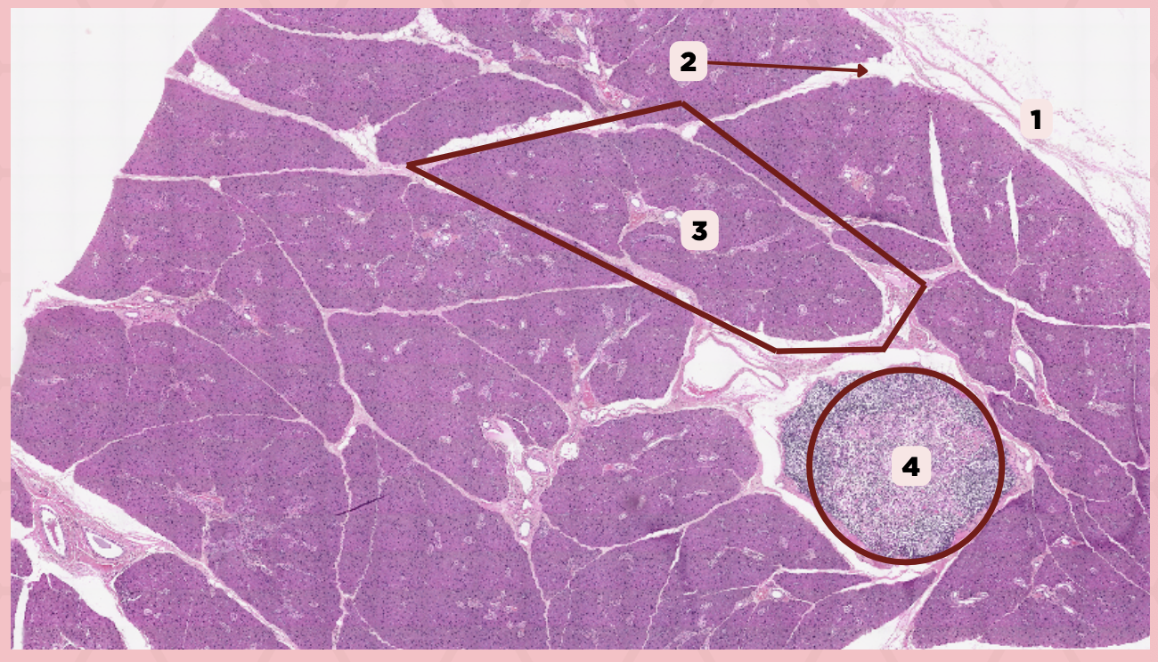
Lymph Node
Identify the structure labeled as 4.

B) Interlobular Ducts
A duct within lobules with stratified cuboidal epithelium in the initial segment and stratified columnar epithelium in the proximal segments.
A) Striated Ducts
B) Interlobular Ducts
C) Intercalated Ducts
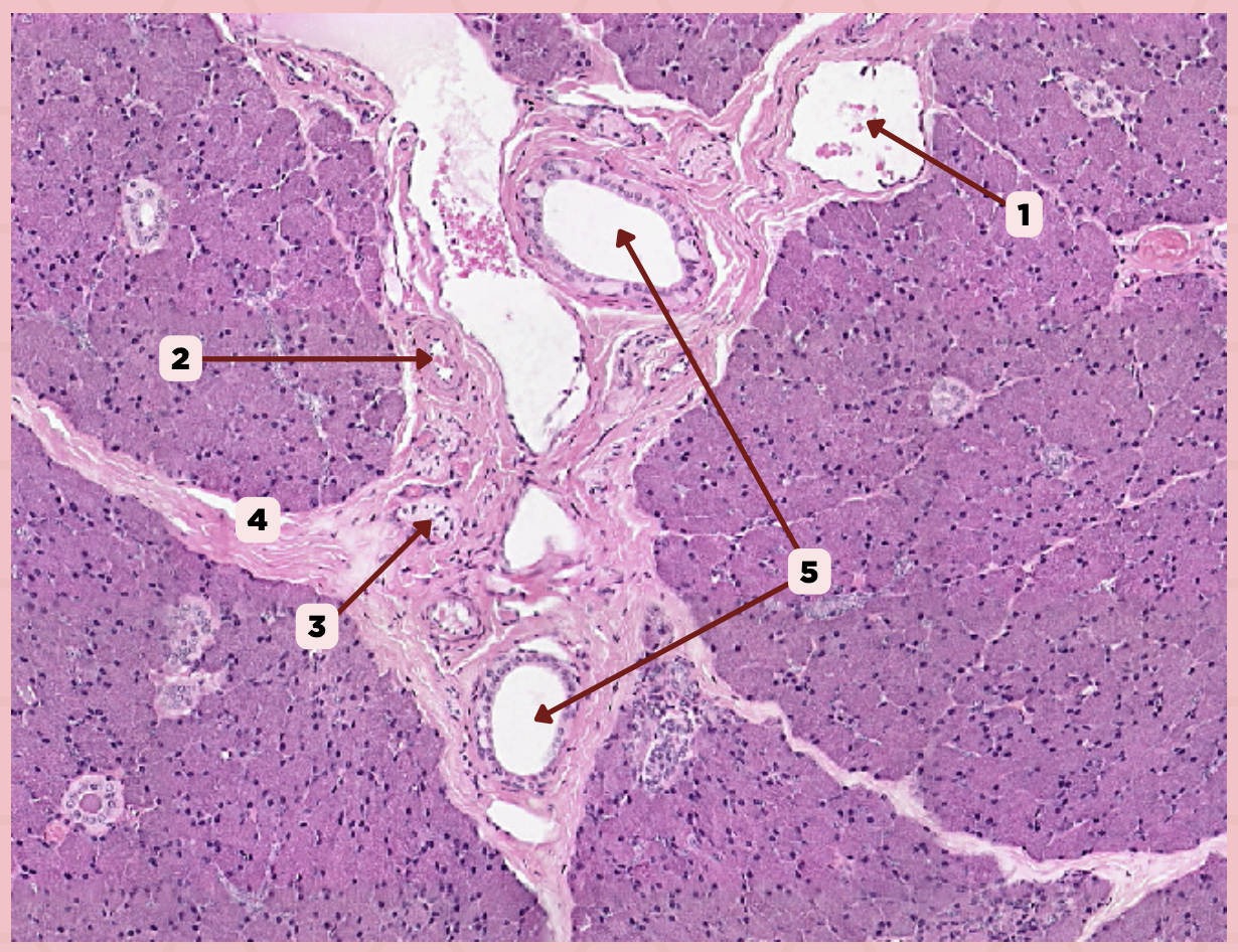
Parotid
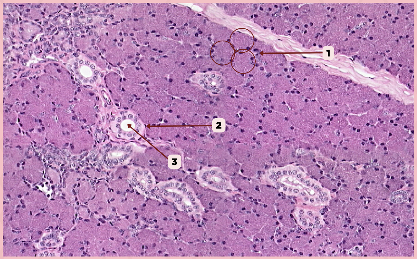
Identify the specimen.

Vein
Identify the structure labeled as 1.

Artery
Identify the structure labeled as 2.

Nerve
Identify the structure labeled as 3.

Interlobular Septa
Identify the structure labeled as 4.

Interlobular Duct
Identify the structure labeled as 5.

A) Myoepithelial Cells
These cells are associated with secretory units of salivary glands that are contractile to help in ejecting secretions
A) Myoepithelial Cells
B) Goblet Cells
C) Adipocytes

A) Striated Ducts
This duct has a wall consisting of a simple cuboidal or columnar epithelium and is formed by the union of intercalated ducts.
A) Striated Ducts
B) Interlobular Ducts
C) Intercalated Ducts

Parotid
Identify the specimen.

Secretory Acini
Identify the structure labeled as 1.
Myoepithelial Cell
Identify the structure labeled as 2.

Secretory Duct
Identify the structure labeled as 3.

C) Adipocytes
The septa delineating the lobes and lobules contain a significant amount of:
A) Loose Connective Tissues
B) Myoepithelial Cells
C) Adipocytes
D) Reticular Tissue

Parotid
Identify the specimen.

Capsule
Identify the structure labeled as 1.
Lobule
Identify the structure labeled as 2.

Lobe
Identify the structure labeled as 3.

Connective Tissue
Identify the structure labeled as 4.

C) Striated Ducts
This duct exhibits intense cytoplasmic eosinophilia and basal striations when seen in routine LM preparations.
A) Intercalated Ducts
B) Excretory Ducts
C) Striated Ducts
D) Typical Serous Cells
The parotid gland is purely serous therefore its secretory units are exclusively formed by:
A) Nonkeratinized Cells
B) Myoepithelial Cells
C) Adipocytes
D) Typical Serous Cells

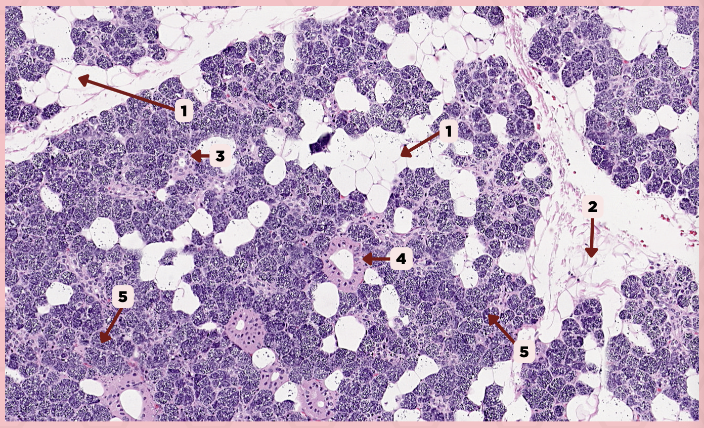
Parotid
Identify the specimen.

Adipocyte
Identify the structure labeled as 1.

Connective Tissue
Identify the structure labeled as 2.

Intercalated Duct
Identify the structure labeled as 3.

Striated Duct
Identify the structure labeled as 4.

Serous Gland
Identify the structure labeled as 5.

B) Wharton’s Duct
This is the main excretory duct found in the submandibular gland
A) Stensen’s Duct
B) Wharton’s Duct
C) Ducts of Rivinus
D) Sublingual Duct of Bartholin
C) Lingual Frenulum
The Wharton’s duct opens into the oral cavity underneath the tongue beside the:
A) Superior Labial Frenulum
B) Inferior Labial Frenulum
C) Lingual Frenulum

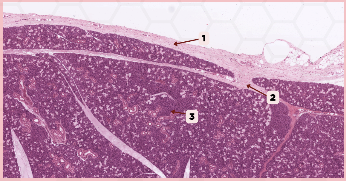
Submandibular
Identify the specimen.

Capsule
Identify the structure labeled as 1.

Septa
Identify the structure labeled as 2.
