Lec 4: The Eye, Optic Nerve and the Visual Pathways
1/64
There's no tags or description
Looks like no tags are added yet.
Name | Mastery | Learn | Test | Matching | Spaced | Call with Kai |
|---|
No analytics yet
Send a link to your students to track their progress
65 Terms
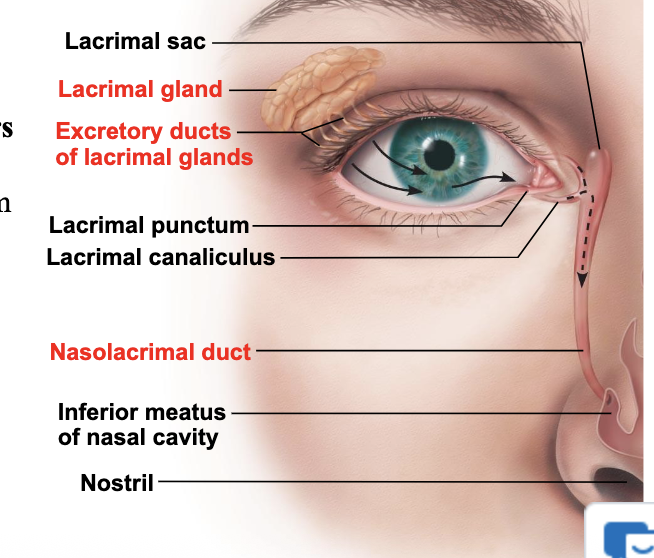
what is Lacrimation?
– Activated by Parasympathetic fibers in the Facial Nerve (CN VII)
– Reduced by Sympathetic fibers from the Superior Cervical/Sympathetic Ganglion in the neck
Excess “tears” drain into
nasal cavity
The __ refracts/focuses light through the Pupil (hole in iris) onto the lens
Cornea
Lens projects and focuses light images onto the
retina
Lens is attached, via ligaments, to the muscles of the
ciliary body
__ in the retina are activated by photons of light
Photoreceptors
Sensory signals, originating at the photoreceptors, are transmitted out of the eye through the
optic nerve
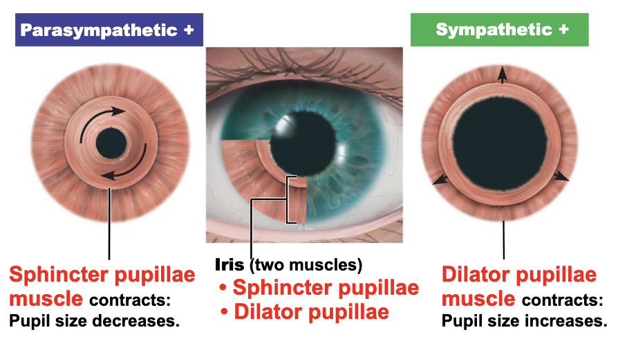
iris muscles
sphincter pupillae muscle contracts:
pupil size decreases
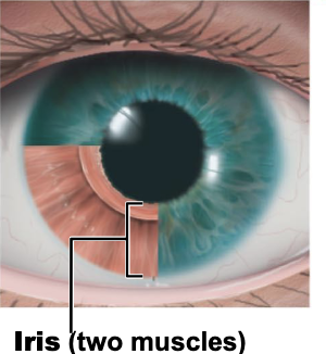
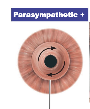
what is two muscles of Iris
• Sphincter pupillae
• Dilator pupillae
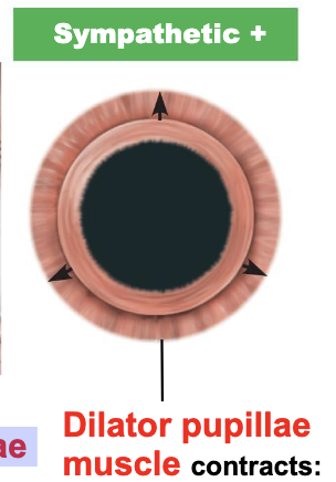
dilator pupillae muscle contracts:
pupil size increaes
Colored part of eye function:
regulates amount of light entering eye by dilation/constriction of Pupil
Near vision and bright light
sphincter pupillae (circular muscles) contract
pupils constrict
parasympathetic CN III
Distant vision and dim light
• dilator pupillae (radial muscles) contract
• pupils dilate
• sympathetic fibers from the Superior Cervical/Sympathetic Ganglion in the neck
Changes in emotional state
pupils dilate when subject matter is appealing, sexually attractive or requires problem-solving skills
Visual accommodation is the ability of the eye to change focus to maintain a clear image of an object as its distance changes. This process is achieved by the lens of the eye changing shape:
near objects
distant objects
accommodation near object:
The lens becomes more round to focus on nearby objects.
accommodation distant object:
The lens flattens to focus on distant objects.
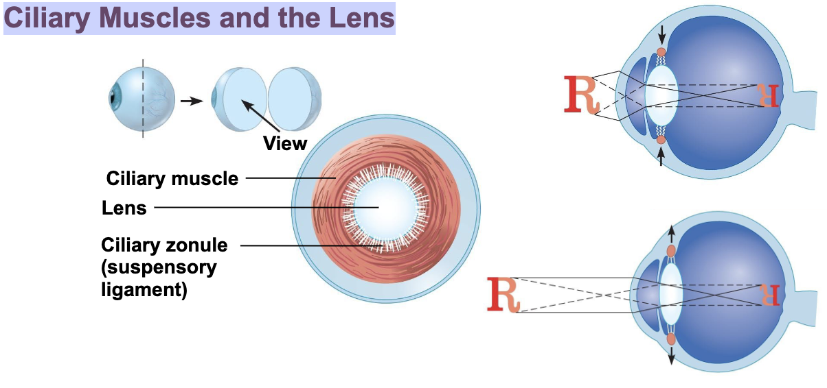
Ciliary Muscles and the Lens
The ciliary muscle and ciliary zonule are arranged sphincter-like
around the lens. As a result,
contraction loosens the ciliary zonule fibers and relaxation tightens them
what is Focusing For Distant Vision (there’s 3)?
far point of vision
pupils dilated
ciliary muscles relaxed
what is Far point of vision?
– Distance beyond which no change in lens shape needed for focusing
20 feet for emmetropic (normal) eye
Cornea and lens focus light precisely on retina
ciliary muscles relaxed:
Lens stretched flat by tension in ciliary muscles
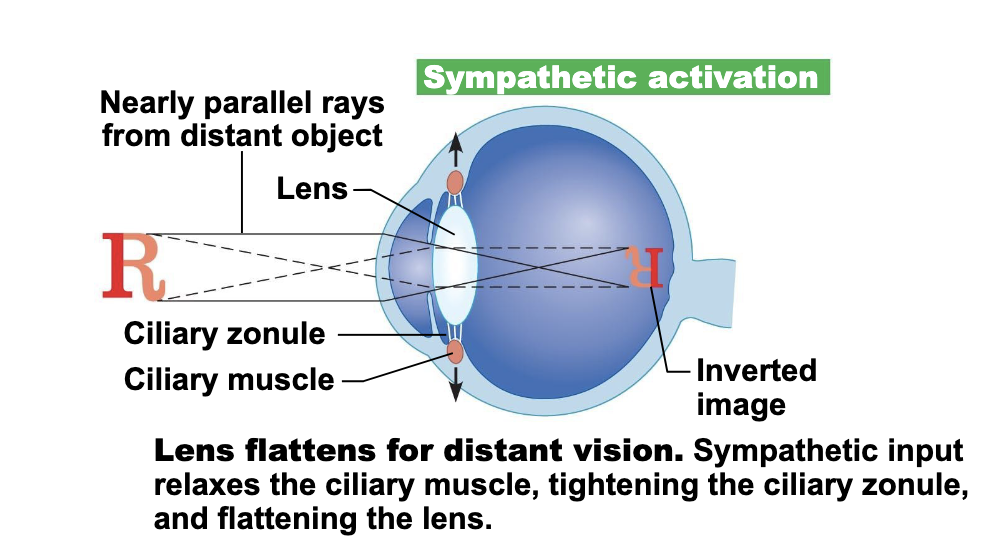
Lens flattens for distant vision.
Sympathetic input relaxes the ciliary muscle, tightening the ciliary zonule, and flattening the lens
Light from close objects diverges it as approaches eye
Requires eye to make active adjustments using three simultaneous processes
Accommodation of lenses – Parasym. CN III
Constriction of pupils – Parasym. CN III
Convergence of eyeballs – Med. Rectus, CN III
Accommodation of lenses –
Parasym. CN III
Constriction of pupils –
Parasym. CN III
Convergence of eyeballs –
Med. Rectus, CN III
what is accommodation?
Changing lens shape to increase refraction
Near point of vision
Closest point on which the eye can focus
Presbyopia—loss of accommodation over age 50
what is constriction ?
Accommodation pupillary reflex constricts pupils to prevent most divergent light rays from entering eye
what is convergence ?
Medial rotation of eyeballs toward object being viewed
Medial Rectus muscle (CN III)
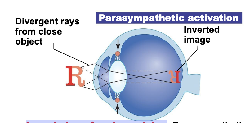
Lens bulges for close vision
Parasympathetic input contracts the ciliary muscle, loosening the ciliary zonule, allowing the lens to bulge.
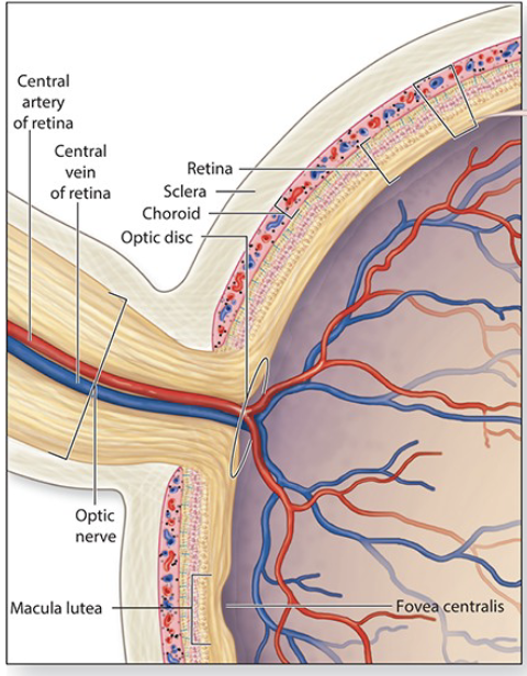
what is optic disc?
the internal location, in the posterior and medial part of the eye, where the optic nerve (and retinal blood vessels) exits the eye
The __ has the highest concentration of photoreceptors
macula
The center of the macula is called the
fovea centralis
what are Photoreceptors?
Rods
cones
what are rods?
Activated in dim/dark conditions
A night-vision photoreceptor
Detect white, gray and black
Highest concentration in the periphery of the retina
what is cones?
Activated in bright light
Detect color
3 types
Activated by different colors
Red/orange, Yellow/Green & Blue
Highest concentration is in the macula
Signals from rods and cones (eventually) exit the eye via the
axons of ganglion cells
• The axons of ganglion cells, not photoreceptors, form the optic nerve
what is Fundoscopy of optic disc?
Increased intraocular pressure results in edema at the optic disc
Axons and blood vessels passing through the optic disc get compressed
Vision can be lost
Retinal arteries appear pale due to reduced blood flow
what is optic nerve?
• From the retina to the optic chiasm
• Injury to the optic nerve results in blindness in the ipsilateral eye
From Optic Chiasm to deeper brain features
Fibers from the nasal portion of each eye cross to the contralateral side of the brain at the optic chiasm
Carrying image signals from the lateral visual field
Fibers from the nasal side of one eye and the temporal side of the others eye (1/2 the visual field) come together at the Lateral Geniculate Nucleus of the Thalamus
optic tract
Fibers from the nasal portion of each eye cross to the contralateral side of the brain at the
optic chiasm
Fibers from the nasal side of one eye and the temporal side of the others eye (1/2 the visual field) come together at the __ of the Thalamus
Lateral Geniculate Nucleus
Compression of the optic chiasm by pituitary adenoma will result in
• deficits in lateral vision in both eyes
• Bitemporal Hemianopia
what is Optic Radiations?
A collection of axons that carry visual information from the LGN to the primary visual cortex.
Ensures visual information reaches the correct area in the primary visual cortex for interpretation
the optic radiations is divided into 2 pathways:
Upper/Dorsal (parietal) pathway
carry information from the Inferior retinal quadrants
Lower (temporal) pathway (Meyer’s loop)
transmits information from the Superior quadrants.
what does Upper/Dorsal (parietal) pathway carry?
carry information from the Inferior retinal quadrants
what does Lower (temporal) pathway (Meyer’s loop) transmit?
transmits information from the Superior quadrants.
Along the Calcarine sulcus of the Occipital lobes of the brain
Processes basic features like edges, orientation, and movement..
Visual information from the right visual field is processed in the left visual cortex, and vice versa
Visual Information is sent on to other parts of the brain for higher processing and identification
primary visual cortex
what are parts of Visual Processing/Interpretation?
parietal lobe processing of vision
temporal lobe processing of vision
frontal lobe processing of vision
what is parietal lobe processing of vision?
• Where is it?
• How fast and in what direction is it moving?
what is temporal lobe processing of vision?
• What/who does it look like?
• What are it’s features?
what is frontal lobe processing of vision?
Combines all visual processing information to make conscious identification
Some optic tract fibers (~5%) go straight to the
superior colliculus
Afferent nerve fibers headed for the superior colliculus have three essential functions:
alignment of the head
control of pupillary diameter
adjusting to near vision accommodation
optic tract: alignment of the head
• Turning your head towards visual/auditory stimuli
• Activates the Tectospinal Tract
optic tract: control of pupillary diameter
Each superior colliculus receives signals from both eyes – unilateral stimulation produces a bilateral response (through CN III)
optic tract: Adjusting to near vision accommodation
The lens changes shape to focus on a near object (through CN III)
Visual information from both eyes reaches the Pretectal Nucleus in the Superior
Colliculus
Pretectal nucleus send information to both Oculomotor nuclei to cause constriction of both pupils
Only injuries to the optic nerve or chiasm can produce an Afferent Pupillary Defect
Pupillary Light Reflex
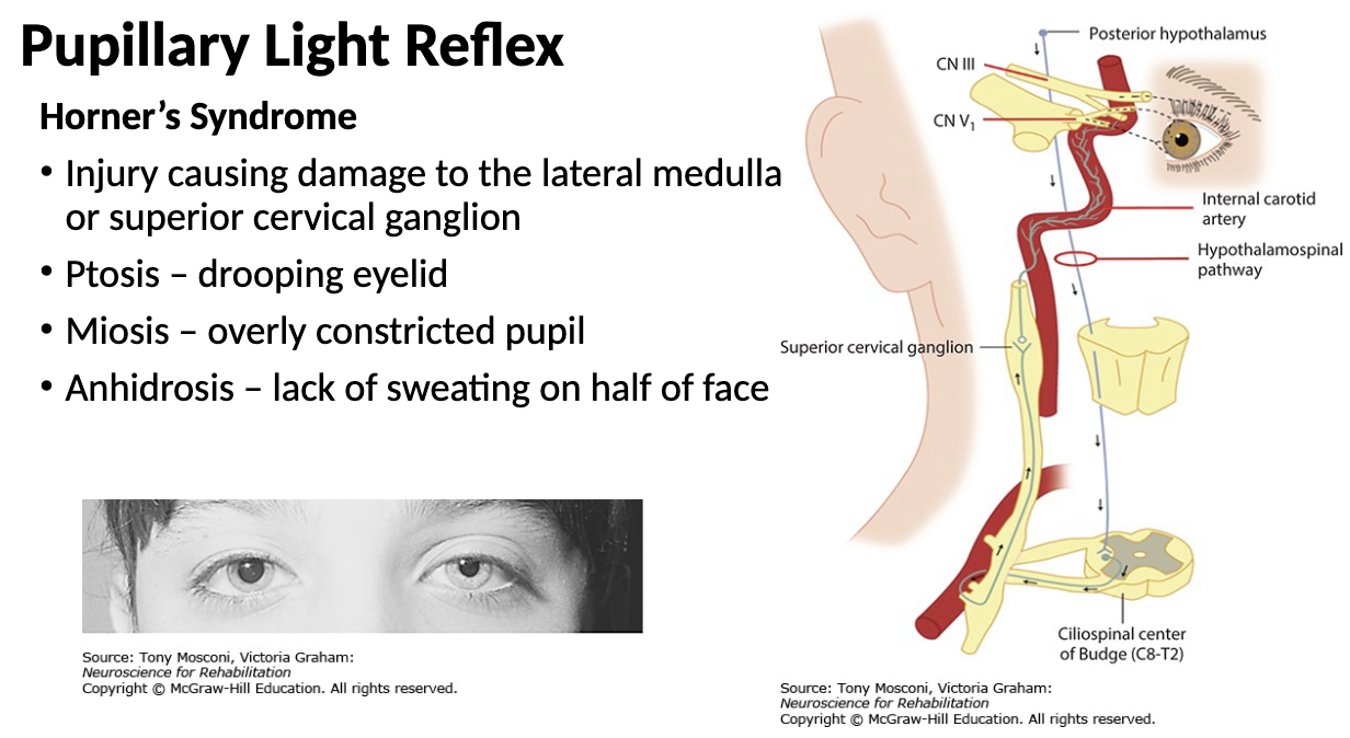
what is Horoner’s Syndrome?
Injury causing damage to the lateral medulla or superior cervical ganglion
• Ptosis – drooping eyelid
• Miosis – overly constricted pupil
• Anhidrosis – lack of sweating on half of face
what is ptosis?
drooping eyelid
what is miosis?
overly constricted pupil
what is anhidrosis?
lack of sweating on half of face