Shiva PC2: Benign Soft Tissue Pathology
1/68
There's no tags or description
Looks like no tags are added yet.
Name | Mastery | Learn | Test | Matching | Spaced |
|---|
No study sessions yet.
69 Terms
Soft tissue tumors
• Derived from mesenchymal tissues
• Majority of benign soft tissue tumors are reactive
• Due to low-grade chronic irritation
• Some are neoplastic
• Fibrous
• Adipose
• Vascular
• Lymphatic
• Neural
• Muscle
• Uncertain Origin
Soft tissue pathology categories
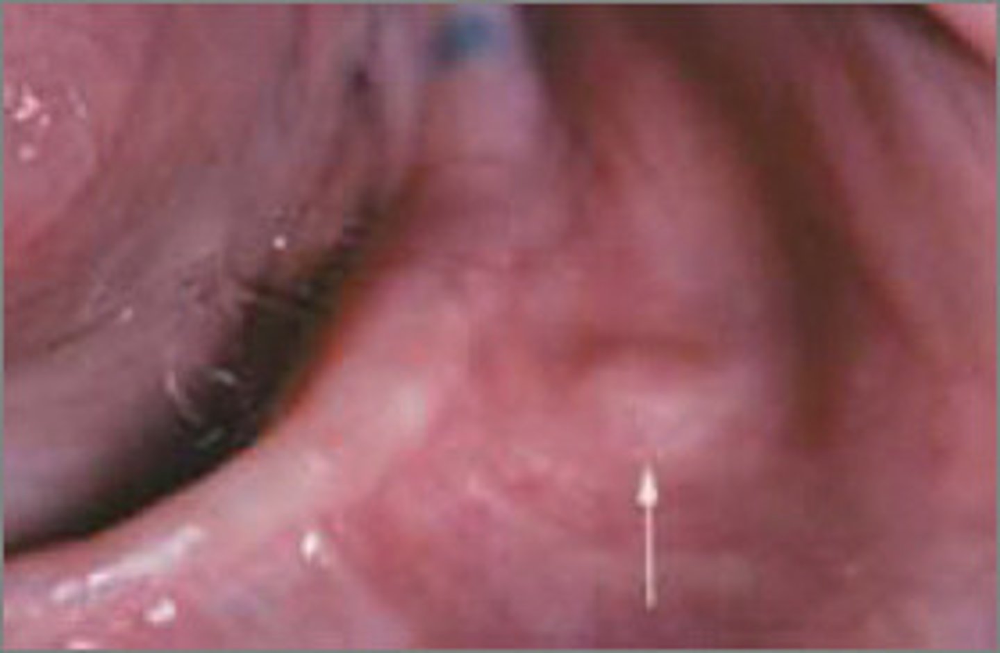
Fibroma
• Most common benign soft
tissue mass
• Results from irritation or trauma
• Reactive lesion
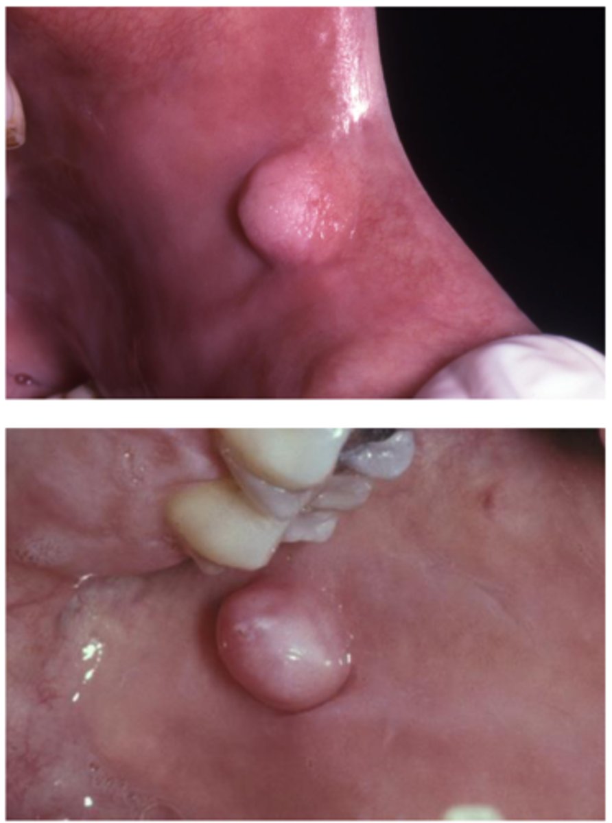
Fibroma
• Well-localized
• Dome-shaped
• Asymptomatic (unless secondary
ulceration)
• Smooth surface
• Often pink but may be ulcerated
if traumatized
• Sessile or pedunculated
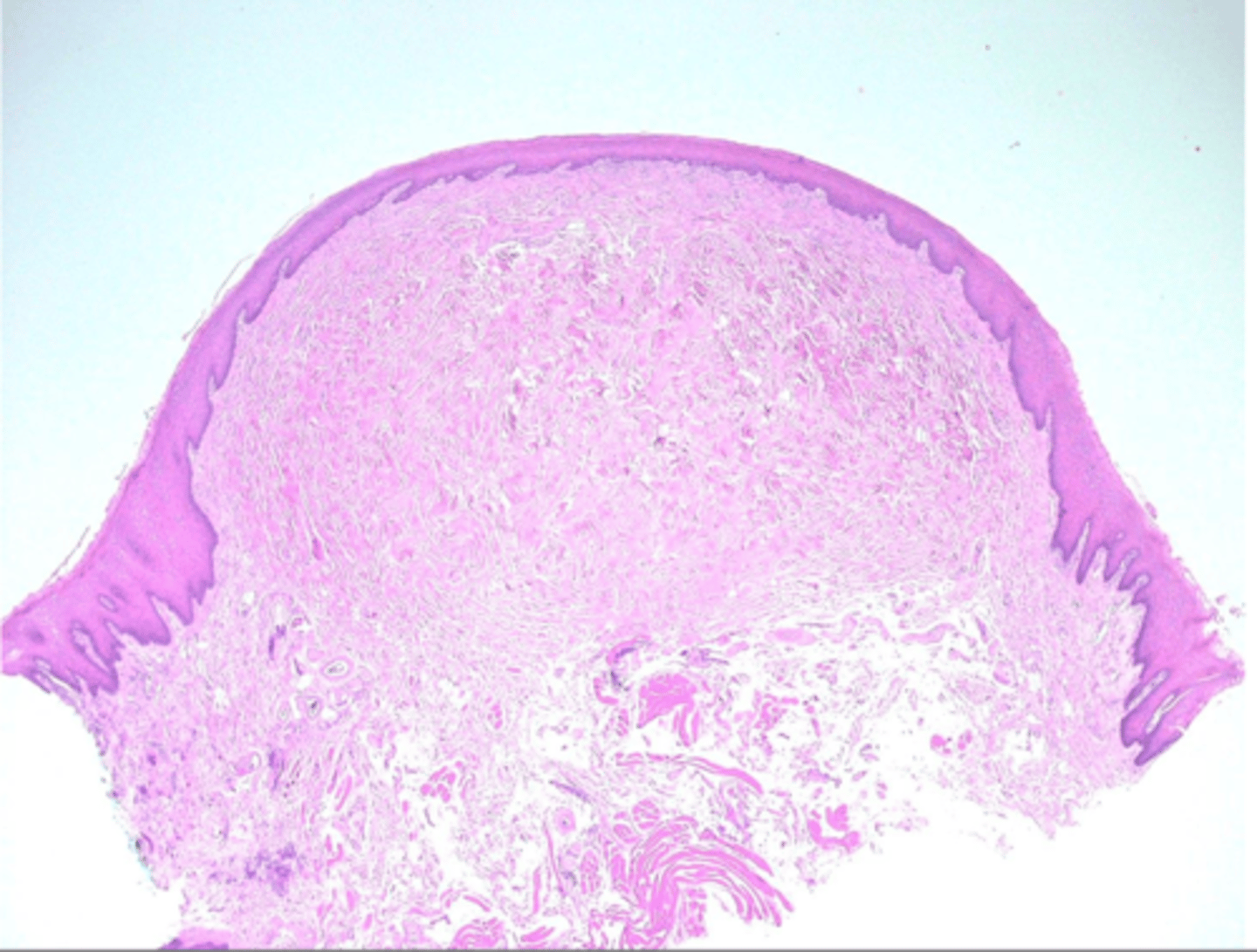
Fibroma (histology)
Nodular mass of fibrous connective tissue covered by stratified
squamous epithelium
• Surface may exhibit hyperkeratosis from secondary trauma
Frenal tag (type of fibroma)
• Fibrous hyperplasia
• Most common on the maxillary
labial frenum
• Small, asymptomatic, exophytic
growths
• Can be diagnosed clinically so no
treatment is necessary
Surgical excision, biopsy, Malignancies can mimic
benign entities!
Fibroma - treatment
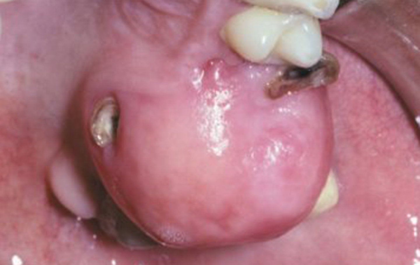
Epulis fissuratum
Reactive hyperplasia due to an
ill-fitting denture or alveolar
resorption
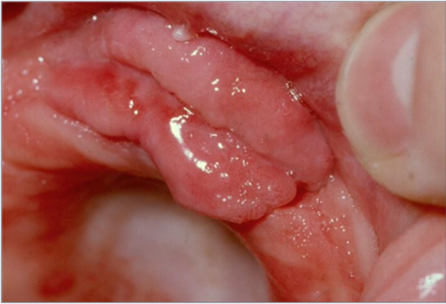
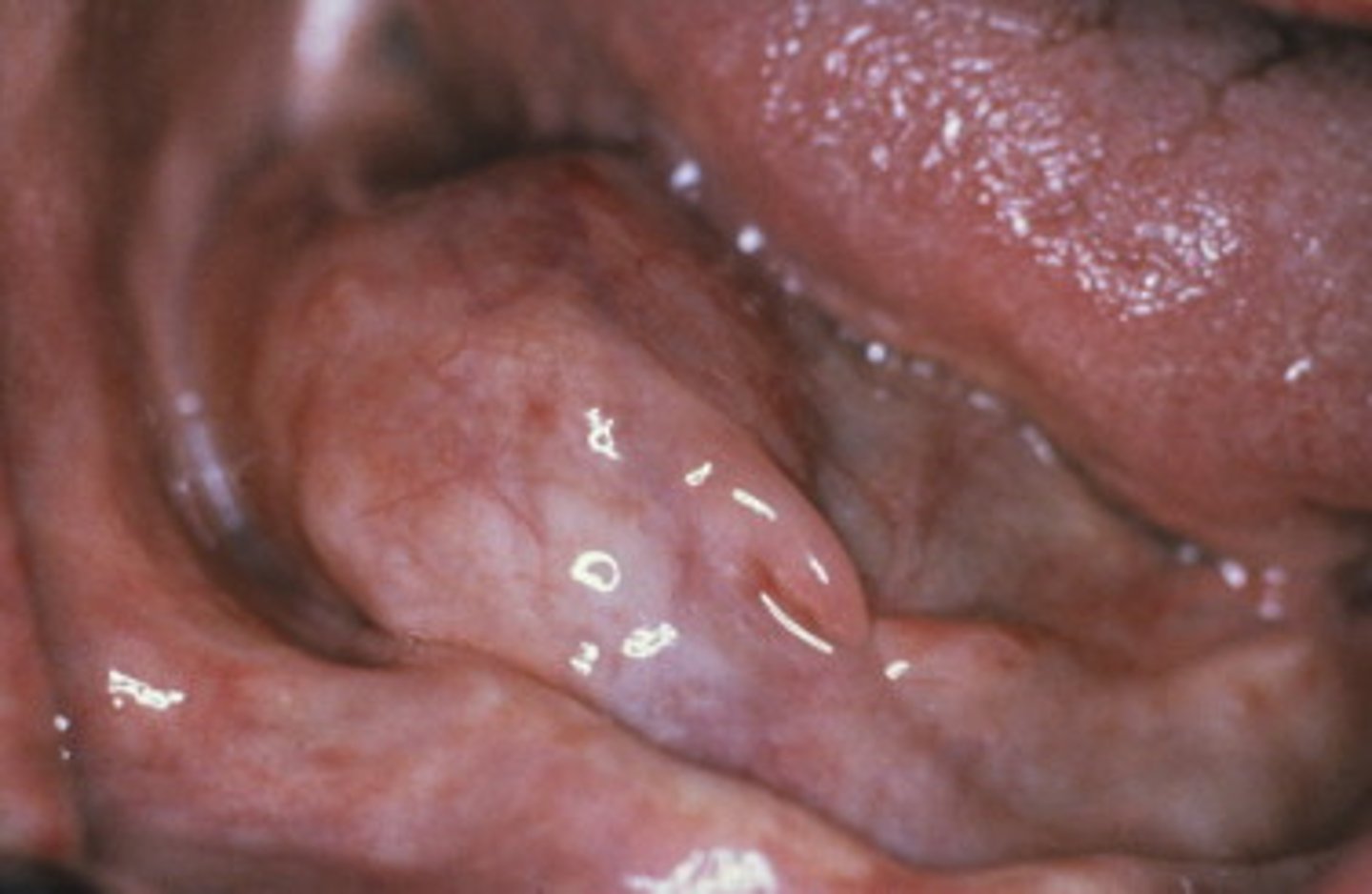
Epulis fissuratum
• Found in the vestibules
• Redundant folds
• Rubbery
• May be ulcerated
• Overgrowth of tissue along the
border of a denture
Epulis fissuratum
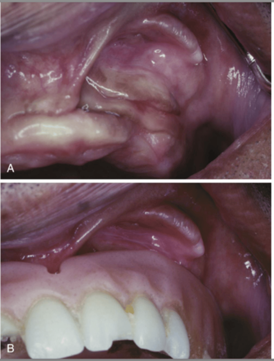
Excision, remake denture
Epulis fissuratum - treatment
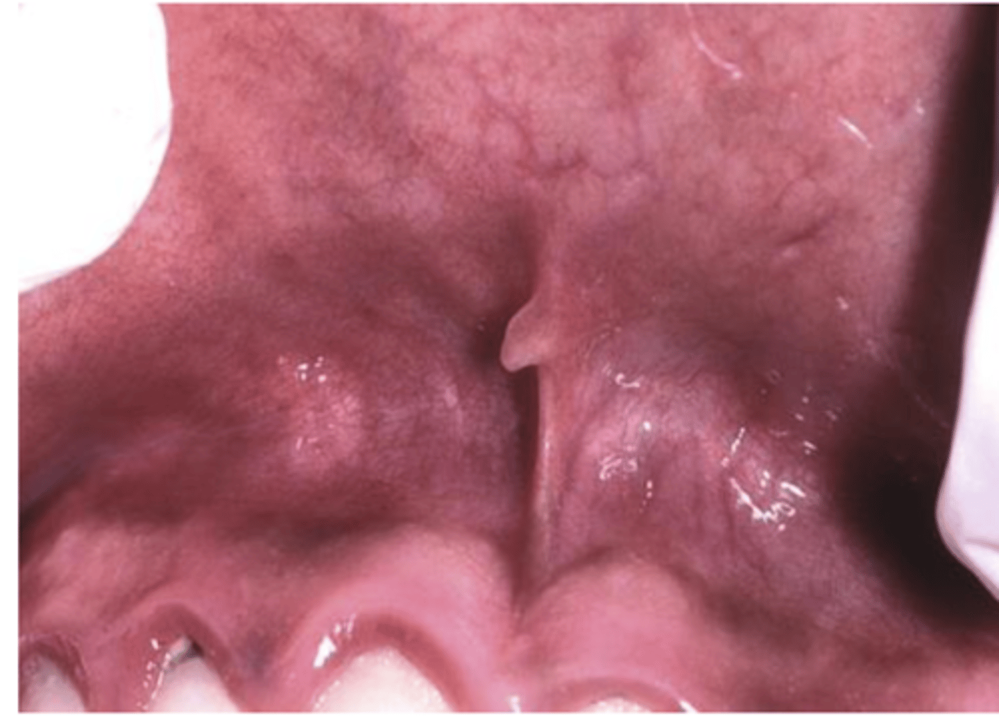
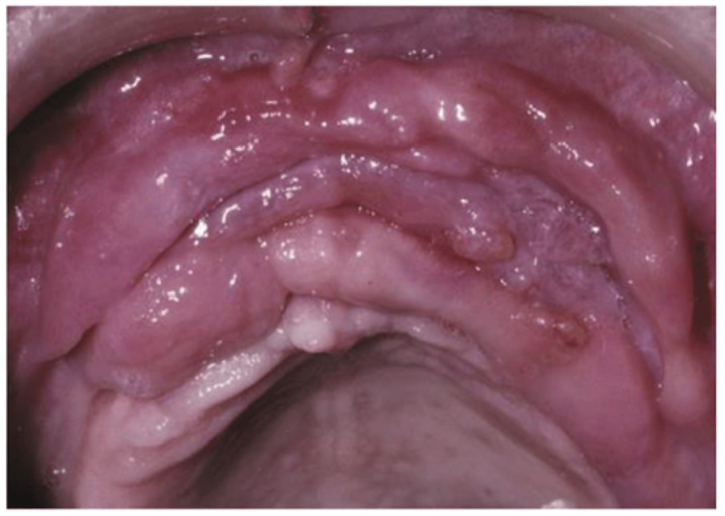
Inflammatory papillary hyperplasia
• Reactive tissue growth related to
• An ill-fitting denture
• Poor denture hygiene
• 24-hour denture wearing
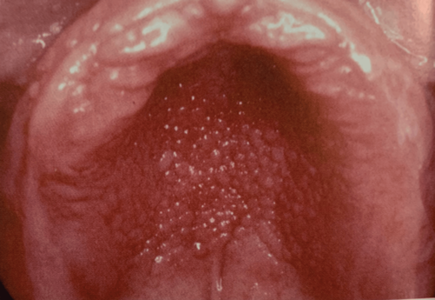
mouth-breathers
Inflammatory papillary hyperplasia may also
be seen in patients who are ...
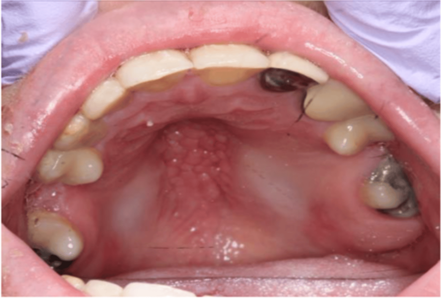
Inflammatory papillary hyperplasia
• Usually occurs on the hard
palate beneath a denture base
• Usually asymptomatic
• Mucosa is erythematous and has
a pebbly or papillary surface
removal of dentures at night, Excise excessive tissue if necessary, Antifungal therapy
Inflammatory papillary hyperplasia - treatment
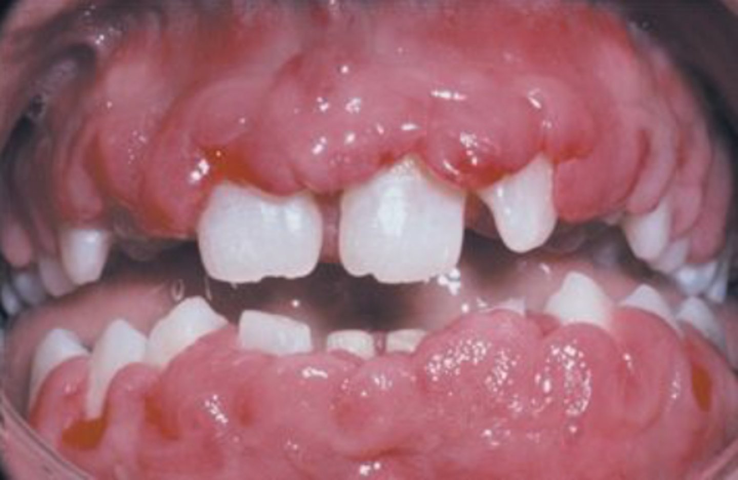
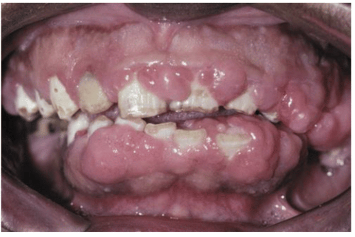
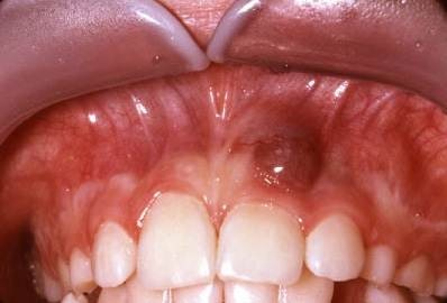
Gingival hyperplasia
• May be hereditary
• May be medication related
• Dilantin (Phenytoin)
• Cyclosporine
• Nifedipine
Dilantin (Phenytoin), Cyclosporine, Nifedipine
3 medications that are linked to medication induced Gingival hyperplasia
Gingival hyperplasia
• Begins in the interdental
papillae
• Gingiva may cover the crowns
• Firm
Discontinuation of the offending medication, Gingivectomy
Gingival hyperplasia - treatment
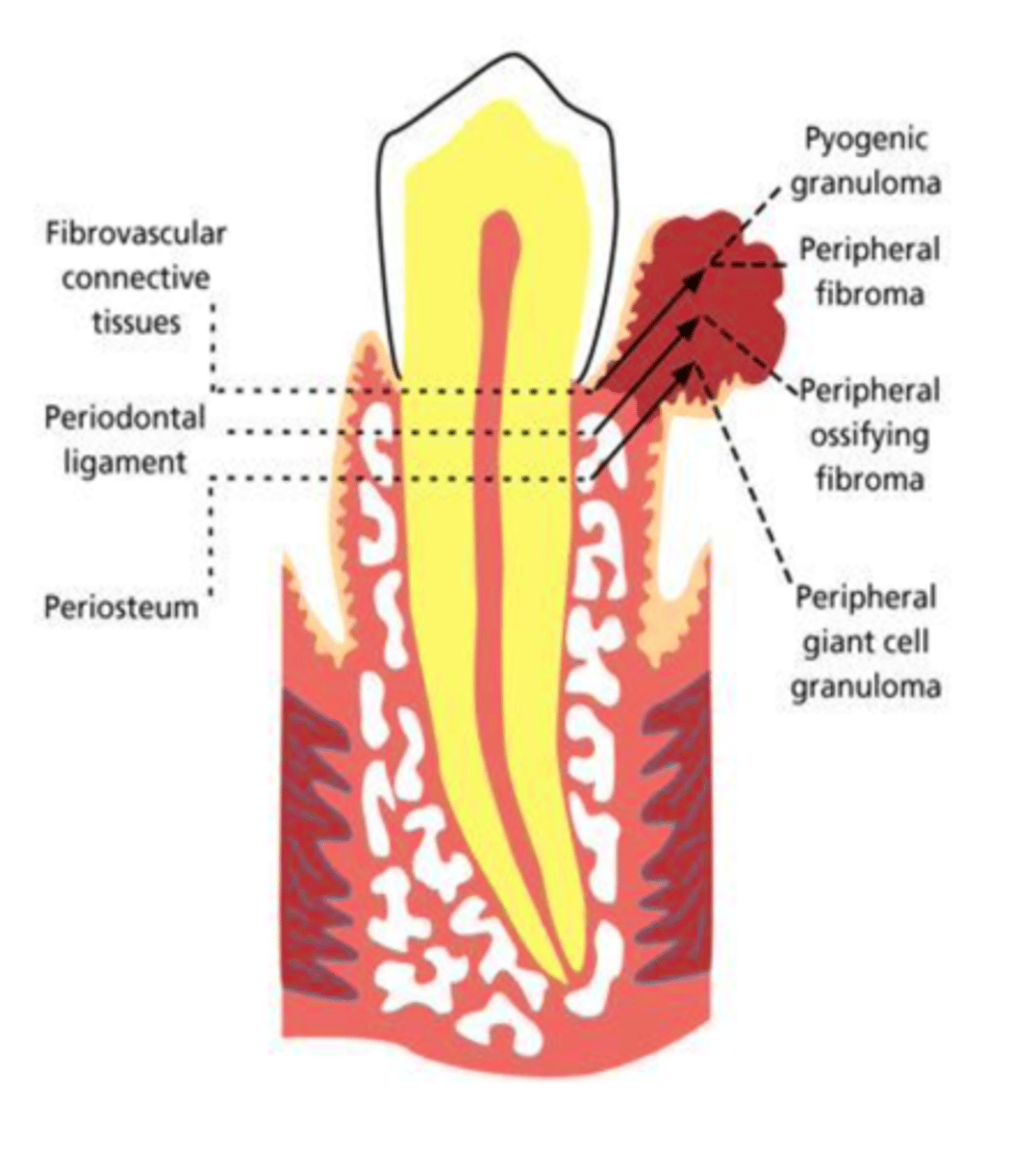
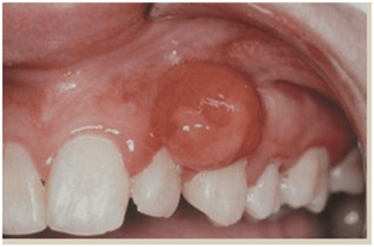
• Pyogenic Granuloma
• Peripheral Giant Cell Granuloma
• Peripheral Ossifying Fibroma
• Peripheral Fibroma
The 4 P’s (Dr. Cohen emphasized these!)
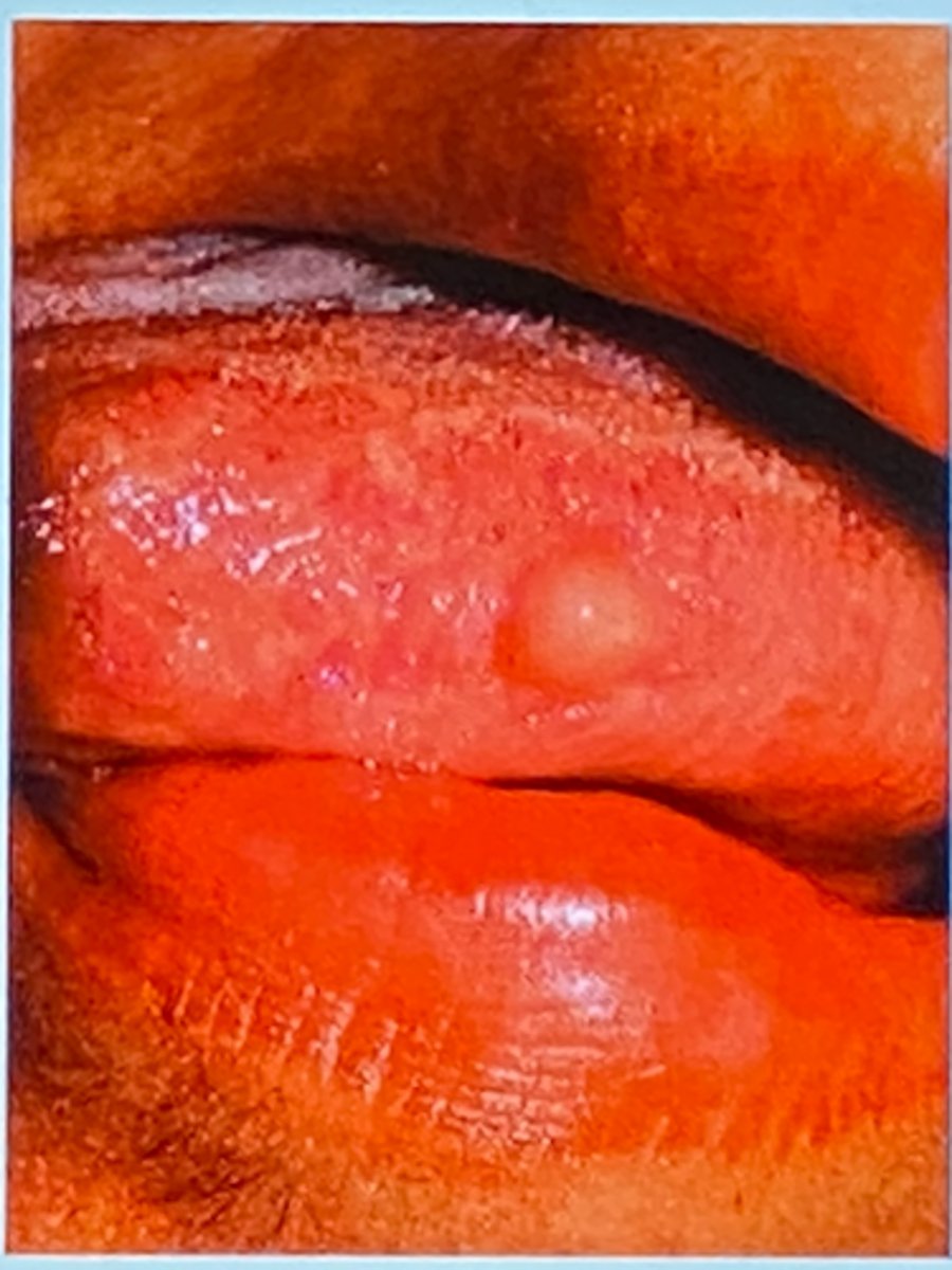
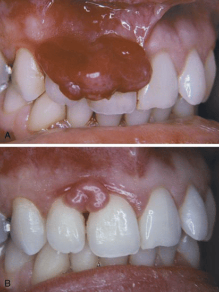
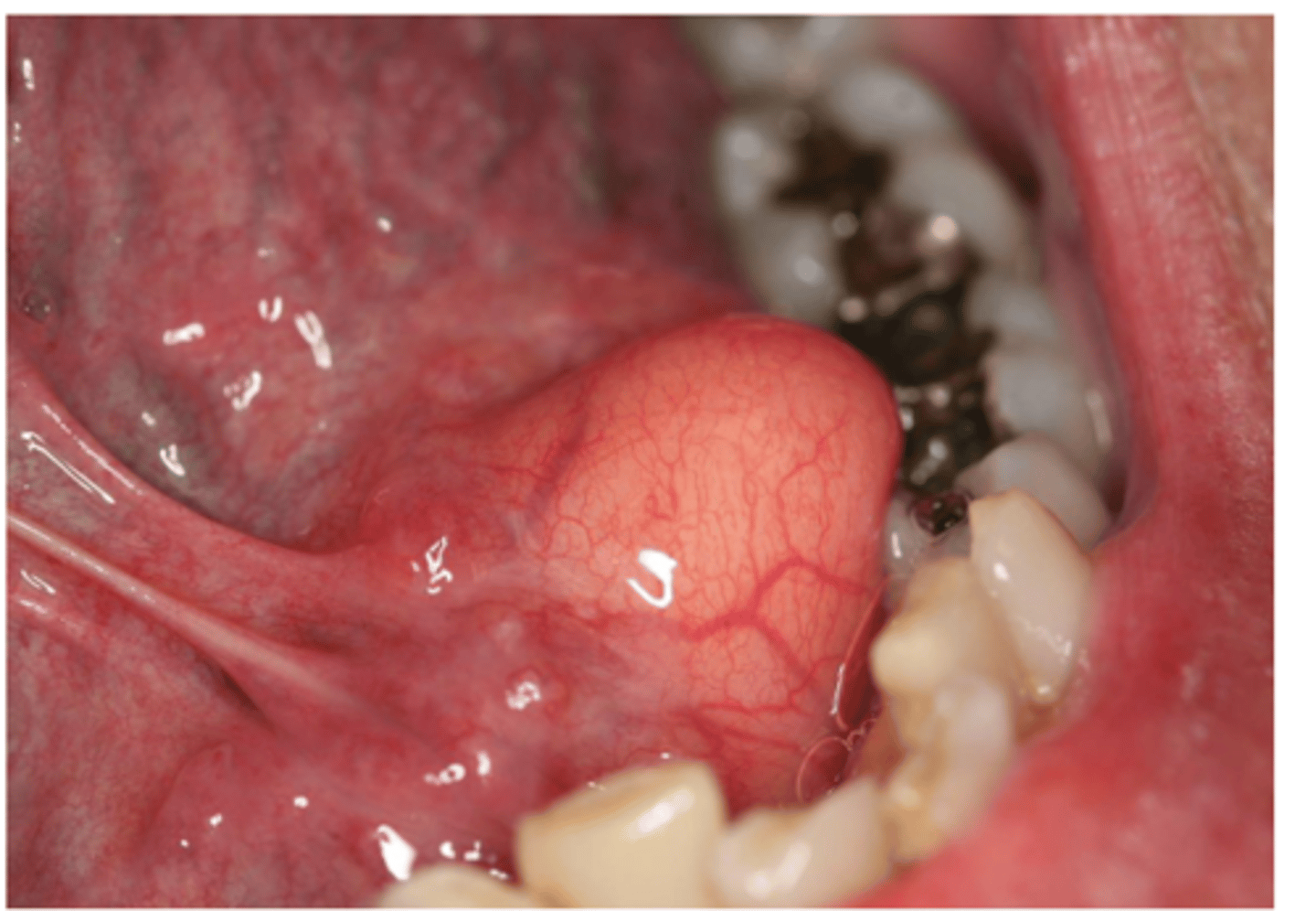
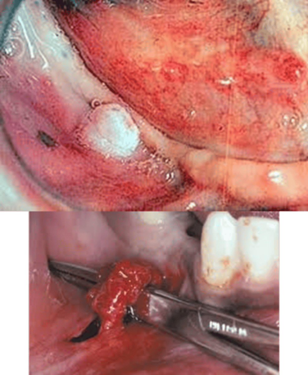
Pyogenic granuloma (AKA Lobular capillary hemangioma)
• Common
• Cause: local irritation or trauma
Pyogenic granuloma
• 75% on the gingiva
• Sometimes called the
“pregnancy tumor”
• Smooth or lobulated vascular
mass
• Pink-red-purple
• Usually ulcerated
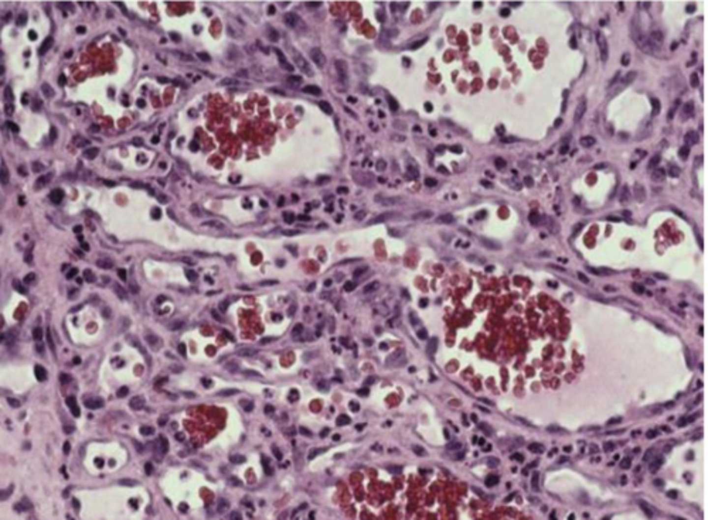
Pyogenic granuloma (histopathology)
• Ulcerated stratified squamous epithelium
• Vascularized fibrous connective tissue
• Mixed inflammatory infiltrate
Surgical excision, biopsy, In pregnant patients defer treatment
Pyogenic granuloma - treatment
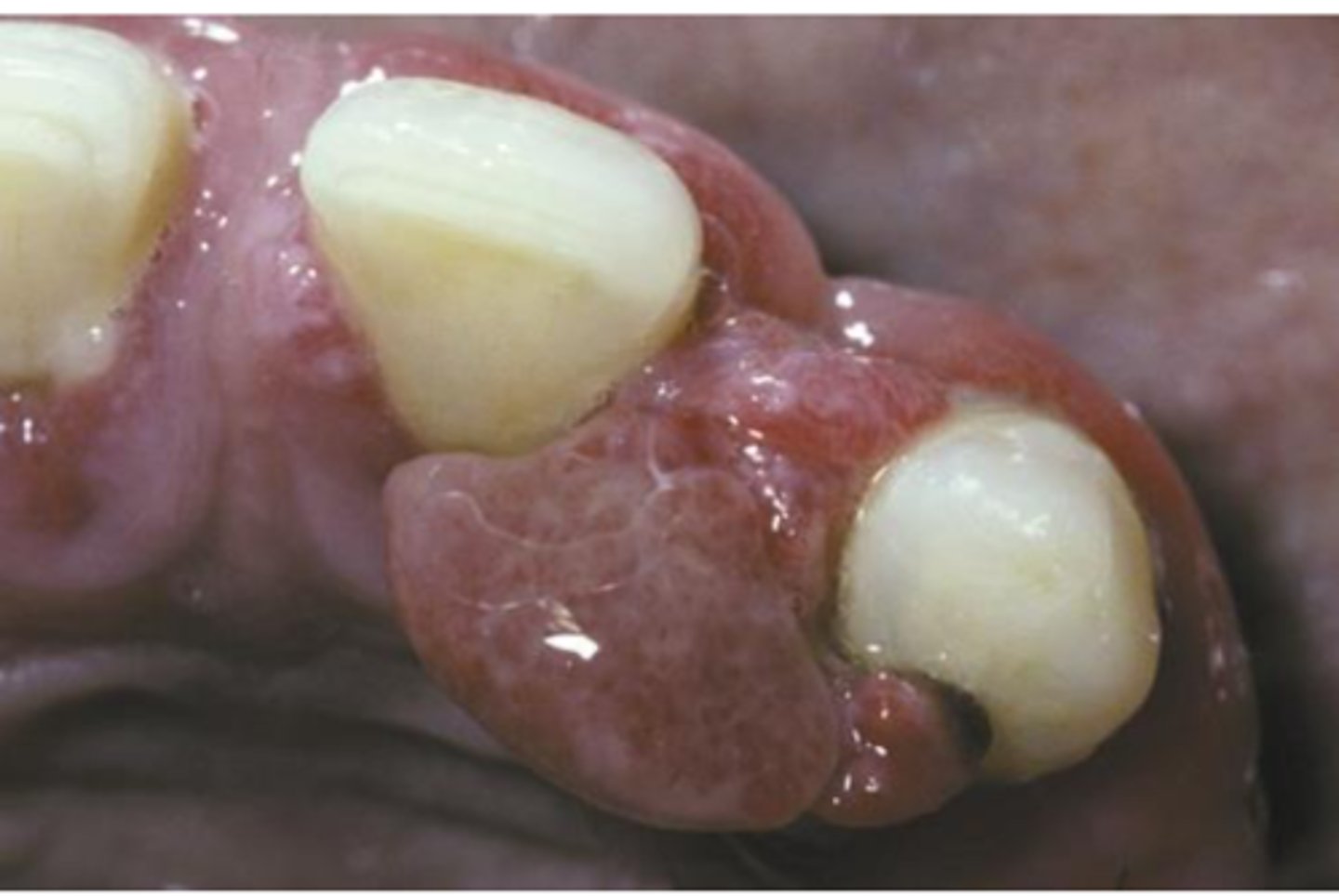
Peripheral Giant Cell Granuloma
• Common
• Reactive lesion caused by local
irritation or trauma
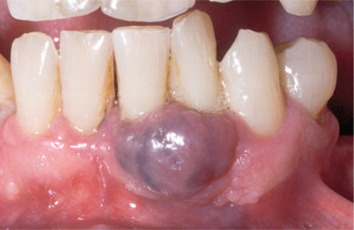
Peripheral Giant Cell Granuloma
• Occurs exclusively on the gingiva
or edentulous alveolar ridge
• Red or red-blue nodular mass
• Frequently ulcerated
• "Cupping" resorption of the
underlying alveolar bone may be
noted
surgical excision down to the underlying bone, biopsy, Adjacent teeth should be carefully scaled
Peripheral Giant Cell Granuloma - Treatment
Peripheral Ossifying Fibroma
• Common
• Reactive lesion caused by local
irritation or trauma
Peripheral Ossifying Fibroma
• Occurs exclusively on the gingiva
• Nodular mass
• Frequently ulcerated
• Red to pink
• Seen most commonly in teens
and young adults
Local surgical excision down to periosteum, biopsy, Adjacent teeth should be thoroughly scaled
Peripheral Ossifying Fibroma - Treatment
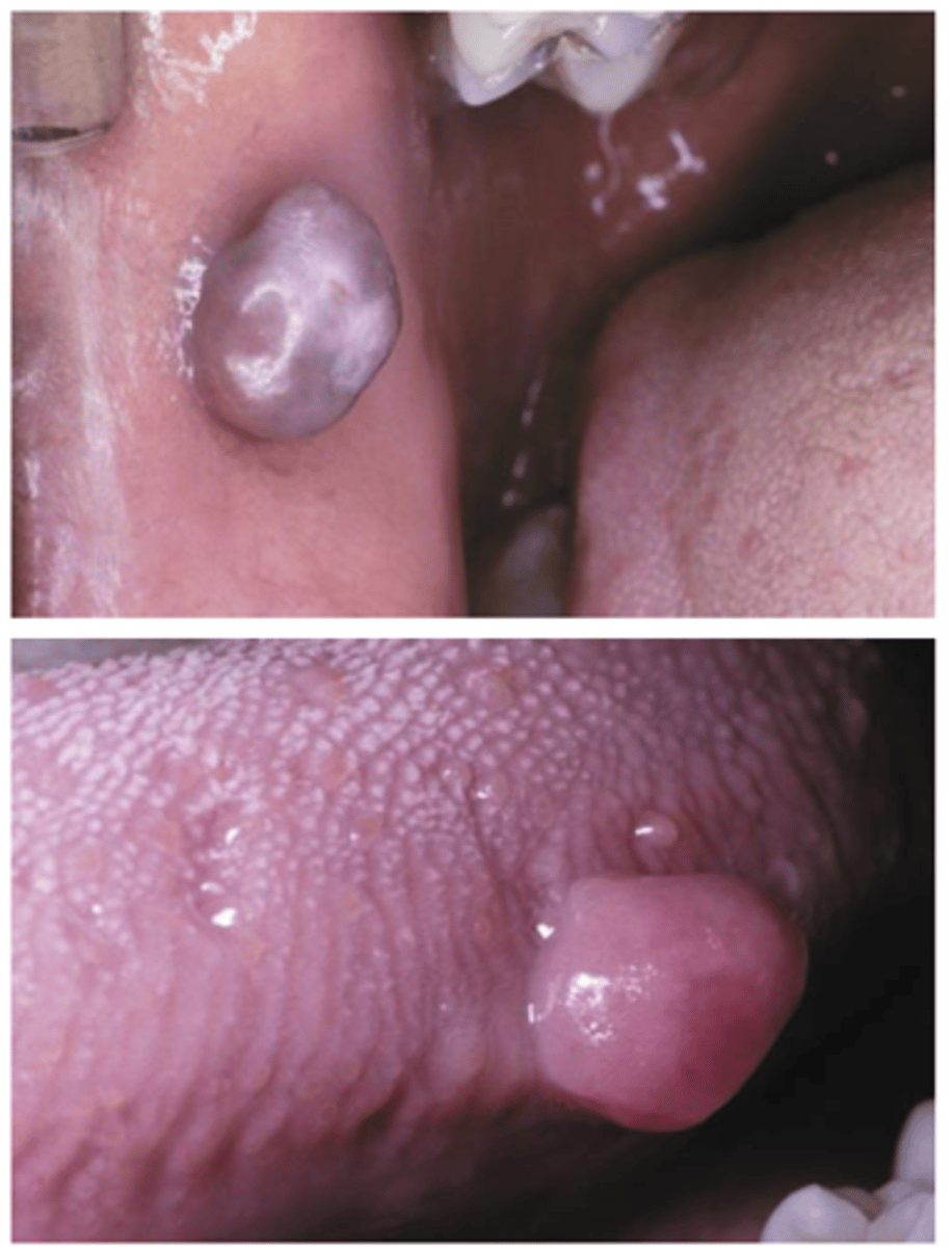
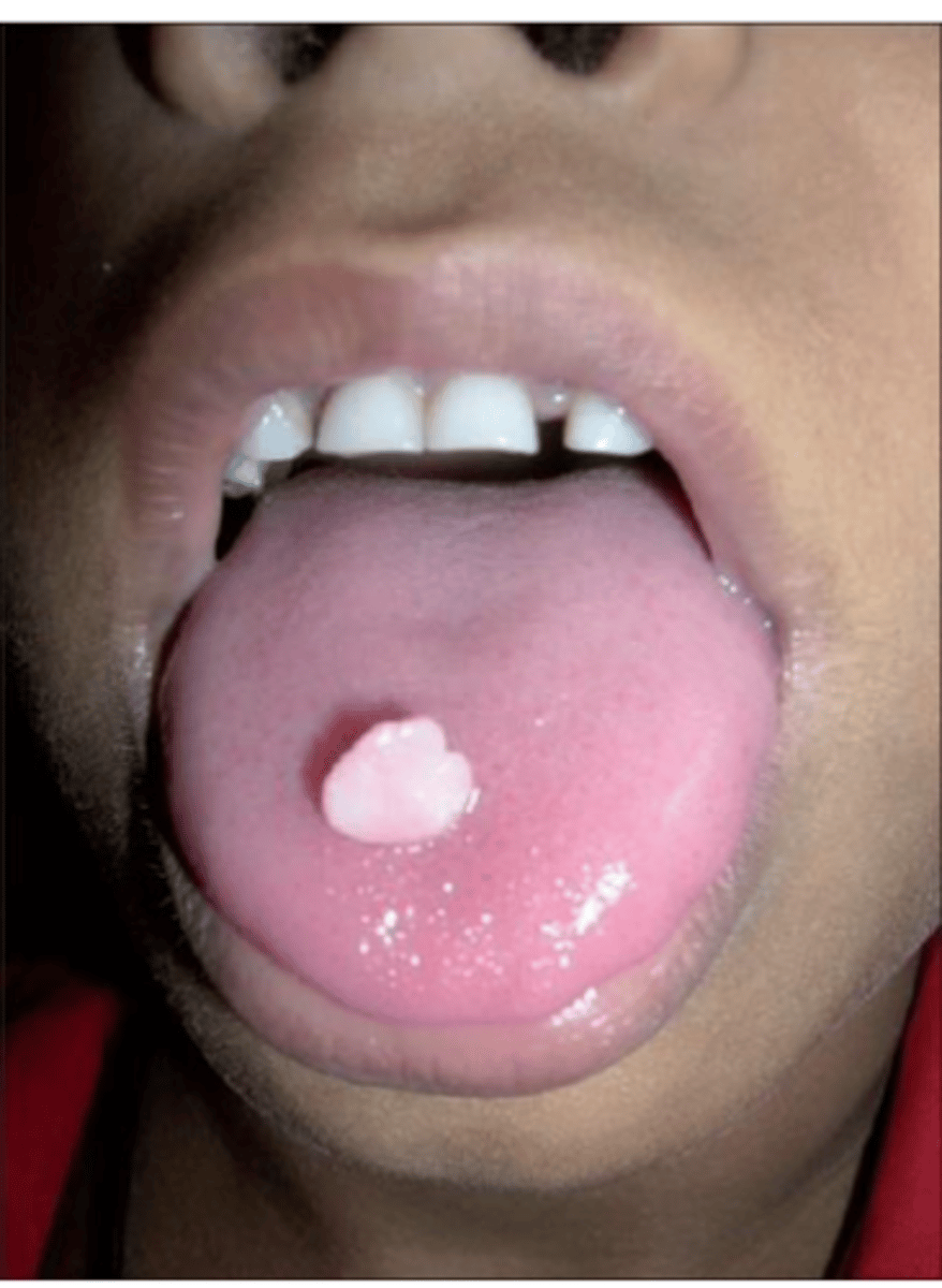
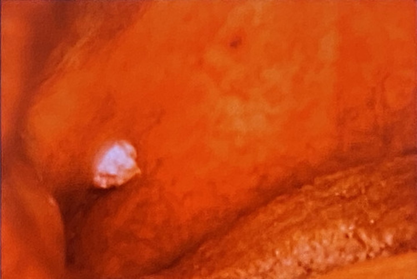
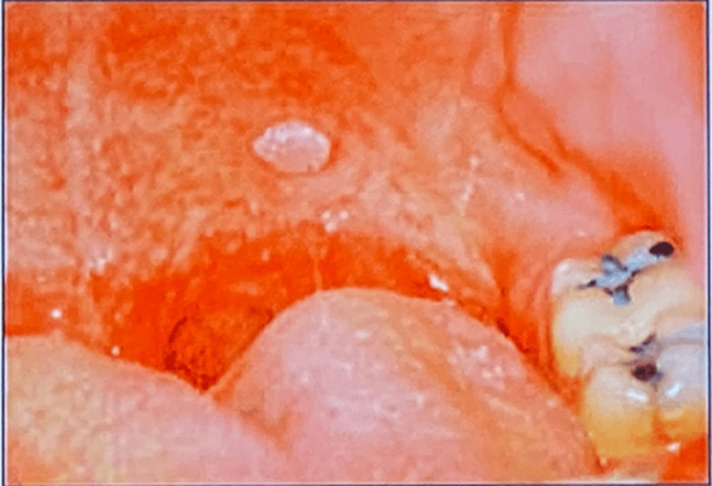
Giant cell fibroma
• Fibrous tumor
• Unlike the traumatic fibroma, it
does not appear to be
associated with chronic irritation
Giant cell fibroma
• Asymptomatic
• The surface of the mass often
appears papillary
• More common in young patients
• Approximately 50% of all cases
occur on the gingiva
• Tongue and palate also are
common sites
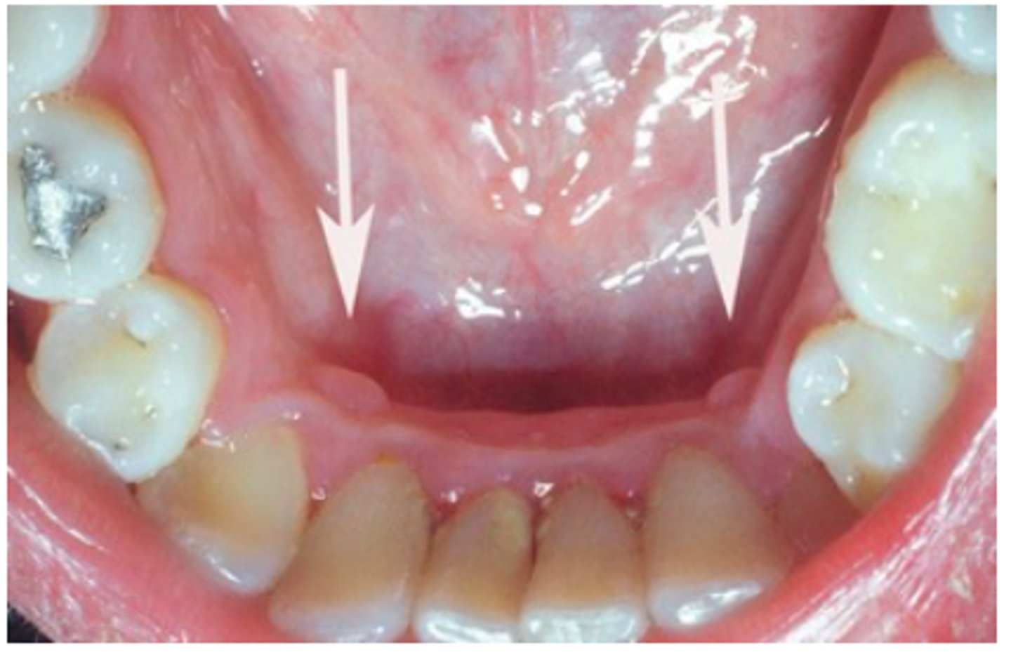
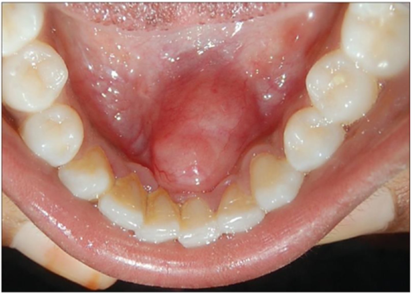
Retrocuspid papilla
• Papillary-like growth
• Mandibular gingiva lingual to
canine
• Frequently bilateral
• 25-99% of children/YA
• Involutes with age
• Should be recognized clinically
as a normal anatomic variation
surgical excision, biopsy, recurrence is rare
Giant cell fibroma - Treatment
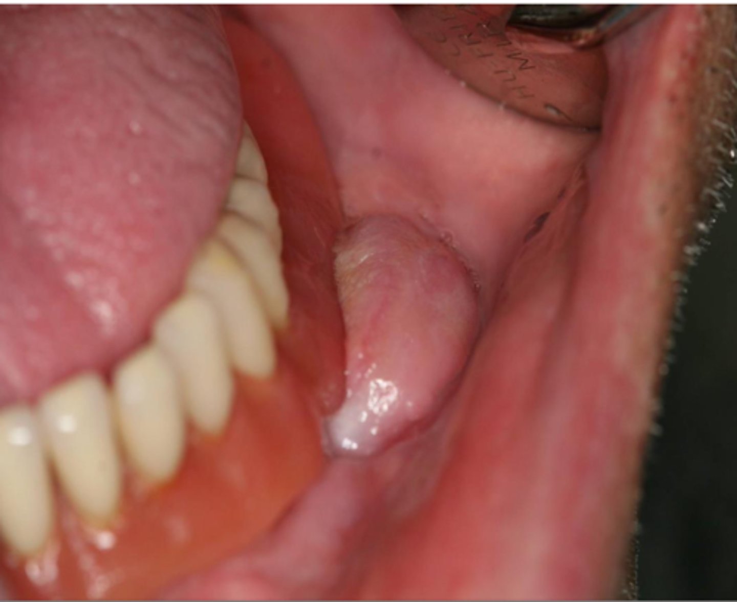
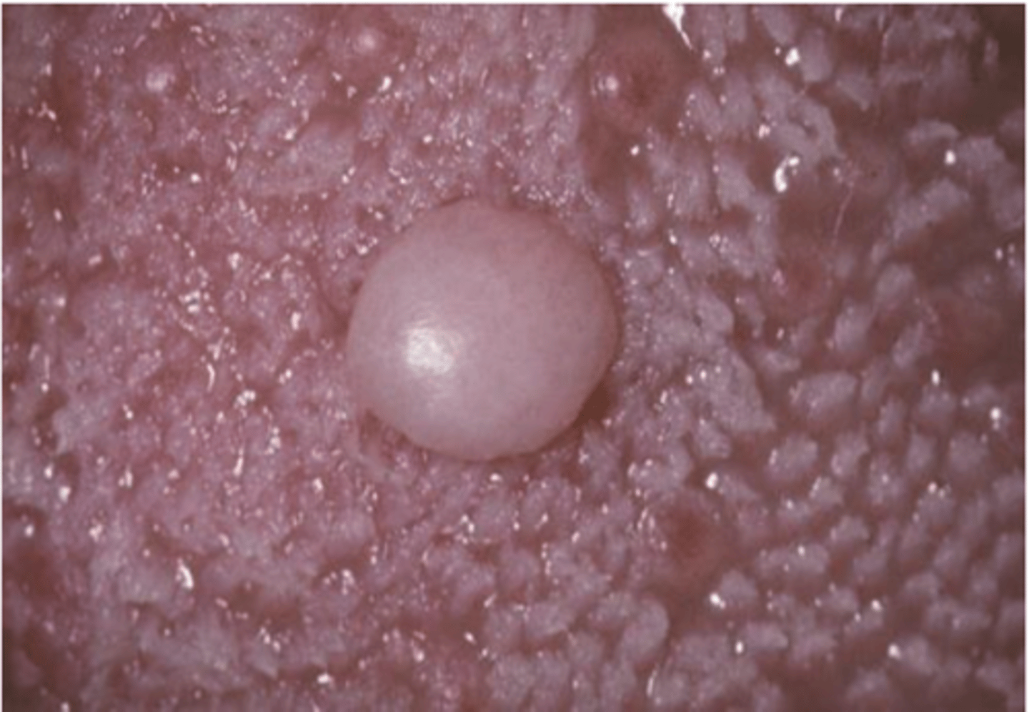
Granular cell tumor
• Uncommon
• Derived for Schwann cells
• Cytoplasm is granular because of
lysosomes
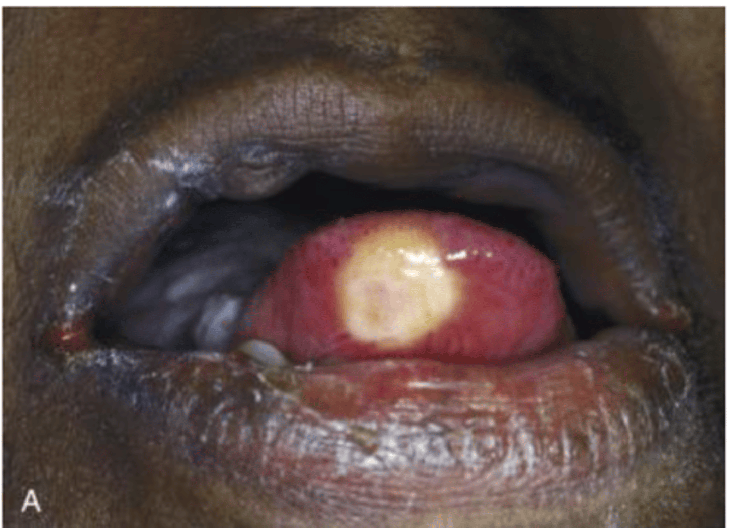
Granular cell tumor
• Most commonly found on the
dorsal tongue
• Asymptomatic sessile nodule
• Pink to yellowish in color

Conservative local excision, biopsy, recurrence is rare
Granular cell tumor - treatment
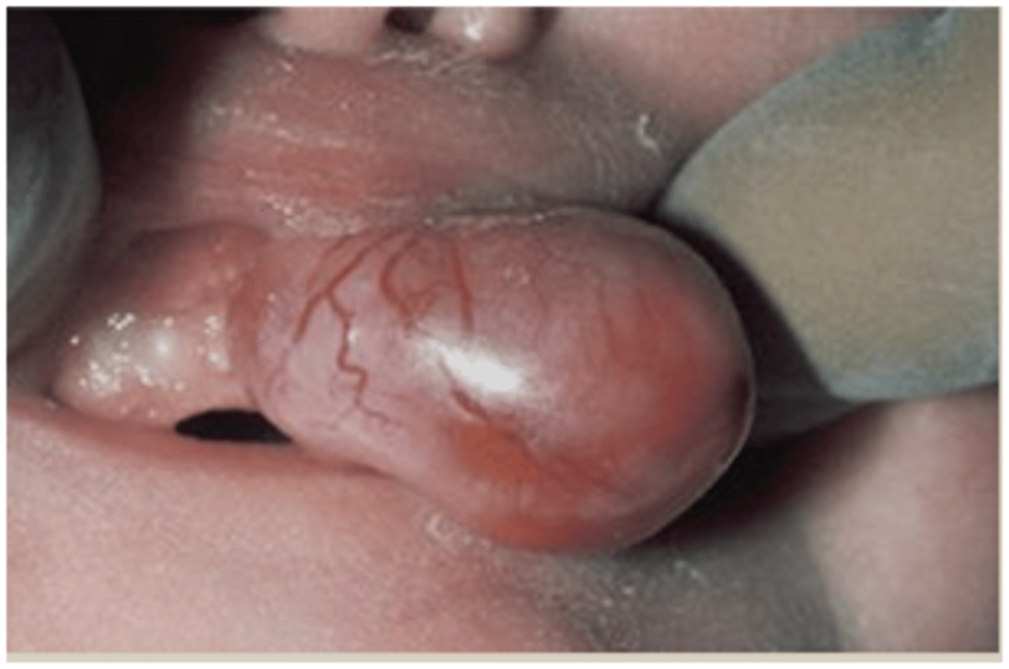
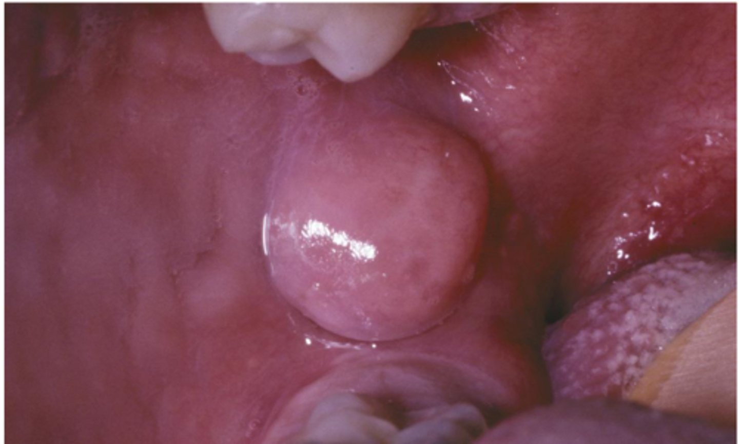
Congenital epulis (Congenital granular cell lesion)
• Uncommon
• Occurs almost exclusively on the
alveolar ridges of newborns
• Bears a microscopic
resemblance to the granular cell
tumor
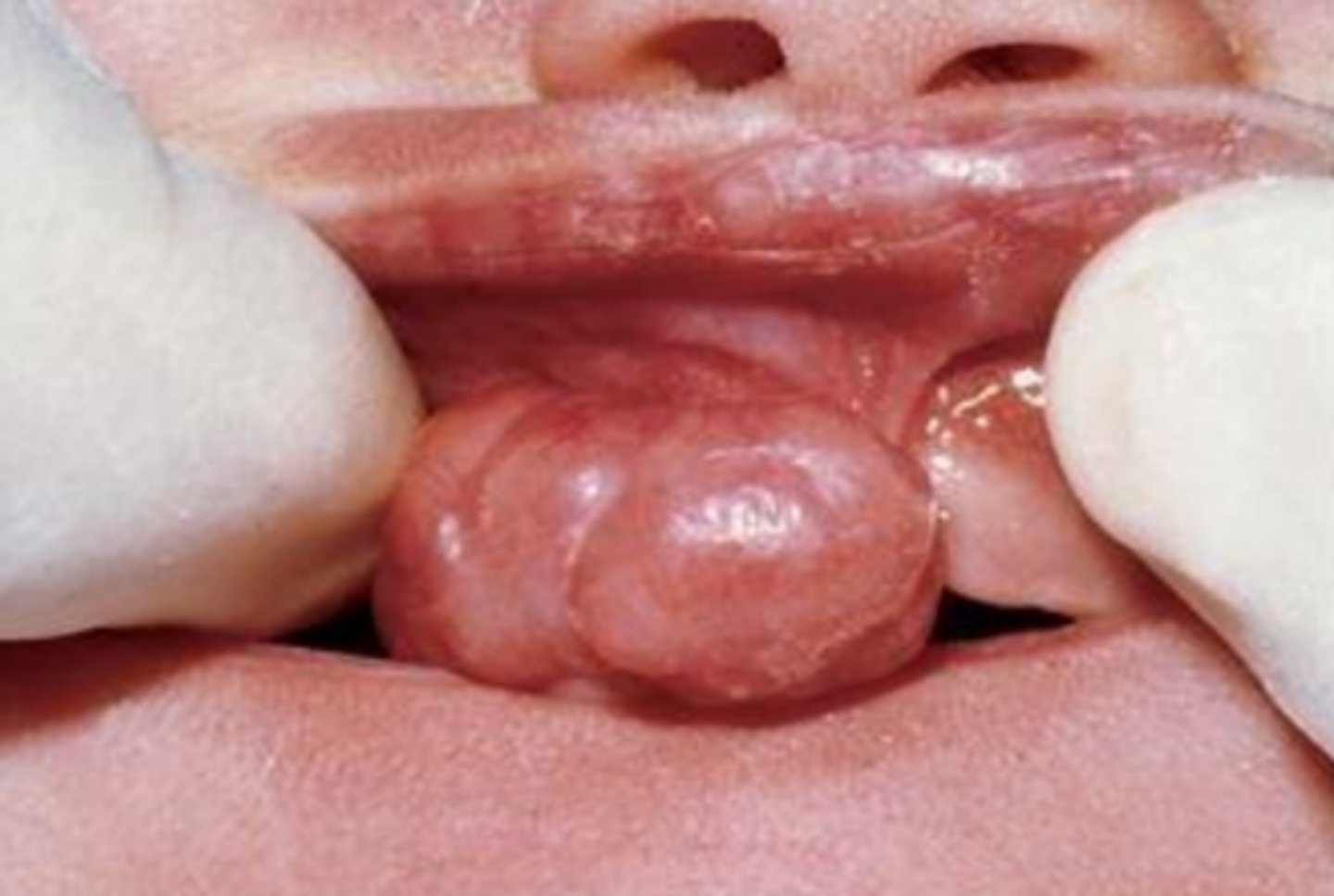
Congenital epulis
• Pink-to-red, smooth-surfaced,
polypoid mass on the alveolar
ridge of a newborn
• More common on the maxilla
than the mandible
• 90% occur in females
Surgical excision, biopsy, no reports of recurrence
Congenital epulis - treatment
Lipoma
• Benign tumor of fat
• 4% of mesenchymal tumors of
oral cavity
Lipoma
• Soft, doughy, smooth-surfaced
nodular
• Asymptomatic
• Pink to yellow in color
Conservative local excision, biopsy, Recurrence is rare
Lipoma - treatment
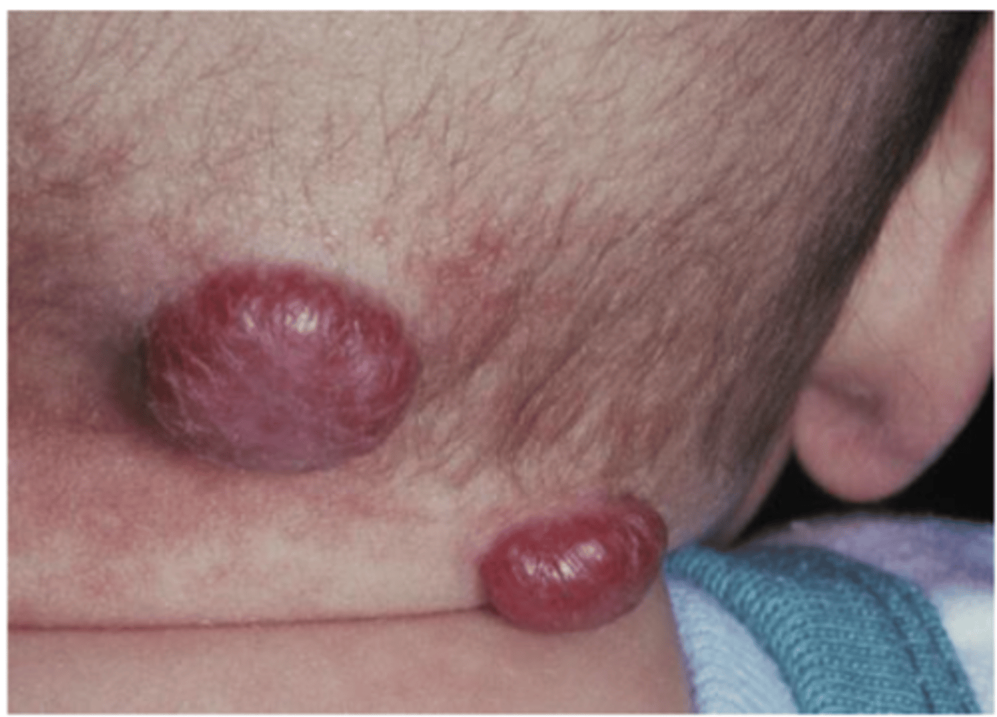
Hemangioma
• Congenital lesion
• Most common tumor of infancy
• Vascular proliferation
• Often red/blue
• Early rapid growth, followed by
slow involution
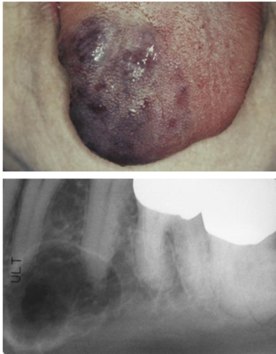
Vascular malformation
◦ Most common sites in the head
and neck:
◦ Lips, tongue, buccal mucosa, or
palate
◦ Deep red or blue compressible
lesion
◦ Lesions can in the soft tissue or
central (intraosseous) in location
spontaneous remission, surgery, embolization, sclerosing agents, cryotherapy, laser , DO NOT BIOPSY (refer it)
Vascular malformation - treatment
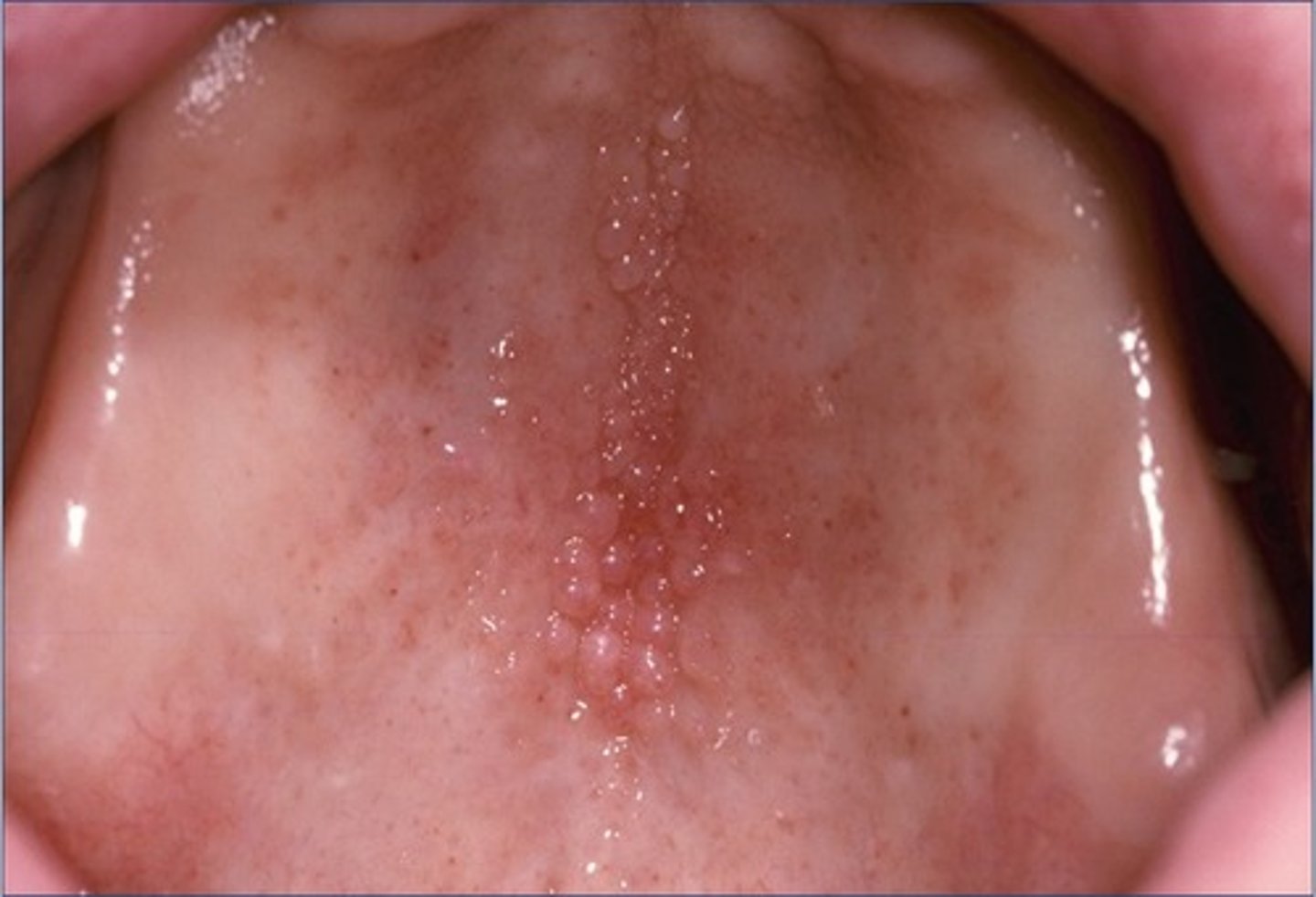
Lymphangioma
• Benign, hamartomatous
tumorlike growths of lymphatic
vessels
• Likely represent developmental
anomalies that arise from
sequestrations of lymphatic
tissue that do not communicate
normally with the rest of the
lymphatic system

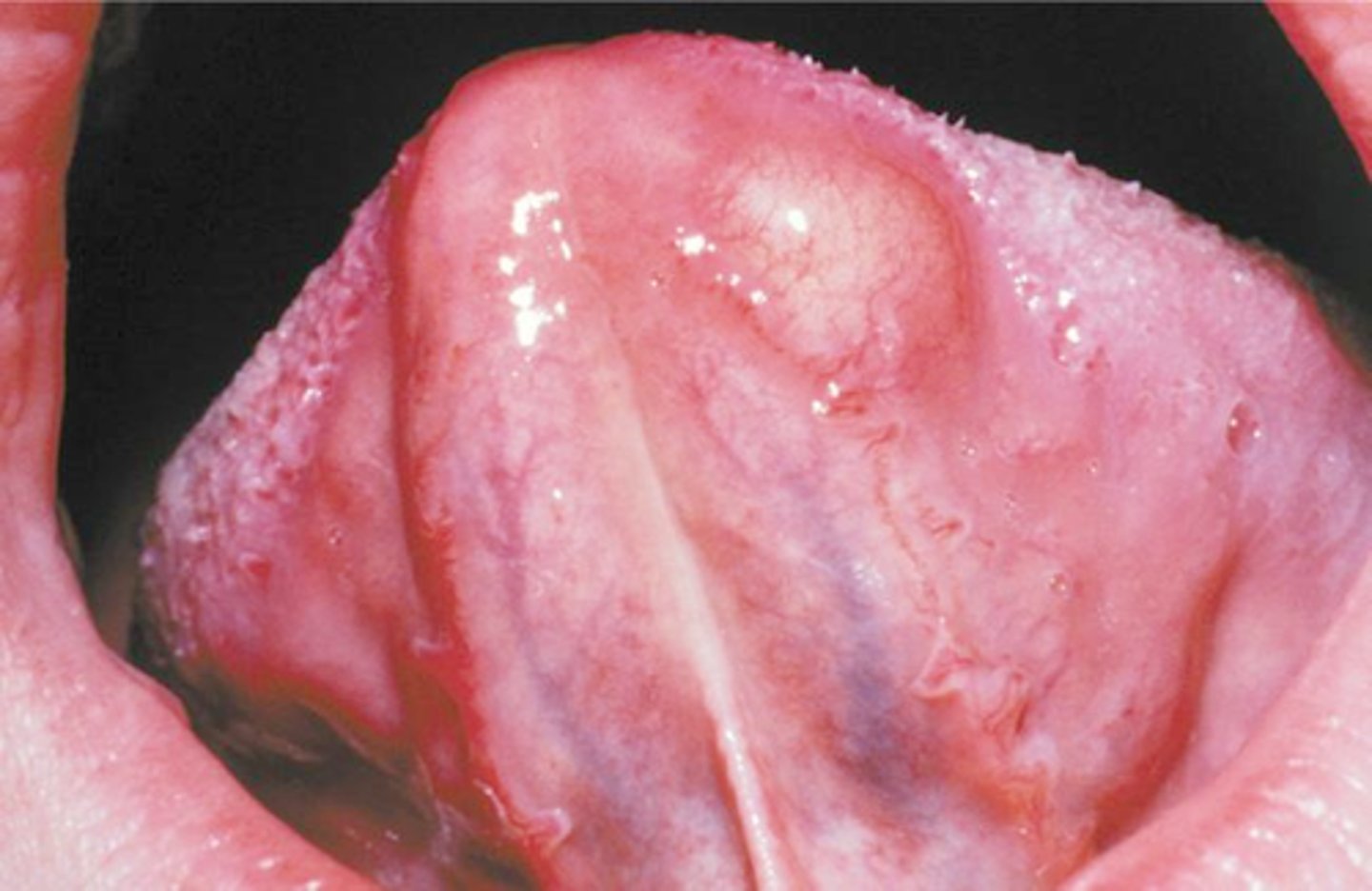
Lymphangioma
• Predilection for the head and
neck
• Oral lymphatic malformations
are most frequent on the
anterior two thirds of the
tongue, where they often result
in macroglossia
• Demonstrates a pebbly surface
that resembles a cluster of
translucent vesicles
Surgical excision, Sclerotherapy, May recur
Lymphangioma - treatment
Neurofibroma
• Most common peripheral nerve
neoplasm
• Can be a solitary tumor or be a
component of
neurofibromatosis*
Neurofibroma
• Slow-growing, soft, painless
lesions that vary in size from
small nodules to larger masses
• Can be in the soft tissue or
central (in bone)
Neurofibroma
inferior alveolar nerve canal looks extremely expanded
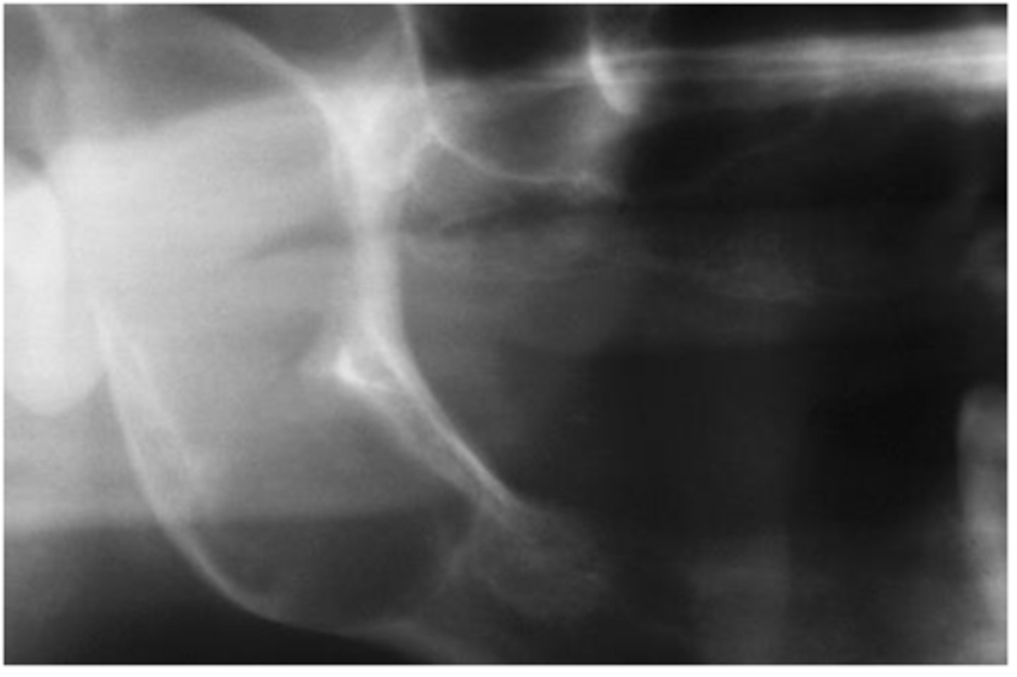
Surgical excision
Neurofibroma - treatment
Schwannoma
• Benign neural neoplasm of
Schwann cell origin
• Uncommon
Schwannoma
• Slow-growing, encapsulated
tumor that typically arises in
association with a nerve trunk
• Asymptomatic
• The tongue is the most common
location for oral _____
• Can arise centrally
Surgical excision
Schwannoma - treatment
Traumatic neuroma
• Reactive proliferation of neural
tissue after transection or other
damage of a nerve bundle
Traumatic neuroma
• Smooth-surfaced, nonulcerated
nodules
• Most common in the mental
foramen area, tongue, and lower
lip
• A history of trauma often can be
elicited
• May be intraosseous
• About 1/3 are painful
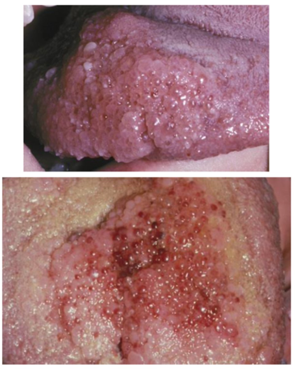
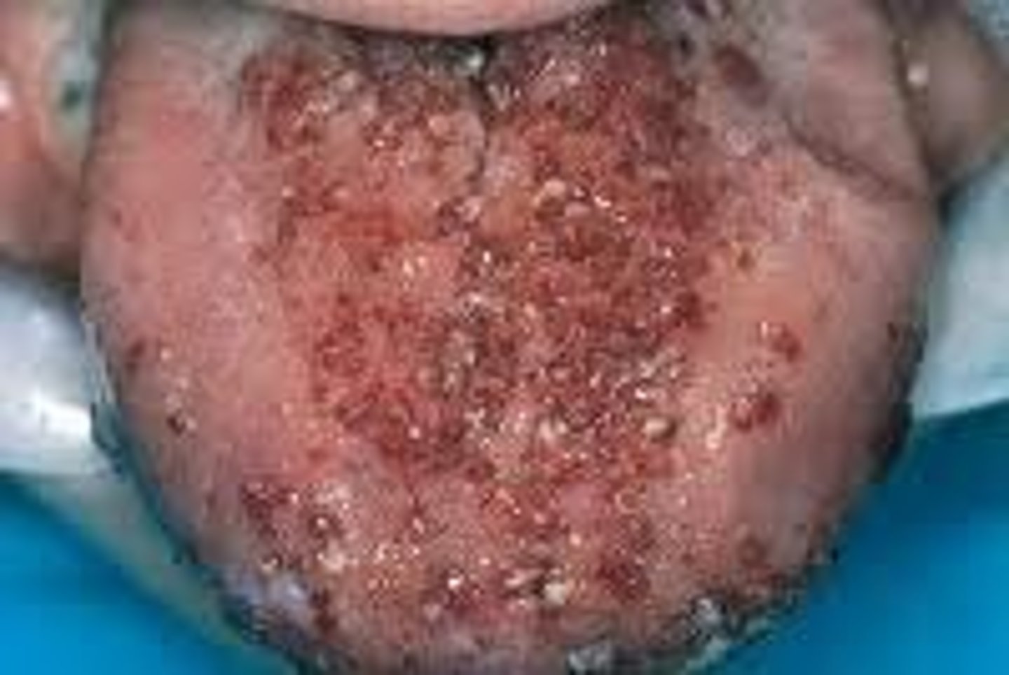
Squamous papilloma
Verruca vulgaris
Condyloma acuminatum
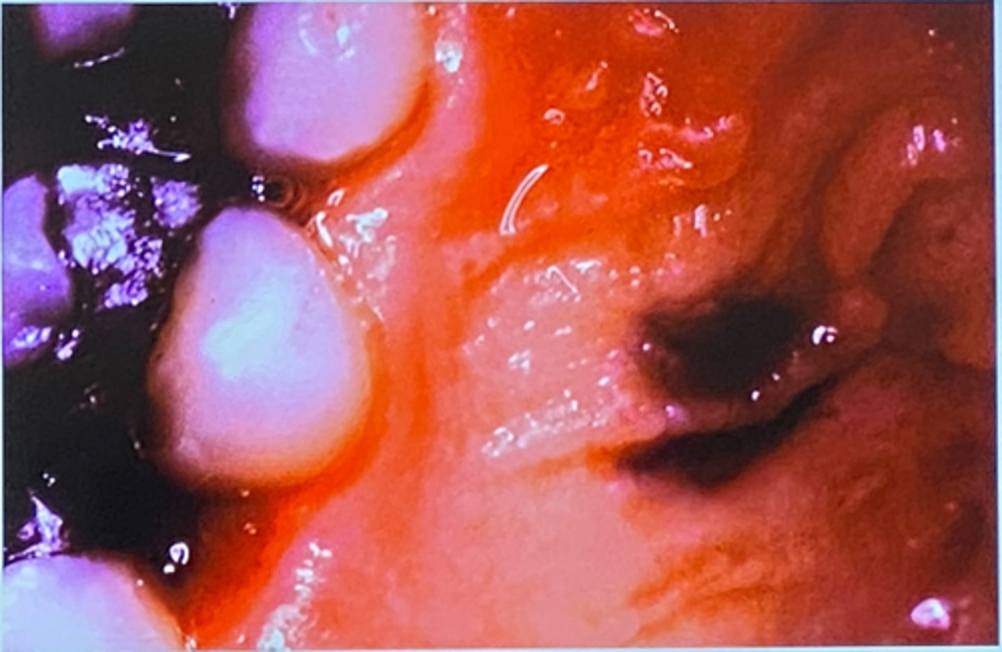
What is your differential for the lesion pictured?
1. Squamous papilloma
2. Verruca vulgaris
3. Giant cell
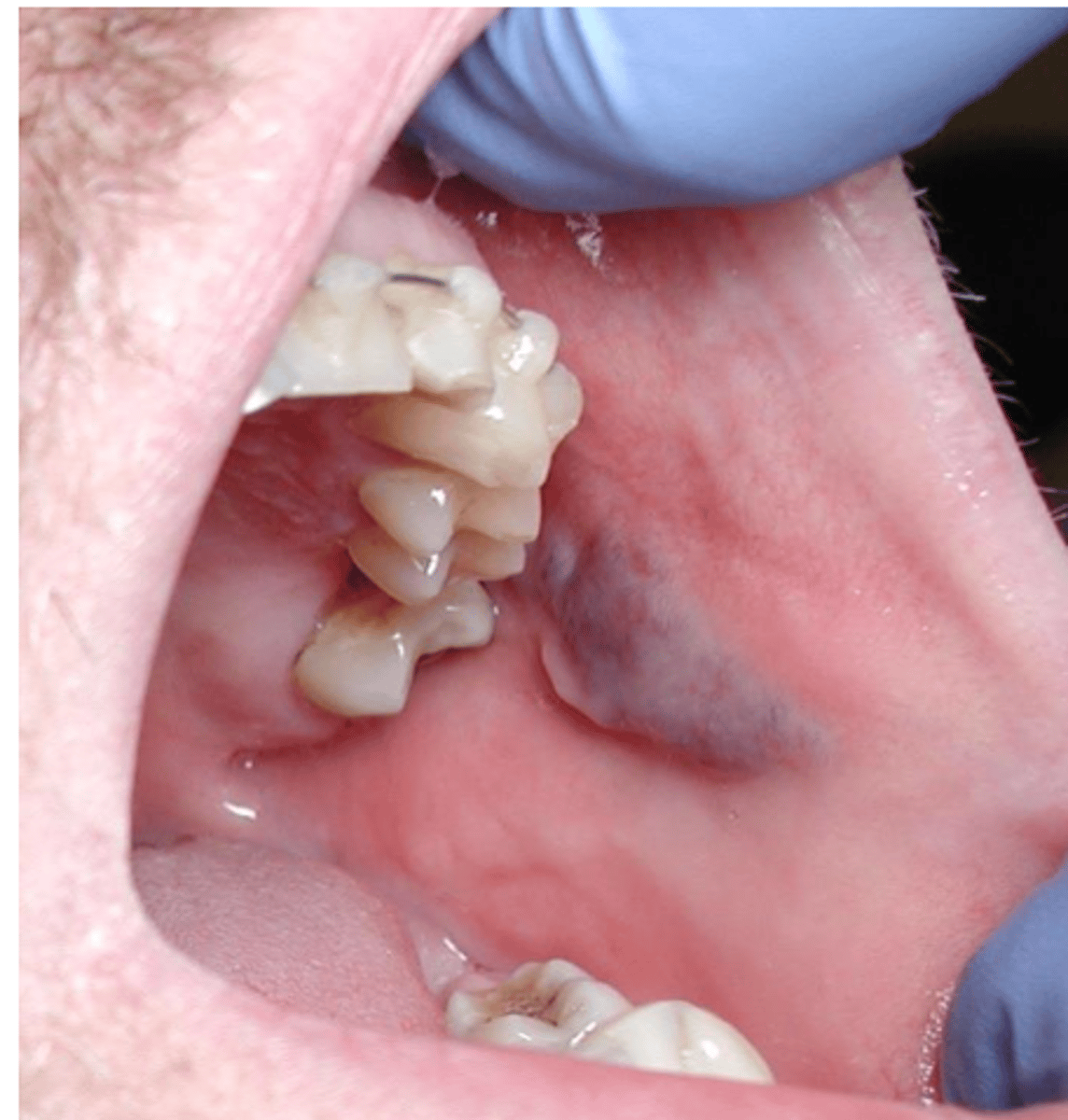
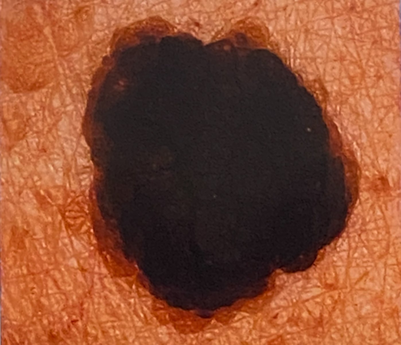
What is you differential for the lesion pictured?
1. oral melanotic macul
2. Acquired melanocytic nevus
3. Blue nevus
What is you differential?
seborrheic keratosis
Diagnosis:
-74-year-old female
-Admits to a long history of sun exposure
-CC: "This thing on my face is so ugly"
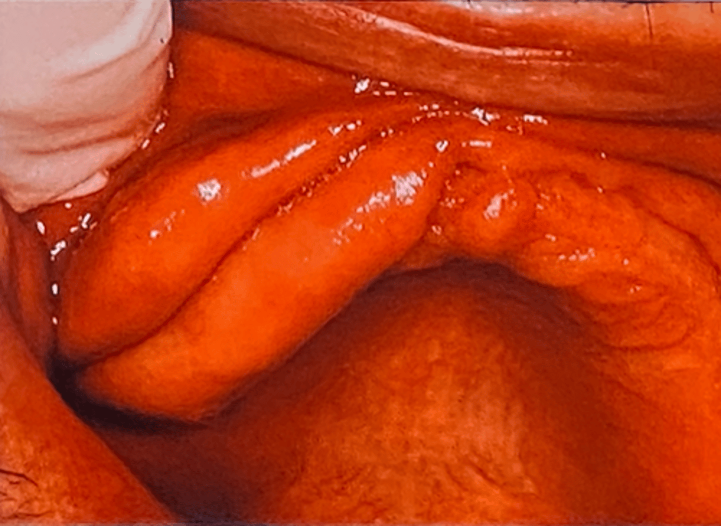
Epulis Fissuratum
-64-year-old male
-Non-contributory medical and social history
-Are there any questions you'd like to ask before you answer the question above?
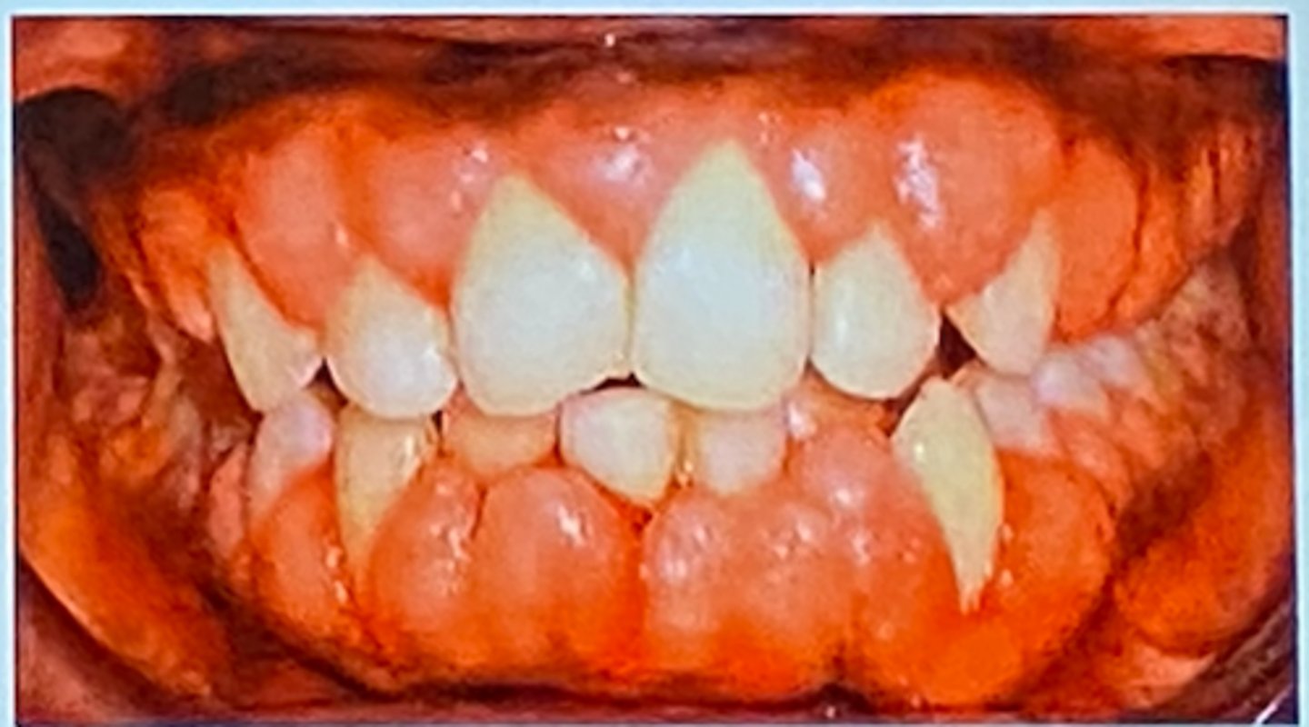
Gingival hyperplasia, ask questions about medications (Dylantin), frequent hygiene and gingivectomy
Diagnosis and treatments
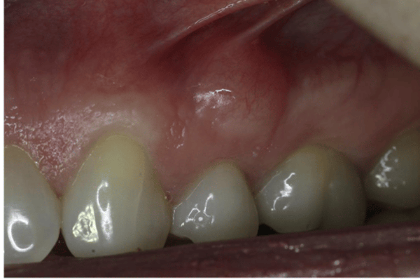
1. Giant Cell Granulomas
2. Pyogenic granuloma
3. Peripheral Ossifying Fibroma
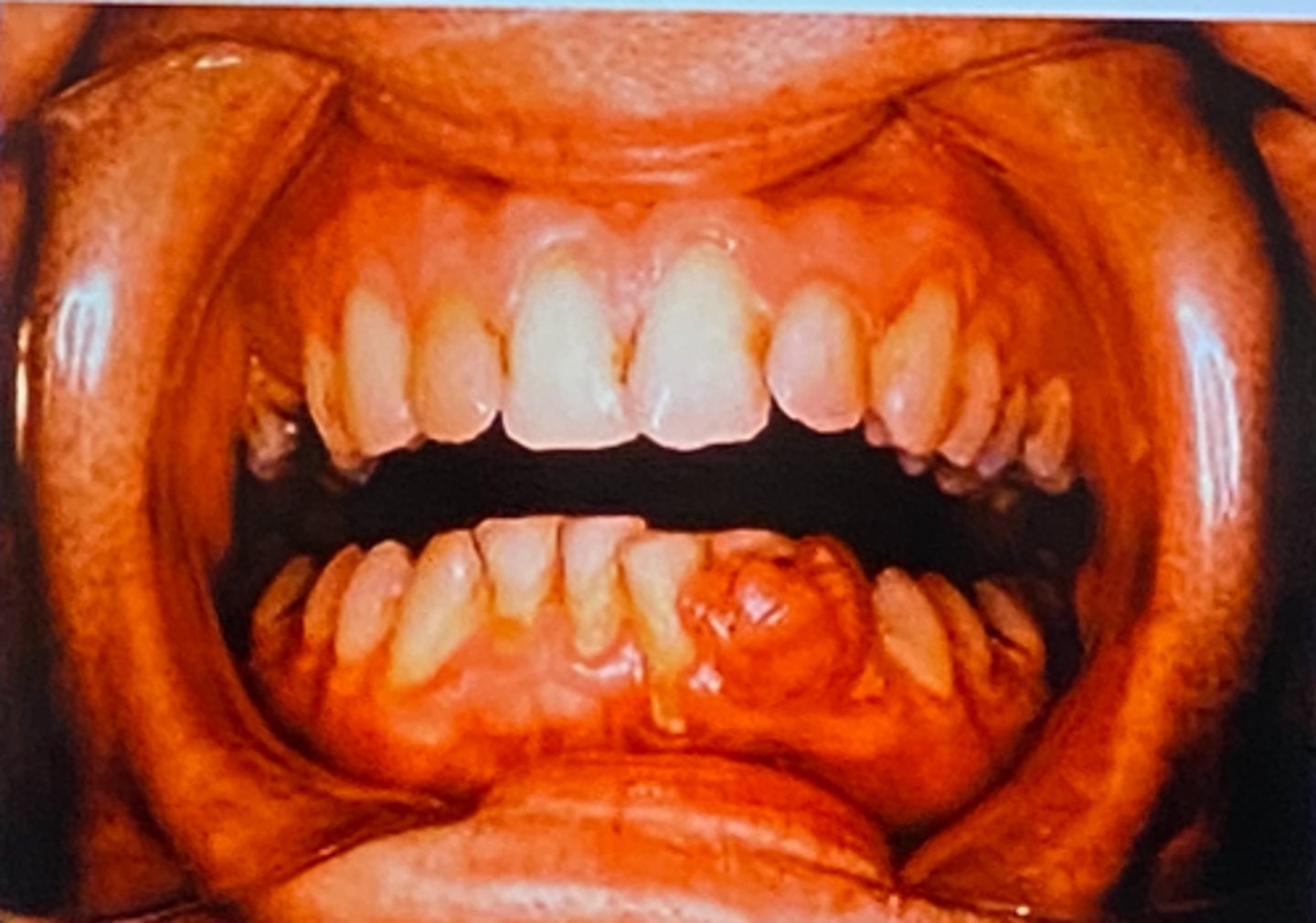
peripheral giant cell granuloma, peripheral Ossifying Fibroma
Which of the 4 P's occur exclusively on the gingiva?
Lipoma
Fibroma
Schwanomma