Cell bio - Vesicle transport and secretory pathway - Chap 15
1/10
Name | Mastery | Learn | Test | Matching | Spaced | Call with Kai |
|---|
No analytics yet
Send a link to your students to track their progress
11 Terms
1. Understand that proteins that are transported into the ER are then packaged and transported in vesicles for transport to the cell surface (plasma membrane) or to other organelles
Know some examples of proteins that are secreted and membrane proteins that are transported this way.
Secreted proteins: Harmons, insulin, and digestive enzymes (that can go into the Gastrointestinal track)
Membrane proteins: GPCR, RTK, Ion channel, and K+/Na pump,
2. Know that vesicles originate from the ER, Golgi and plasma membrane and all carry protein cargo and that all plasma membrane proteins (like receptors) and any secreted proteins are shipped via vesicles.
Vesicles can only be made from membranes places
3. Be able to give the differing functions of the smooth and rough endoplasmic reticulum and describe the structure of the ER
serves as entry point for proteins destined for other organelles
Rough E.R: Region of the E.R associate with ribosomes and involved in the synthesis of secreted and membrane-bound proteins → protein
Smooth E.R: scanty in most cells but is highly developed for performing particular functions—> the site of steroid hormone synthesis in endocrine cell of the adrenal gland and the site where a variety of organic molecule (alcohols) are detoxified in liver. sequester Ca2+ from the cytosol; the release and reuptake of Ca2+ from the E.R is involved in muscles contraction and other response to extracellular signals
4. Be able to track the general path a proteins takes from the ER until it is secreted from the cell
SECRETORY Pathway: ER —> Golgi —> cell surface—> EXOCYTOSIS
5. Be able to describe how a vesicle forms and how the coat proteins clathrin and COP aid in the formation of the vescicle.
What is the structure of clathrin? How does it form around the vesicle?
membrane bound cargo receptor—> cargo molecules on entracelluar side attaches to receptor—> adaptin is attach to receptor on cytosol side —> a clathrin is attached to adaptin—> clathrin attached to to other clathrin in a sphere shape—> dynamin (GTP-binding protein) cuts the neck of off and form a clathrin coated vesicle—> detaches its adaptin-clathrin and its a naked vesicle
they help shape the membrane into a bud and captures molecules for onward transport
Clatrhin - coats vesicles going from Golgi OUT AND from plasma membrane IN
● COP - coats vesicles going from ER to Golgi
-The structure of a clathrin three-armed shape that connect together to form a basketlike cage
-they are formed by attaching themselves to adaptin that are connected to cargo receptor which are bound to cargo molecules
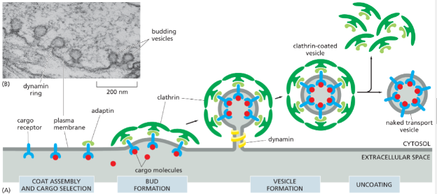
6. Be able to describe how protein cargo is loaded into the clathrin-coated vesicle and the role of the cargo receptor and adaptin.
cargo molecule—>cargo receptor—>adaptin on other side of receptor—>clathrin on adaptin—>more clathrins attached to each other—>dynamin cuts neck off vesicle
Cargo receptors: receptors on the cytosolic side of the membrane that bind to cargo molecules and adaptin to begn the formation of a vesicle
Adaptin: second class of coat proteins- secure clathrin coat to the vesicle membrane and help select cargo molecules for transport—> help capture specific cargo molecules by trapping the cargo receptor that bind them
by this a selected set of cargo molecules , bound to their specific receptors is incorporated into the lumen of each newly formed clathrin-coated vesicle
7. Understand how the transport of proteins in vesicles is specific - that is how protein cargo is shipped to the correct location in a cell - what mechanisms are important. Be able to describe the role of Rab in this process.
The transport vesicles are identified through the help of a Rab protein (Small GTP-binding proteins present on the surface of transport vesicles and organelles that serves as a molecules marker to help ensure that transport vesicles fuse only with the correct membrane)
Rab protein are recognized by corresponding tethering proteins (Filamentous transmembrane protein involved in docking of transport vesicle to target membranes) on the cytosolic surface of the target membrane
Also has SNAREs (One of a family of memebrane proteins responsible for the selective fusion of vesicles with a target membrane inside the cell) —> v-SNARE is on the transport vesicle which interactice with the t-SNARE on the membrane of the organelle firmly locking it into place
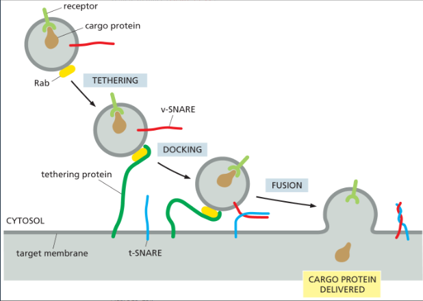
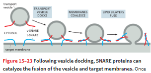
8. Be able to describe the role that the SNARE molecules have in membrane fusion between vesicles and target membranes
SNAREs help with firmly docking the vesicle into place as well as having a central role in catalyzing the membrane fusion required for a transport vesicle to deliver its cargo—> also add the vesicle membrane into the membrane of the organelle (intermix)
when fusion is triggered, the v-SNAREs and the t-SNAREs wrap around each other tightly, thereby acting like a winch that pulls the two lipid bilayers into close proximity squeezing out of any water molecules that remain trapped between the two membranes allowing fo the lipids to flow together
9. Be able to describe protein modification that take place in the ER and Golgi.
○Proteins are cleaved
○ Quaternary proteins assemble
○ Disulfide bonds form
○ Proteins are glycosylated
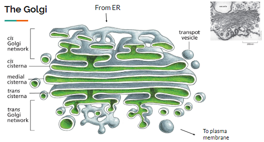
10. Describe the structure of the Golgi and explain how proteins travel to it and through it.
What does the Golgi do to proteins to ensure that they are targeted to the correct place (plasma membrane, lysosome, etc.) in the cell?
5 main parts
cis Goglgi network —> next to the E.R
(Cis Cisterna, Medial & Trans Cisternae)—> in the middle of the golgi
Trans Golgi network
Protein travel from the E.R to the Golgi via vesicles, when they reach the golgi, they fusion with other proteins to form the Cis Cinsterna—> as protein moves through the stacks they are modified by residnet Golgi enzymes—> provides signal for the final destination of the proteins —> movement occurs in waves
Cis Cinsterna becomes a part of the medial cisternae—> new cis cisterna form—> medial cistern becomes the new trans-cisterna: Cis-maturation model
proteins are sorted within trans golgi—> then buds off into vesicles—> miragate to organelles —> lyosomes or cell memebrane
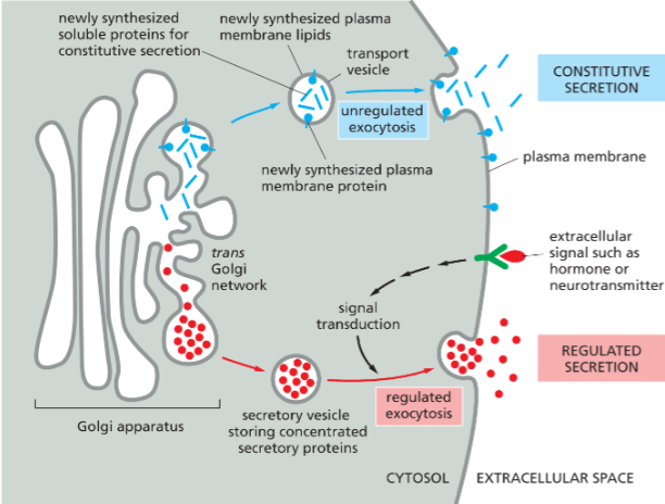
11. Know the difference between constitutive exocytosis and regulated exocytosis.
What are some examples of regulated exocytosis? Why is this important?
constitutive exocytosis: Soluble proteins that are continuously secreted from the cell, also continuously supplies the plasma membrane with newly synthesized lipids and proteins—> doesn’t require signal sequences
regulated exocytosis: Specialized secretory cell; which selected proteins into the trans Golgi network are diverted into secretory vesicles, where the proteins are concentrated and stored until an extracellular signal stimulates their secretion.
Release of insulin from pancreatic B cell; increase in glucose levels—> rapid dissolves into the blood