OTHED 1505 Week 1: Key Terms/ Acquisition Questions
1/72
There's no tags or description
Looks like no tags are added yet.
Name | Mastery | Learn | Test | Matching | Spaced | Call with Kai |
|---|
No analytics yet
Send a link to your students to track their progress
73 Terms
Nervous System
The body's communication network that controls voluntary and involuntary actions, transmitting signals between the brain, spinal cord, and body. Components: CNS (brain + spinal cord) and PNS (cranial + spinal nerves)
Application Question: If someone touches a hot stove and quickly pulls their hand away, which two main divisions of the nervous system are working together to make this response happen?
Answer: The Peripheral Nervous System (PNS) detects the pain and sends it to the Central Nervous System (CNS), which processes the signal and sends a motor command back to the muscles.
Central Nervous System (CNS)
Brain and spinal cord; processes information and initiates responses.
Peripheral Nervous System (PNS)
: All nerves outside the CNS, including cranial and spinal nerves; connects CNS to the body.
If you step on a tack, which division of the nervous system carries the pain message to your spinal cord and brain?
The sensory nerves of the PNS carry the pain message to the CNS.
If your physical therapist says your pain is "distal to the knee," where is the pain located in relation to the knee?
The pain is below or farther away from the trunk than the knee, such as in the shin or foot.
Anterior (ventral)
front of the body
Posterior (dorsal)
back of body
superior (cephalic)
toward the head
Inferior (caudal)
away from the head end or toward the lower part of a structure or the body; below
Medial
Toward the midline of the body
Lateral
Away from the midline of the body
Proximal
Nearer to the trunk of the body
Distal
Farther from the trunk of the body
Frontal Lobe
Motor control, problem solving, planning, speech production (Broca's area).

Parietal Lobe
Sensory processing, spatial awareness.

Temporal Lobe
Hearing, memory, emotion; contains hippocampus & amygdala.
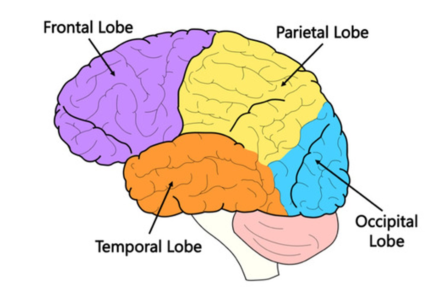
Occipital Lobe
Vision processing.

Insular Cortex
Pain, taste, emotion, homeostasis.
Cerebrum
Largest brain part; higher brain functions.
If someone has difficulty speaking and planning movements, which lobe of the brain is most likely damaged?
The frontal lobe
Neuron
Functional unit of the nervous system that transmits signals.
What is a neuron and its main structures?
A neuron is a nerve cell that sends and receives signals.
• Dendrites: Receive signals.
• Cell body (soma): Processes signals.
• Axon: Sends signals to other neurons or muscles.
• Myelin sheath: Covers axon for faster signal transmission.
• Axon terminals: Release neurotransmitters to communicate.
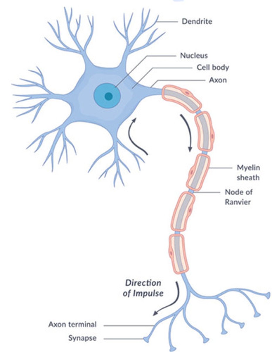
Neuron Structures
Dendrites = receive signals, Cell body (soma) = processes info, Axon = sends signals, Axon terminals = release neurotransmitters.
If a neuron cannot send signals to the next cell because neurotransmitters are not released, which part of the neuron is malfunctioning?
The axon terminals.
White Matter
Myelinated axons; fast signal transmission.
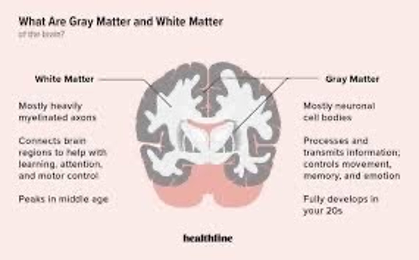
Gray Matter
Neuron cell bodies; information processing.

Explain the difference between white matter and gray matter.
White matter: Made of myelinated axons; transmits signals quickly.
Gray matter: Made of cell bodies and dendrites; processes information.
If a disease causes damage to myelin, which type of matter is affected and how might it impact communication speed in the nervous system?
White matter is affected, causing slower or blocked signal transmission (like in multiple sclerosis).
Cranial Nerve I (name, sensory or motor, function)
Olfactory
Sensory
Smell
Cranial Nerve II (name, sensory or motor, function)
Optic
Sensory
Vision
Cranial Nerve III (name, sensory or motor, function)
Oculomotor
Motor
Eye movement, pupil constriction
Cranial Nerve IV (name, sensory or motor, function)
Trochlear
motor
eye movement (superior oblique)
Cranial Nerve V (name, sensory or motor, function)
Trigeminal
Both
Face sensation, chewing
Cranial Nerve VI (name, sensory or motor, function)
Abductens
Motor
Eye movement (lateral rectus)
Cranial Nerve VII (name, sensory or motor, function)
Facial
Both
Facial expression, taste
Cranial Nerve VIII (name, sensory or motor, function)
Vestibulocochlear
Sensory
Hearing, balance
Cranial Nerve IX (name, sensory or motor, function)
Glossopharyngeal
Both
Swallowing, taste
Cranial Nerve X (name, sensory or motor, function)
Vagus
Both
Parasympathetic control, swallowing, speech
Cranial Nerve XI (name, sensory or motor, function)
Accessory
Motor
Shoulder and neck muscles
Cranial Nerve XII (name, sensory or motor, function)
Hypoglossal
Motor
Tongue movement
Spinal Nerve Pathways : Ascending
Ascending (Sensory): Dorsal root → CNS; carries sensory info to the brain.

Explain the pathway of ascending and descending spinal nerves.
Ascending spinal nerves: Carry sensory information from the body → spinal cord → brain. They enter through the dorsal root.
Descending spinal nerves: Carry motor commands from the brain → spinal cord → body. They exit through the ventral root.
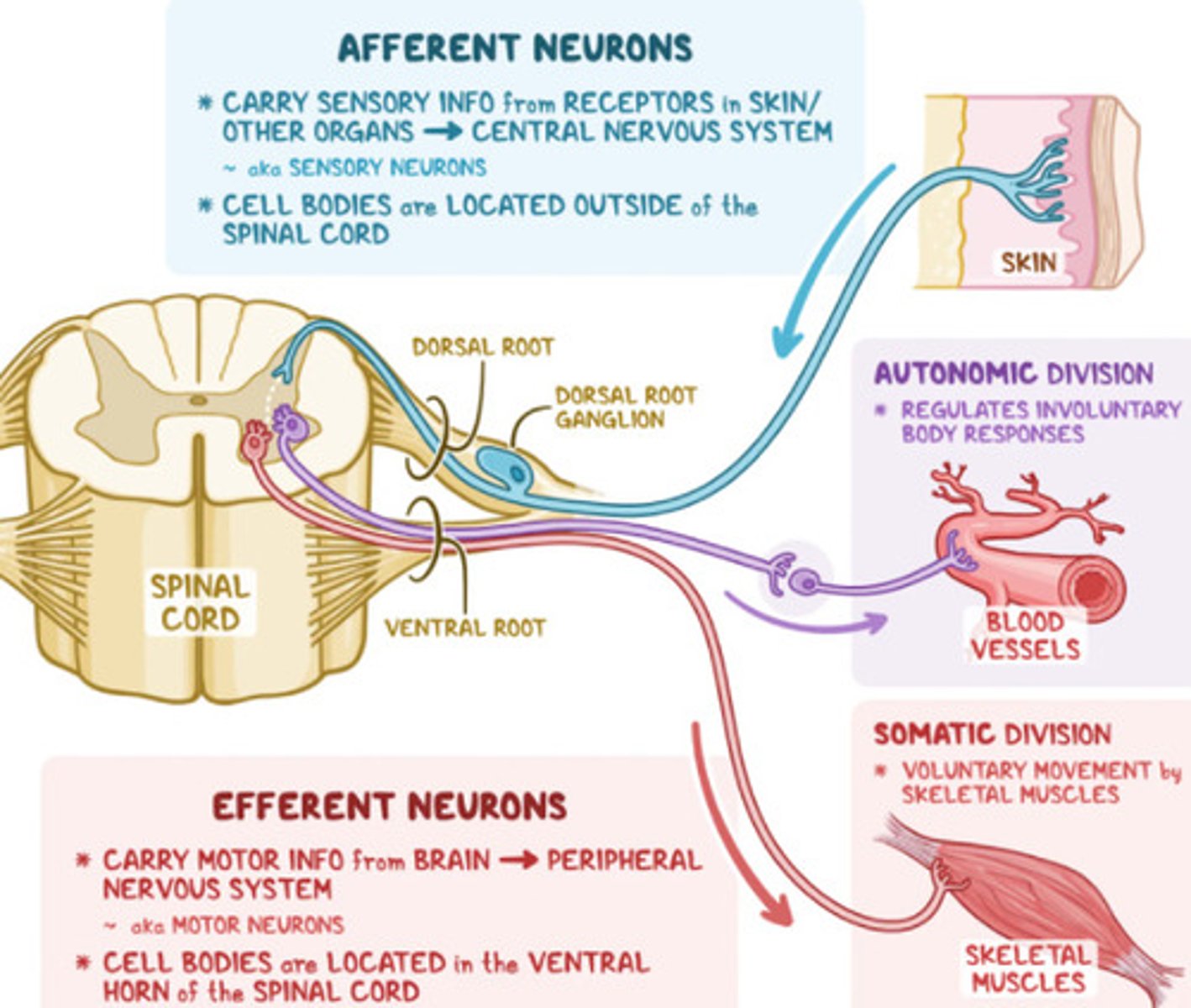
Afferent neurons (sensory neurons)
Carry sensory information from the body's periphery (e.g., skin, eyes, ears) to the central nervous system (brain and spinal cord

Efferent neurons (motor neurons)
Carry motor commands from the central nervous system to the body's muscles and glands

Ascending sensory pathway
The pathway likely damaged if a patient cannot feel sensations in their foot.
Spinal Nerve Pathways: Descending
(Motor): Brain → CNS → ventral root → muscles; carries motor commands.

Dermatomes (definition and function)
Skin area supplied by a single spinal nerve.
Function: Sends sensory info to the somatosensory cortex; helps identify nerve injury levels.
Dermatome mapping
The tool that could help identify which spinal nerve may be injured if a patient has numbness along their thumb and index finger.
Myotomes (definition and function)
Muscle groups supplied by a single spinal nerve.
Function: Receives motor commands from the primary motor cortex; helps assess motor function.
Myotome mapping
The type of mapping that would help identify the affected spinal nerve if a patient cannot lift their wrist.
What is an important component of each spinal cord level?
Cervical: Controls neck, arms, hands, breathing (diaphragm).
Thoracic: Controls trunk and abdominal muscles.
Lumbar: Controls legs and lower body.
Sacral: Controls bowel, bladder, sexual function.
Spinal Reflex Arc / Withdrawal Reflex
Definition: Automatic response to stimuli for protection. Pathway: Sensory receptor → sensory neuron → interneuron → motor neuron → effector (muscle).
Spinal reflex arc
The type of nervous system response that occurs when you accidentally step on a sharp object and immediately lift your foot.
Explain a spinal reflex arc/withdrawal reflex and why it is important.
Reflex arc: Automatic, fast response that doesn't require the brain.
Example: Touching something hot → sensory neuron → spinal cord → motor neuron → pull hand away.
Importance: Protects the body and helps clinicians assess nervous system function.
Upper Motor Neuron (UMN)
Brain → spinal cord; damage causes spastic paralysis (Ex: stroke, multiple sclerosis).
Lower Motor Neuron (LMN)
Spinal cord → muscles; damage causes flaccid paralysis (Ex: Bell's palsy).
Difference between upper motor neurons (UMN) and lower motor neurons (LMN) give an example of each
UMN: Located in brain and spinal cord; damage causes spasticity and hyperreflexia.
Example: Stroke, Multiple Sclerosis.
LMN: Connect spinal cord to muscles; damage causes flaccidity and hyporeflexia.
Example: Bell's Palsy, Peripheral Nerve Injury.
Difference between upper motor neurons (UMN) and lower motor neurons (LMN) + examples.
UMN: Located in brain and spinal cord; damage causes spasticity and hyperreflexia.
Example: Stroke, Multiple Sclerosis.
LMN: Connect spinal cord to muscles; damage causes flaccidity and hyporeflexia.
Example: Bell's Palsy, Peripheral Nerve Injury.
Upper Motor Neuron (UMN) damage
The type of motor neuron that is damaged if a patient has spastic paralysis in their right arm after a stroke.
Corticospinal tract
Motor: Voluntary movement - Descending, sends signals from brain to muscles
Spinothalamic tract
Sensory: Pain & temperature -Carries pain, temperature, and touch sensations to the brain
Dorsal Column-Medial Lemniscus tract
Sensory: Fine touch & proprioception - Ascending, Carries vibration, pressure, and proprioception information to the brain.
Limbic System
Emotion & memory
Hippocampus
Memory formation
Amygdala
Emotion processing.
Brainstem
Heart rate, breathing, reflexes.
Thalamus
Sensory relay center.
Hypothalamus
Homeostasis, hormone regulation.
Dorsal Root Ganglia
Sensory neuron cell bodies outside CNS.
Motor Pathways
Descending; brain → muscles.
Sensory Pathways
Ascending; body → brain.
Motor pathway damage
The pathway affected if a patient cannot move their leg but can still feel touch in it.