lab exam histo 7.1 circulatory system
1/60
There's no tags or description
Looks like no tags are added yet.
Name | Mastery | Learn | Test | Matching | Spaced | Call with Kai |
|---|
No analytics yet
Send a link to your students to track their progress
61 Terms
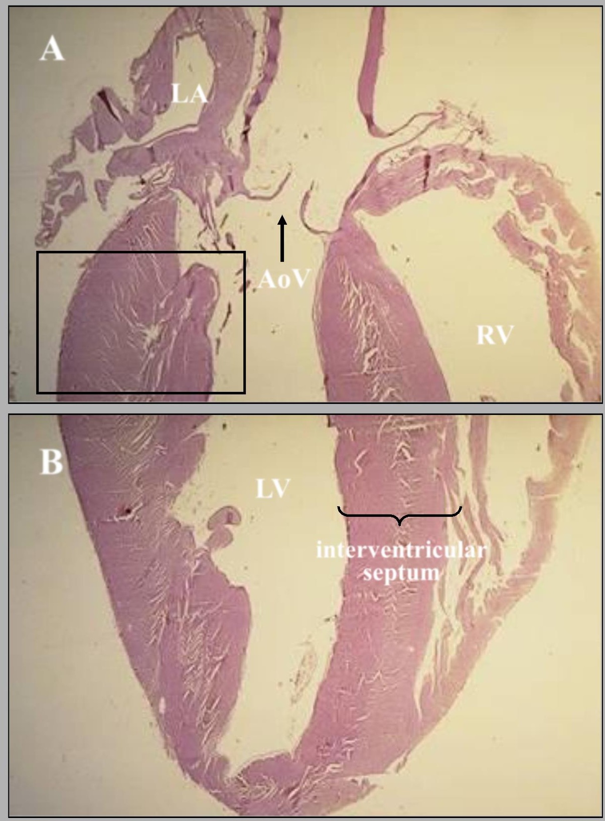
name of this slide? state the parts
composite figure of a sagittal section of feline heart. The parts are: AoV-Aortic valve, LA-Left atrium, LV/RV-Left/ Right ventricles, and the Interventricular septum.
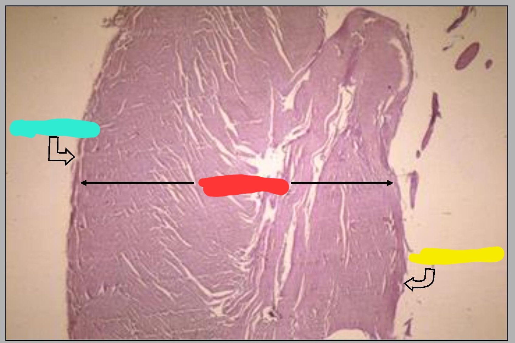
What part of feline heart is this? What are the parts shown in this picture?
Wall of left ventricle.
Cyan-Epicardium, Red-Myocardium, Yellow-Endocardium. This is arranged from external to the internal surface.
Comprises the bulk of the heart wall.
Myocardium
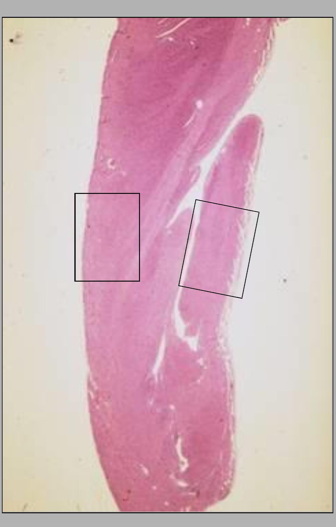
Name of this slide? What is on the left side and the right side?
Wall of the canine right ventricle. Epicardium is on the left side and Endocardium on the right side.
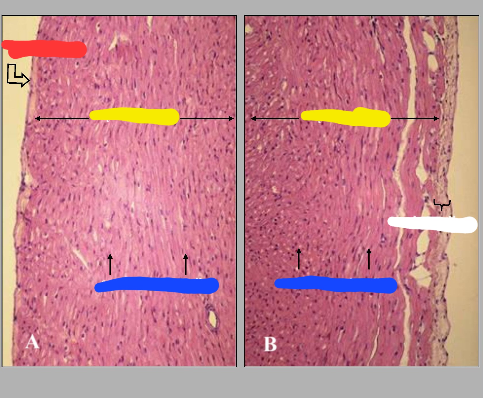
What is the name of this slide? What are the parts called?
Heart wall.
Red-Epicardium
Yellow-Myocardium
Blue-Cardiac muscle cells
White-Endocardium
Is lined by mesothelium on the outside
Epicardium
Comprises the bulk of the ventricular wall and is composed of cardiac muscle.
Myocardium
Is lined by endothelium on the inside
Endocardium
The pale staining connective tissue layer beneath the endothelium
Subendothelial layer
Name this slide.
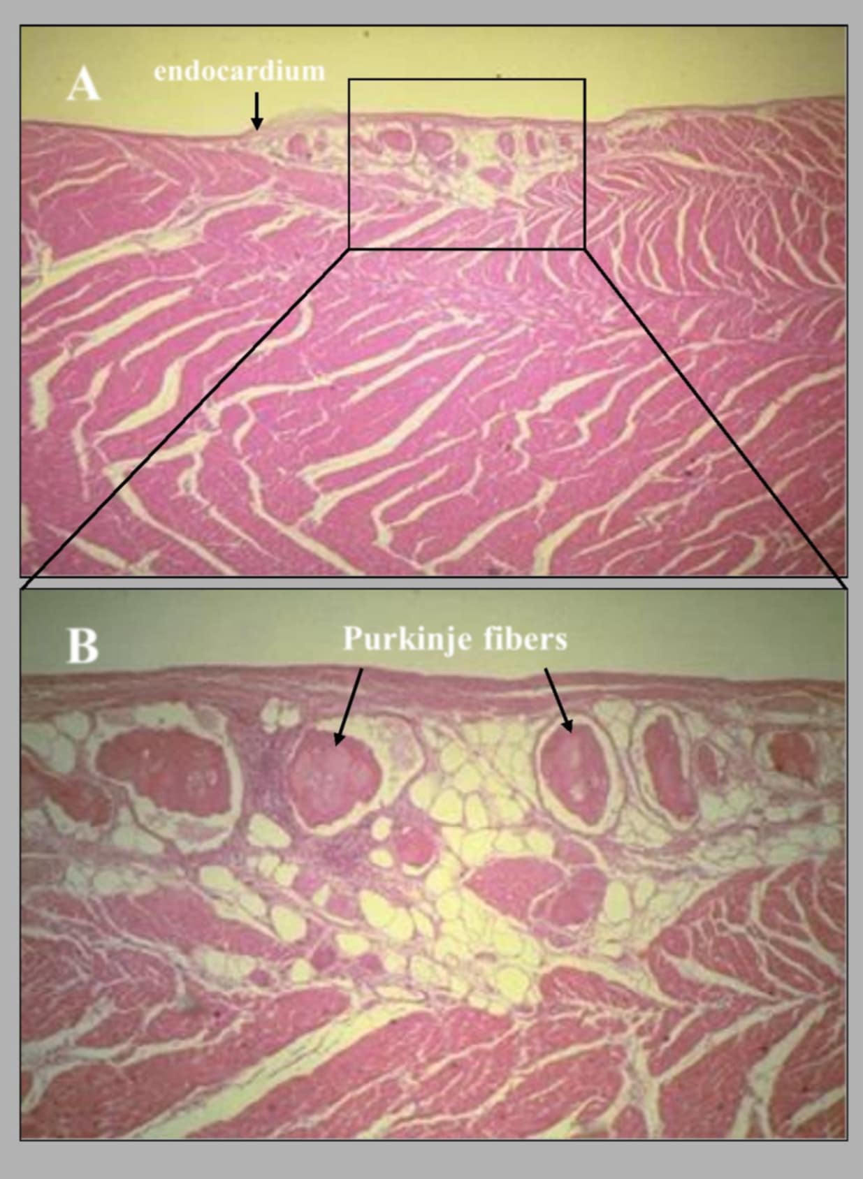
Section of ventricular wall of ovine heart
Name the species that owns this heart.
Ovine
are a special type of cardiac muscle fibers that comprise the atrioventricular bundle or bundle of His. They have a larger diameter than ordinary cardiac muscle fibers; they function in impulse conduction.
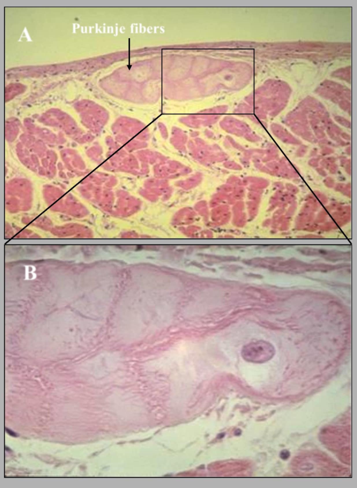
Purkinje fibers
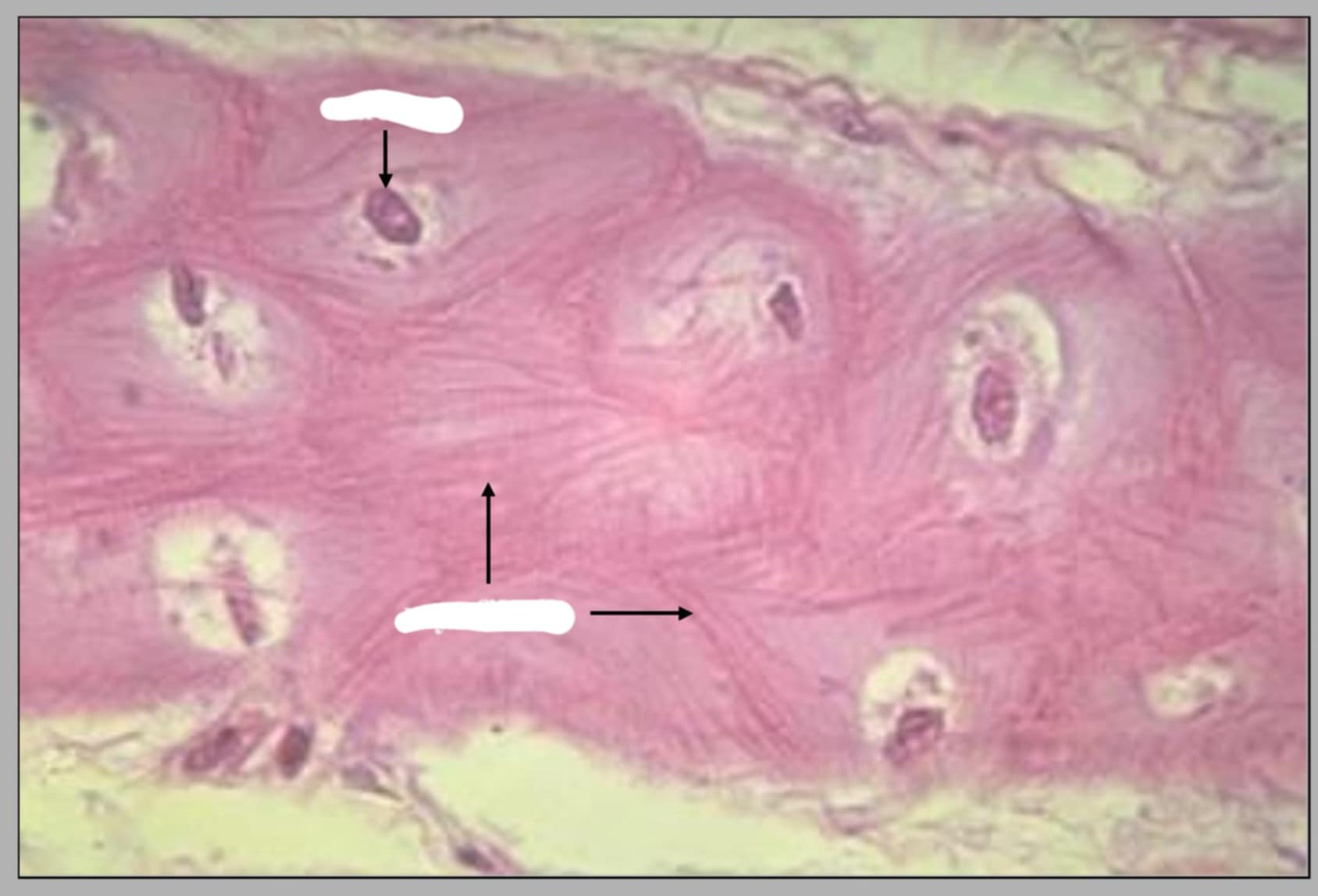
This is another view of purkinje fibers in ovine heart. What are the parts seen in this slide?
Nucleus-top and Myofibrils-bottom.
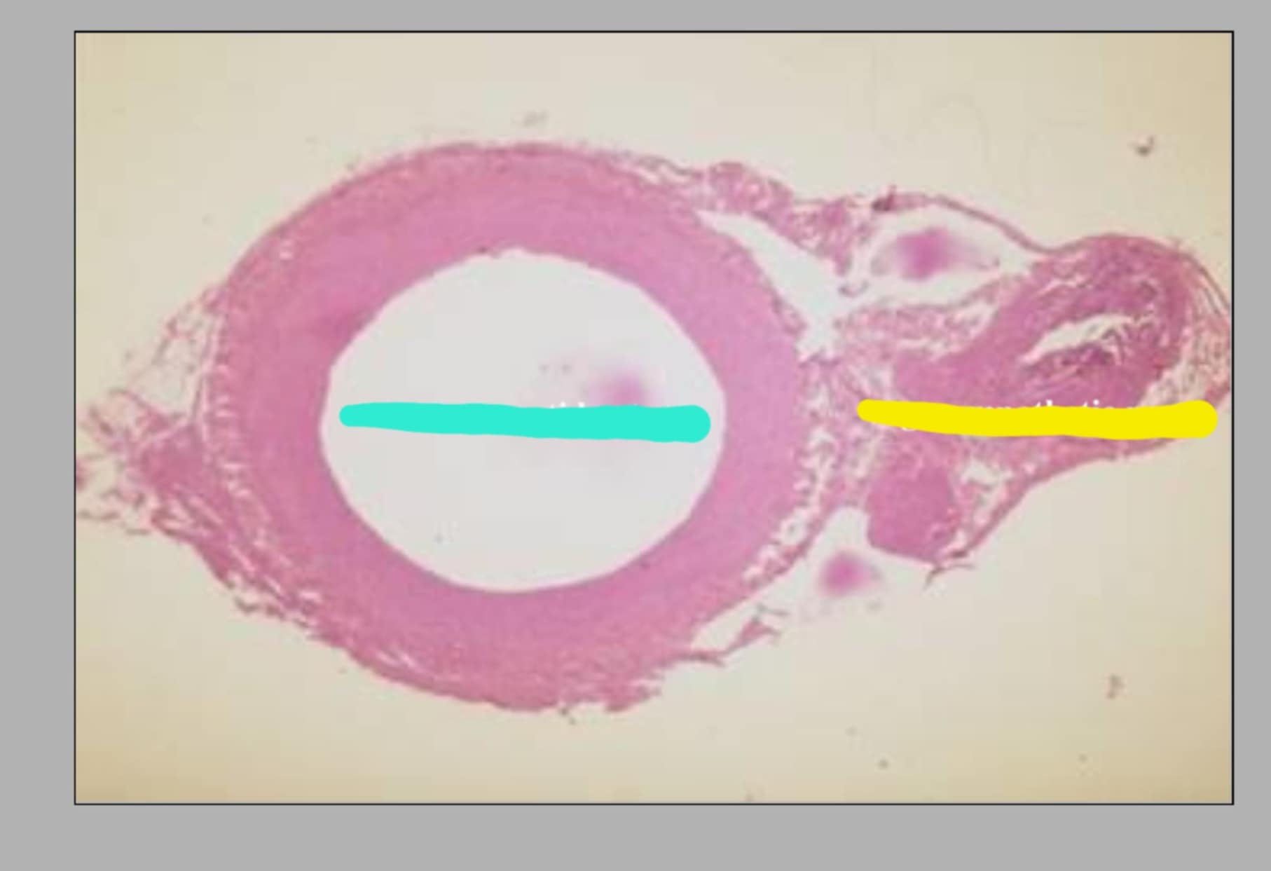
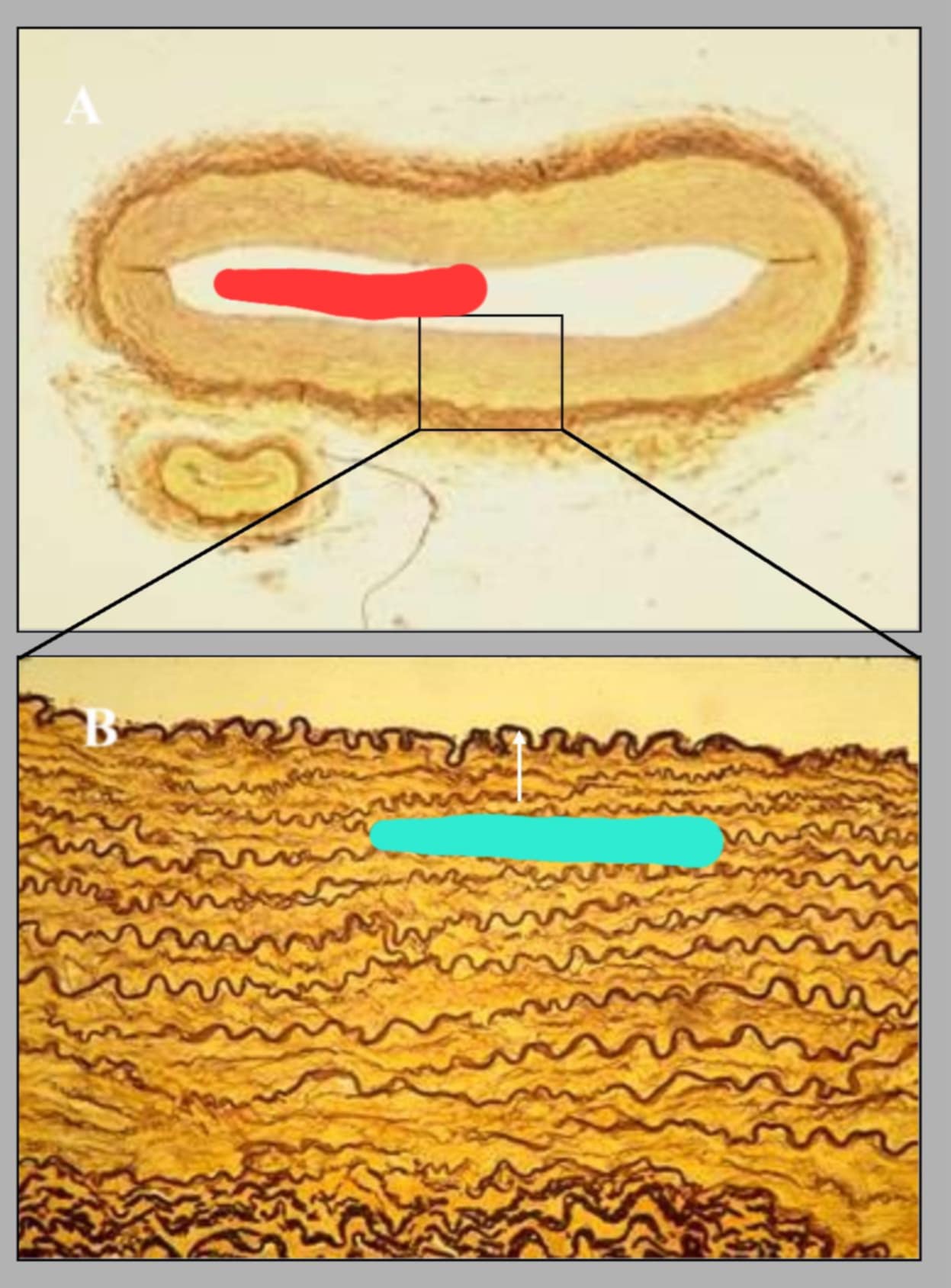
Name of this slide? State the parts of the colored parts.
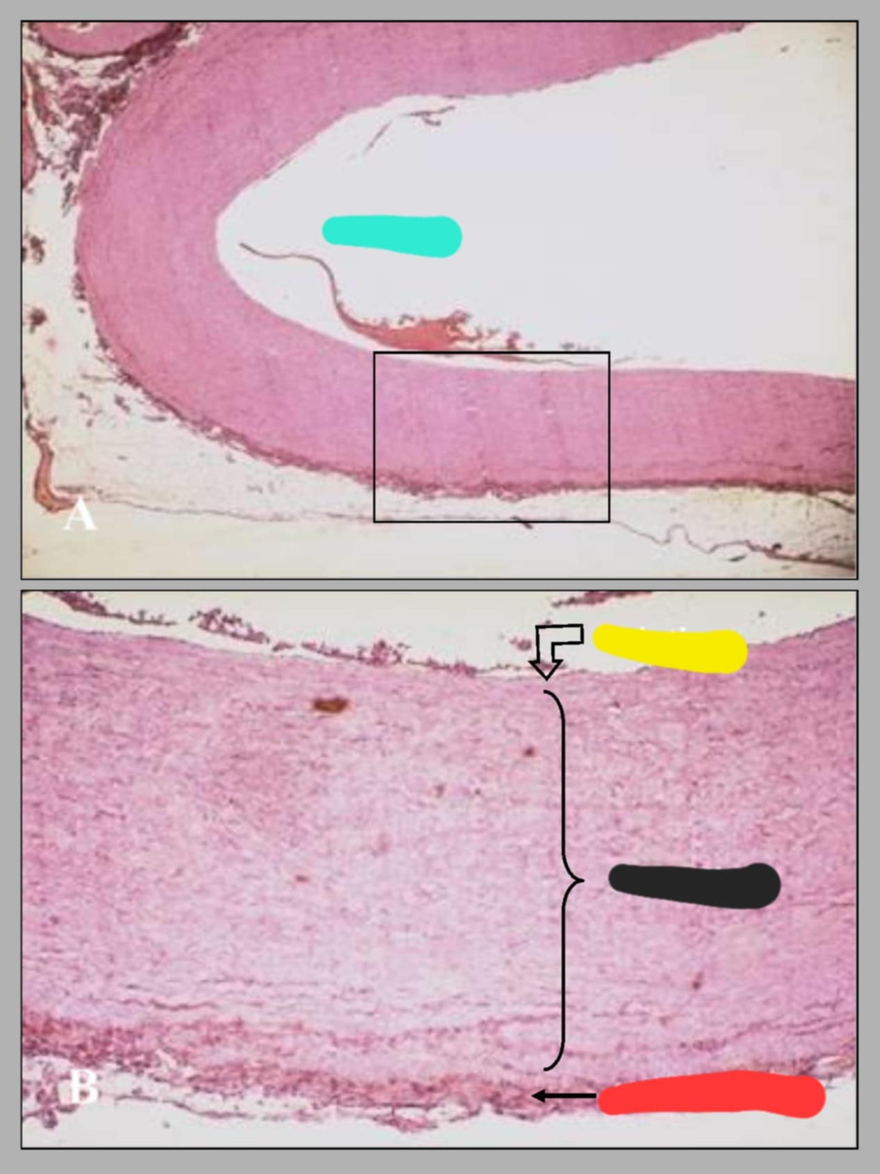
Cross section of canine aorta.
Cyan-Aorta, Yellow-T. intima, Black-T. media, and Red-T. adventitia
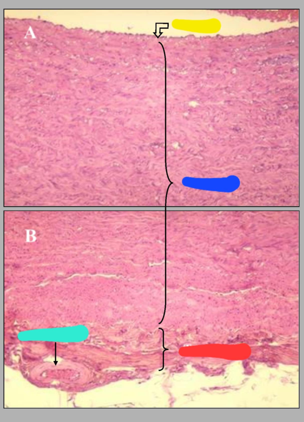
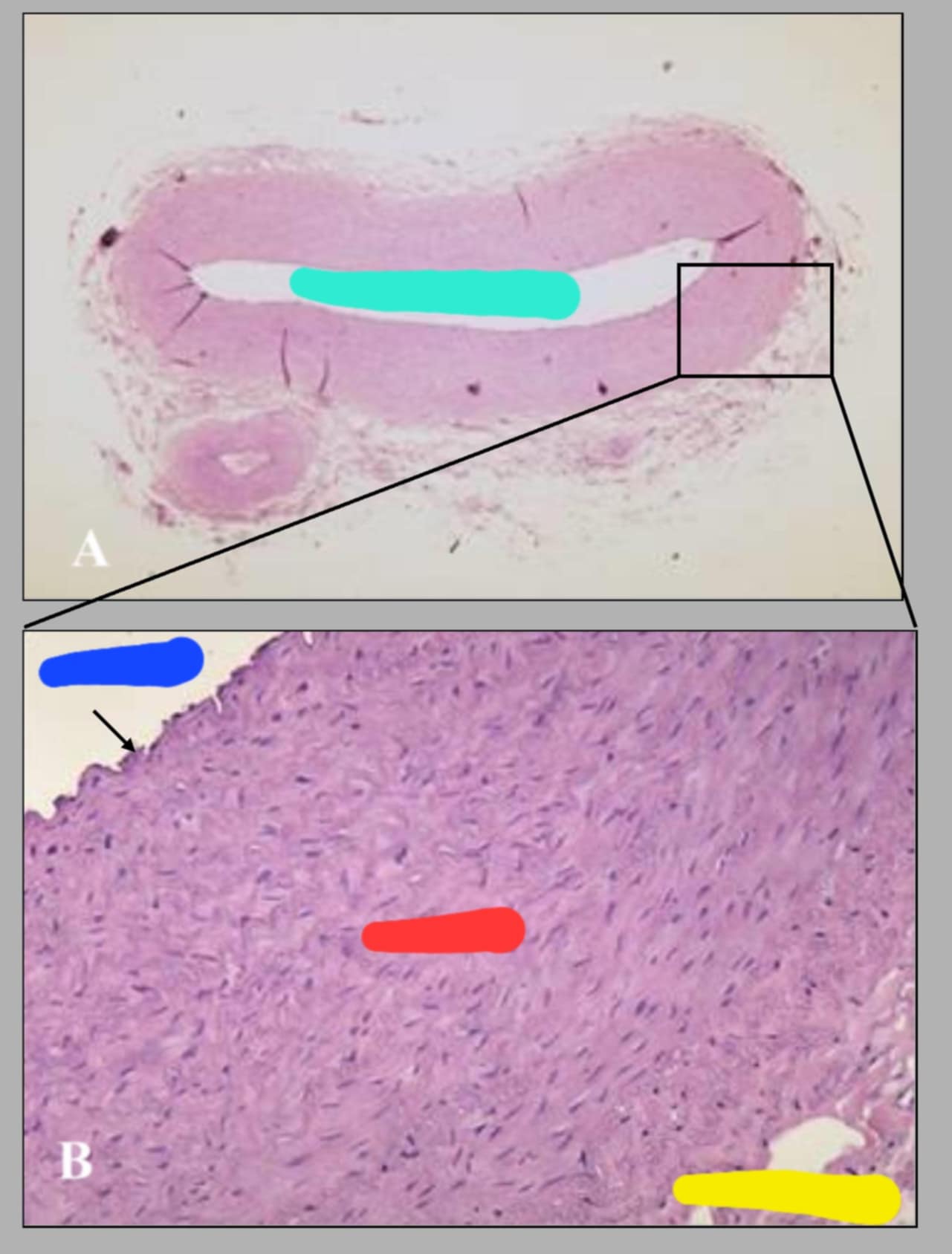
Name of the slide. State the parts.
Wall of canine aorta.
Yellow-T. intima, Blue-T. media, Red-T. adventitia, and Cyan- Vasa vasorum
The wall of the aorta is composed of 3, which are:
tunica intima, tunica media(which comprise the bulk of the arterial wall, and the tunica adventitia.
The small blood vessels that supply blood to the outer wall of bigger blood vessels
Vasa vasorum
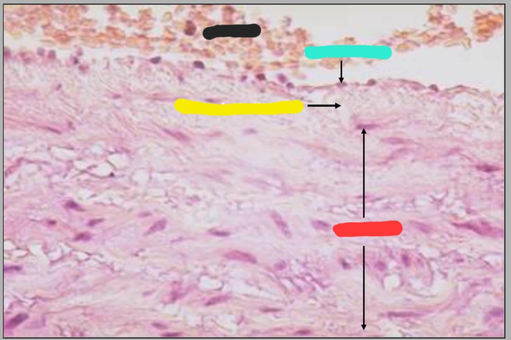
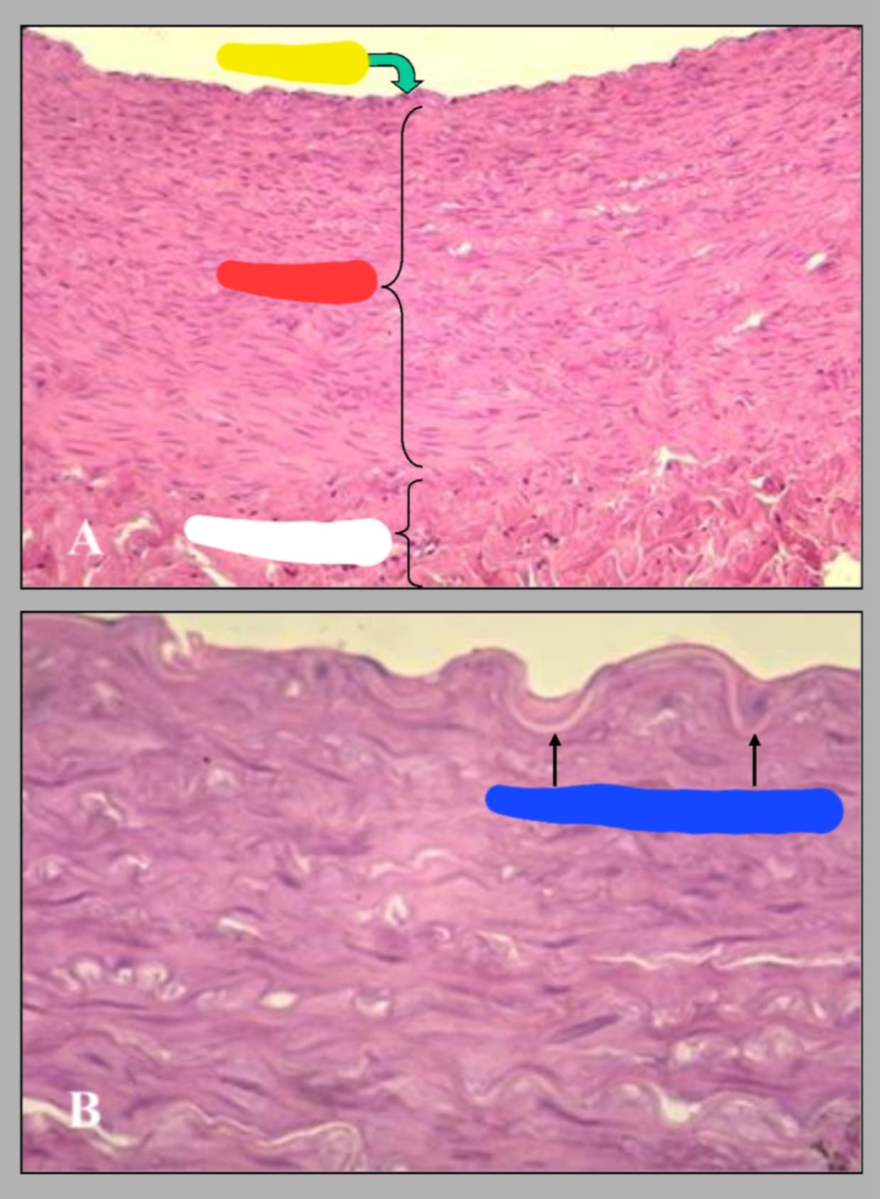
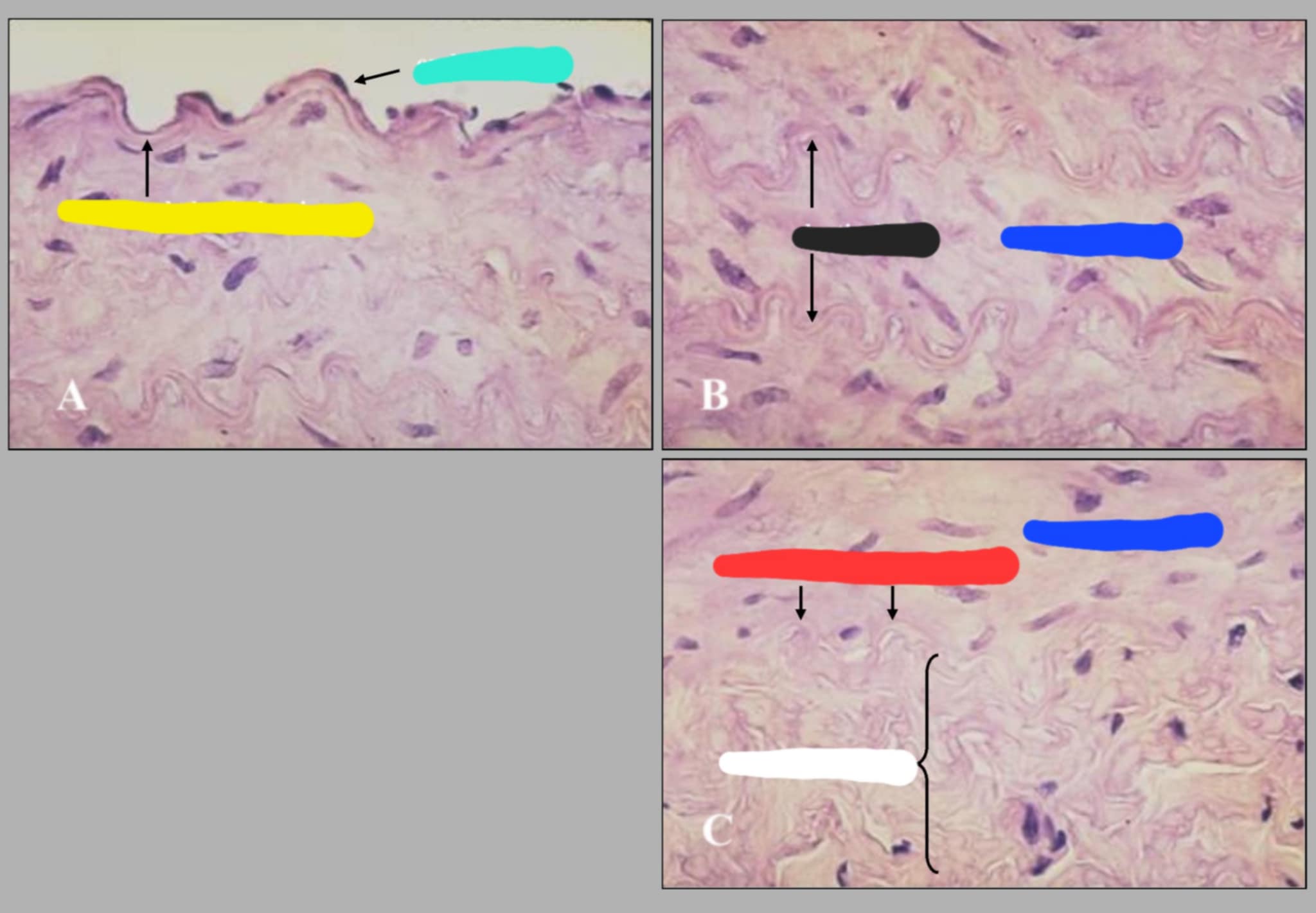
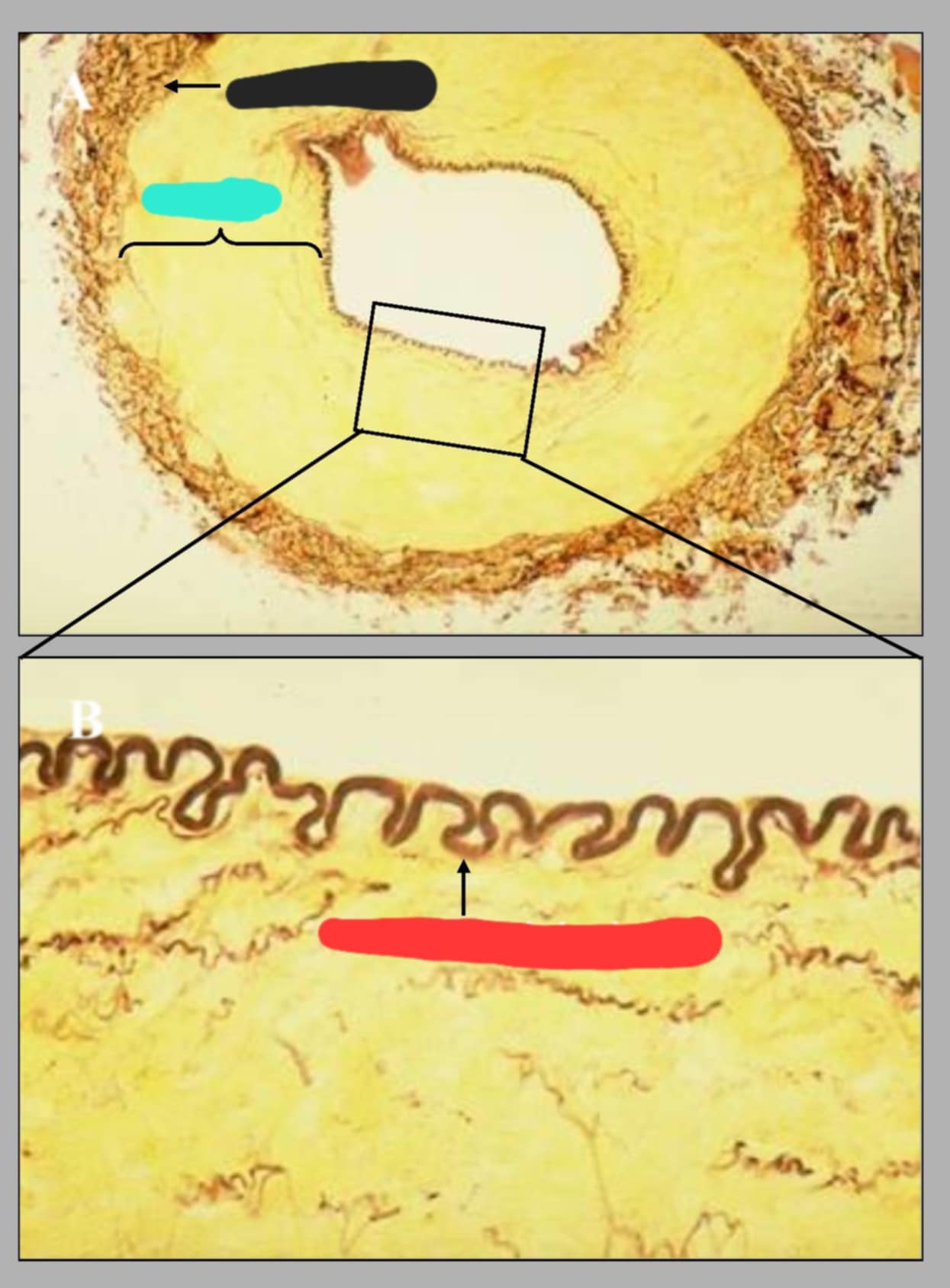
What are the parts that can be seen in this slide of Tunica intima and tunica media of aorta?
Black-RBC, Cyan-Endothelium, Yellow-Subendothelial CT, Red-T. media
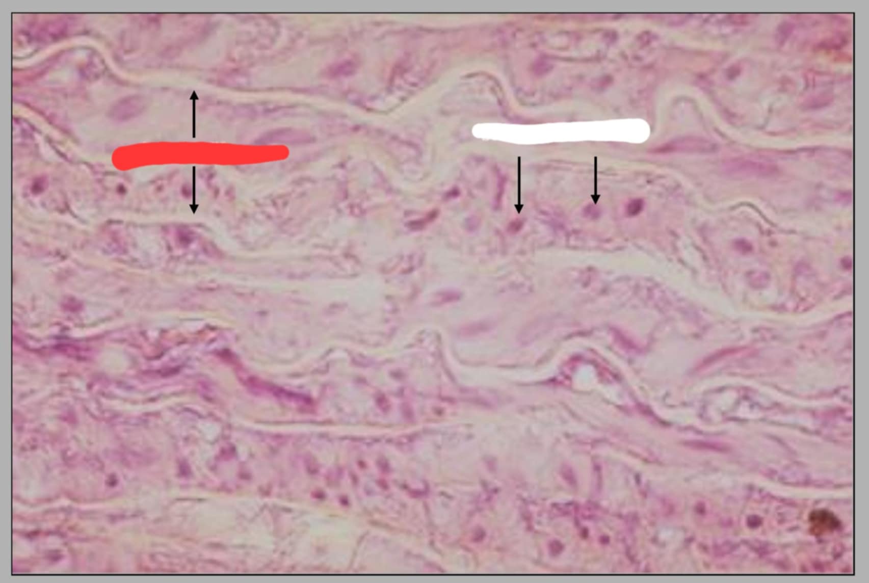
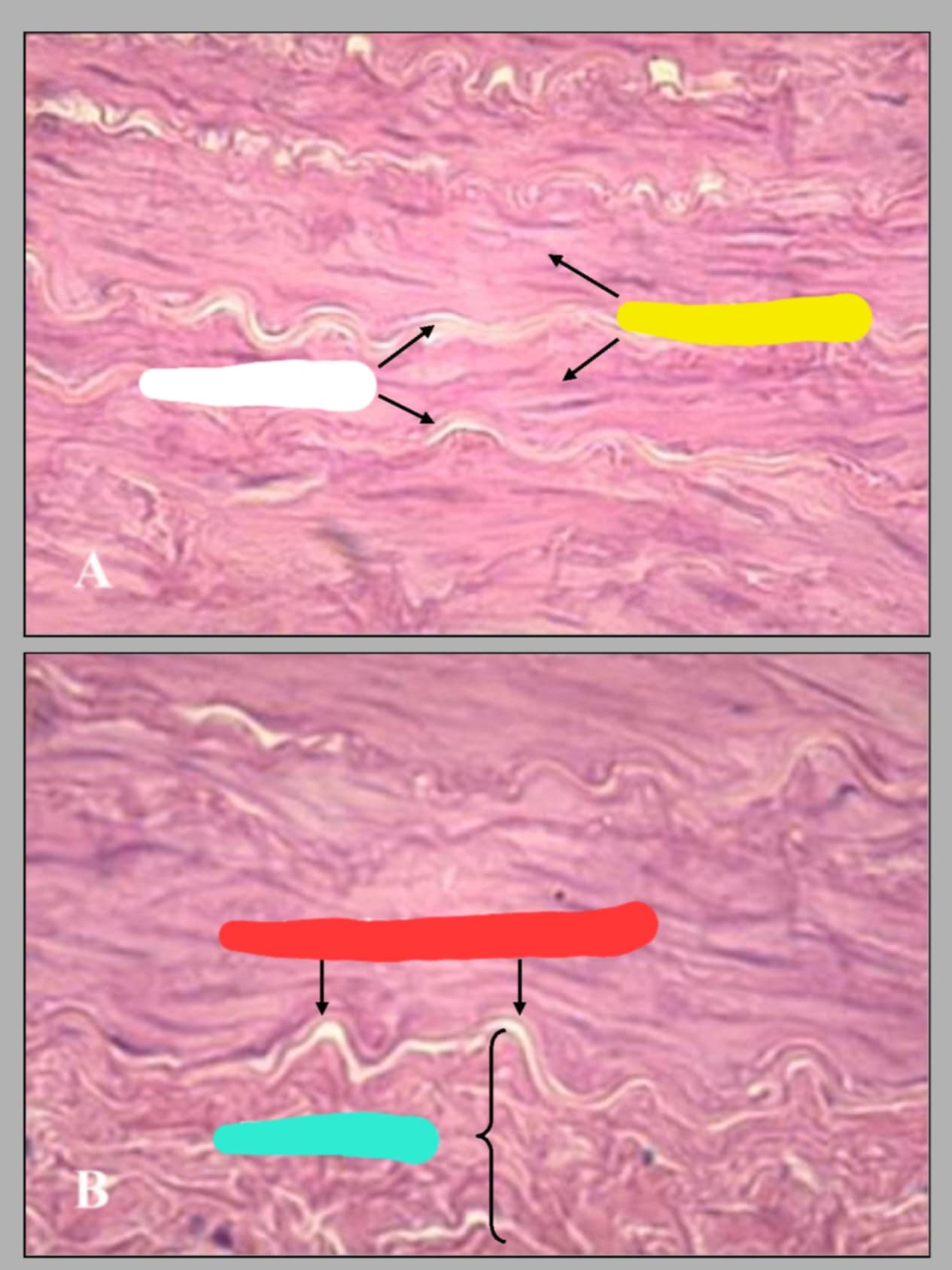
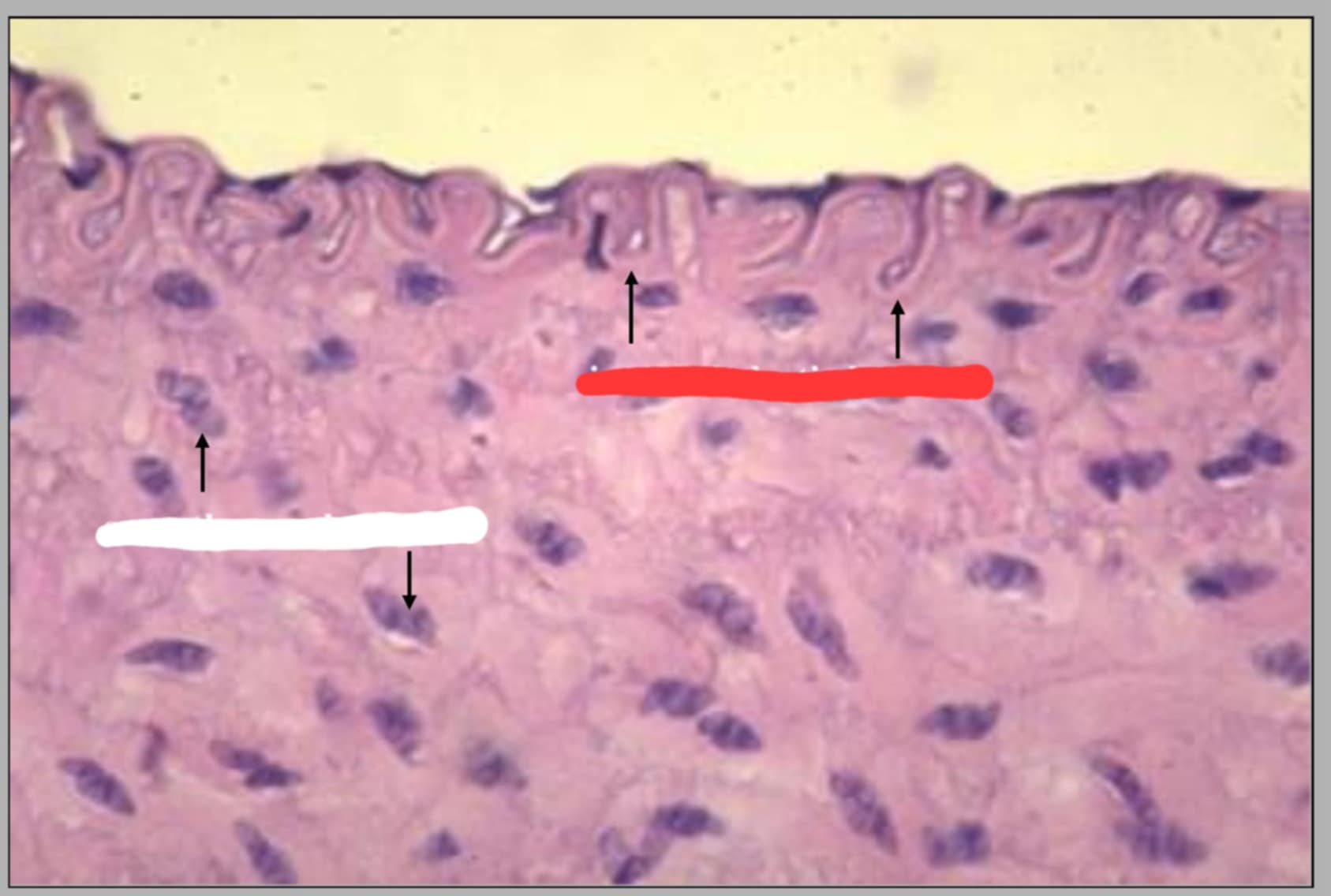
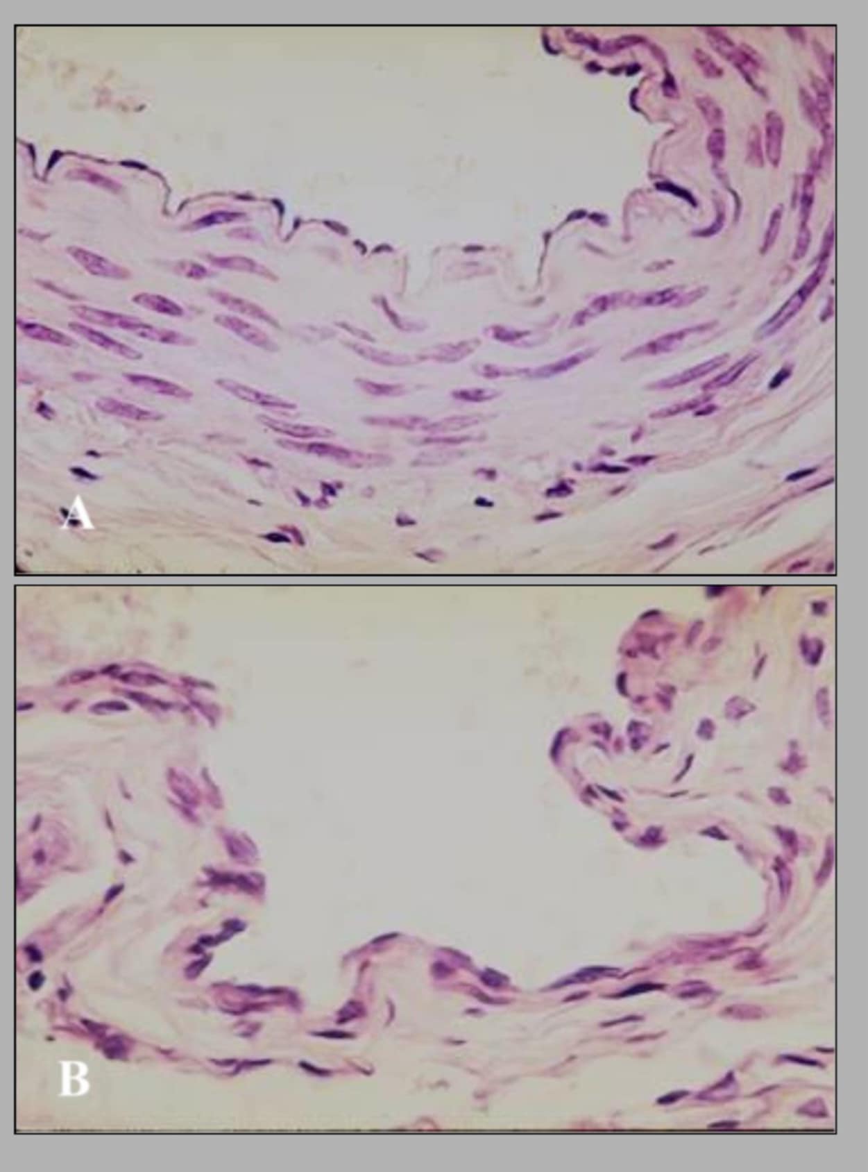
Name of this slide and state the parts.
Tunica media of aorta. Red-Elastic fibers, White-Smooth muscle
is lined by endothelium (simple squamous epithelium)
Tunica intima
is poorly defined in large elastic arteries like the aorta
internal elastic lamina
is composed of cicularly ot spirally arranged smooth muscle and elastic fibers.
tunica media of aorta
are the refractile wavy lines between the smooth muscle cells
Elastic fibers
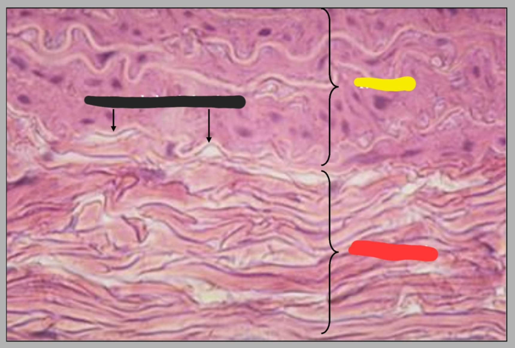
State the parts of this slide.
Yellow-tunica media
Black-external elastic lamina
Red-tunica adventitia
is composed of connective tissue elements. the connective tissue fibers are primarily collagen and elastic fibers. the external elastic lamina may be seen between the tunica media and tunica adventitia.
tunica adventitia
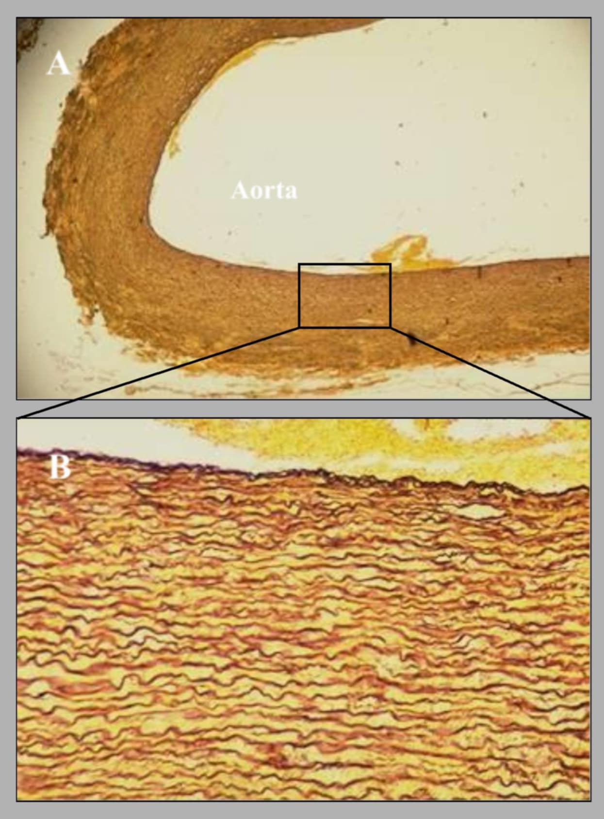
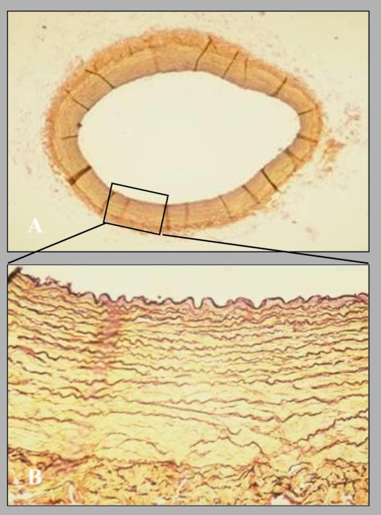
wall of aorta, canine, elastic fiber stain. this cross section of the aorta was stained with ______ to demonstrate the presence of elastic fibers.
orcein
-are characterized by the abundance of elastic fibers in their wall, thus they are also called Elastic Arteries.
Large arteries like Aorta
help the arteries withstand the great force of blood pumped by the heart; they also act as an auxillary pump (via elastic recoil)
Elastic fibers
this is a common carotid artery of what species? state the parts shown.
Canine.
cyan-common carotid artery and yellow-vagosympathetic nerve
name this slide and state its parts.
All of common carotid artery.
yellow-t. intima, red-t. media, white-t. adventitia, blue-internal elatic lamina
is demarcated from the tunica media by the internal elastic lamina
Tunica intima
name the parts in this wall of common carotid artery
yellow-smooth muscle, white-elastic fibers, red-external elastic lamina, cyan-t. adventitia
is composed of several concentric layers of smooth muscle, and elastic fibers are present between the smooth muscle layers.
tunica media
is composed of connective tissue elements; it is demarcated from the tunica media by the external elastic lamina.
tunica adventitia
name this slide.
section of common carotid artery, canine, elastic fiber stain
true or false: as arteries become smaller, the amount of elastic fibers decreases but the amount of smooth muscle fibers increases
true
name this slide and state the parts
cross section of brachial artery of canine. the parts are: cyan-brachial artery, blue-t. intima, red-t. media, yellow-t. adventitia
name of this slide? state the parts.
wall of brachial artery.
cyan-endothelium, yellow-internal elastic lamina, black-elastic fibers, blue-smooth muscle, red-external elastic lamina, white-t. adventitia
name this slide. state its parts.
cross section of brachial artery of canine (stained with orcein)
red-brachial artery, cyan-internal elastic lamina
name this slide and state its parts.
cross section of digital artery of canine.
red-tunica adventitia, cyan-tunica media, black-tunica intima
name this slide and state its parts
wall of muscular artery.
red-internal elastic lamina, white-smooth muscle nuclei
name of this slide and its parts.
cross section of digital artery of canine (stained with orcein)
black-t. adventitia, cyan-t. media, red-internal elastic lamina
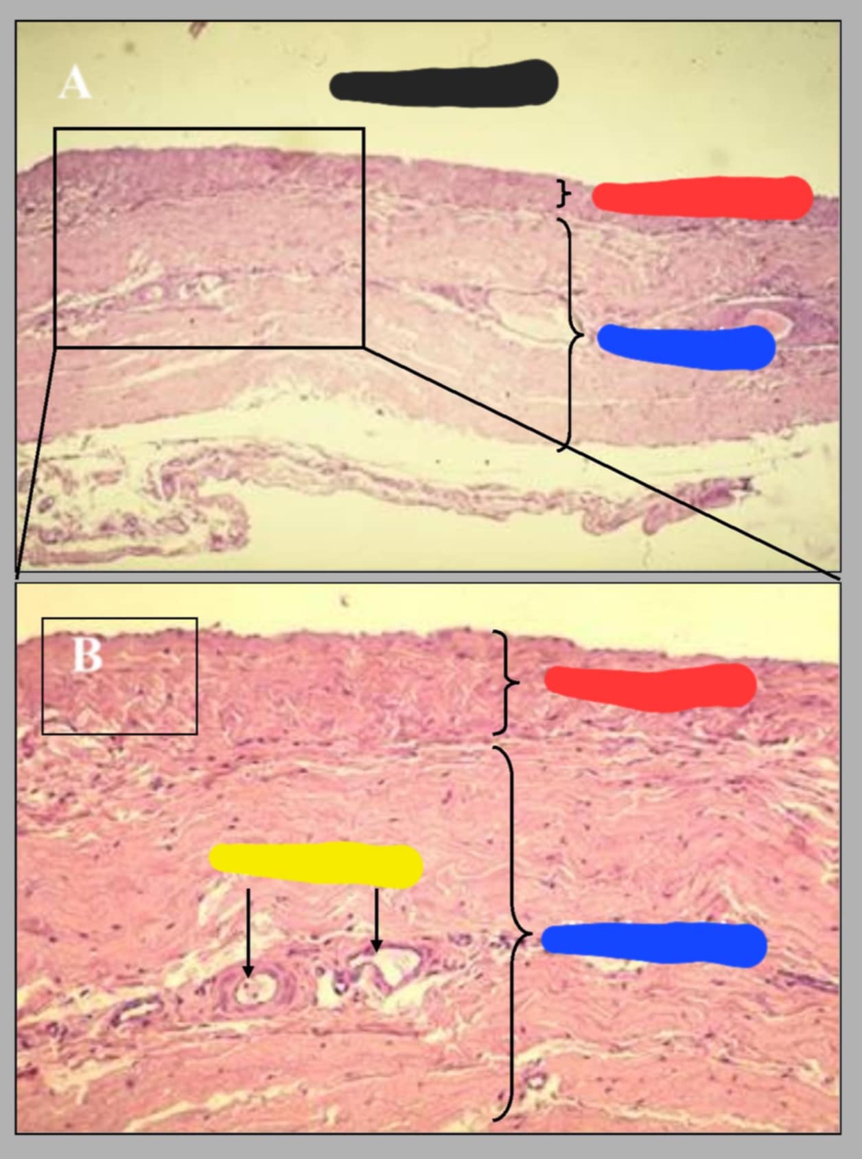
name this slide and its parts
longitudinal section of equine vena cava.
black-lumen of vein, red-t. intima/media, blue-t. adventitia, yellow-vasa vasorum
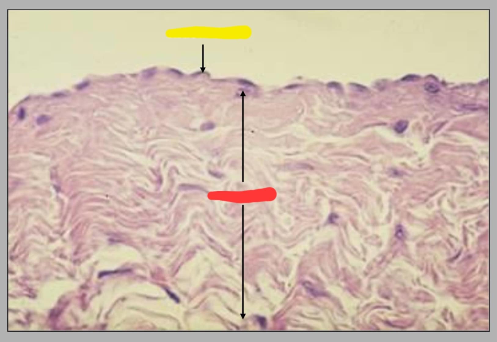
name of the slide and its parts
tunica intima/media of vena cava.
yellow-endothelium, red-t. media
tunica intima is represented by endothelium; the internal elastic lamina is very poorly developed. the tunica media is much thinner than in arteries.
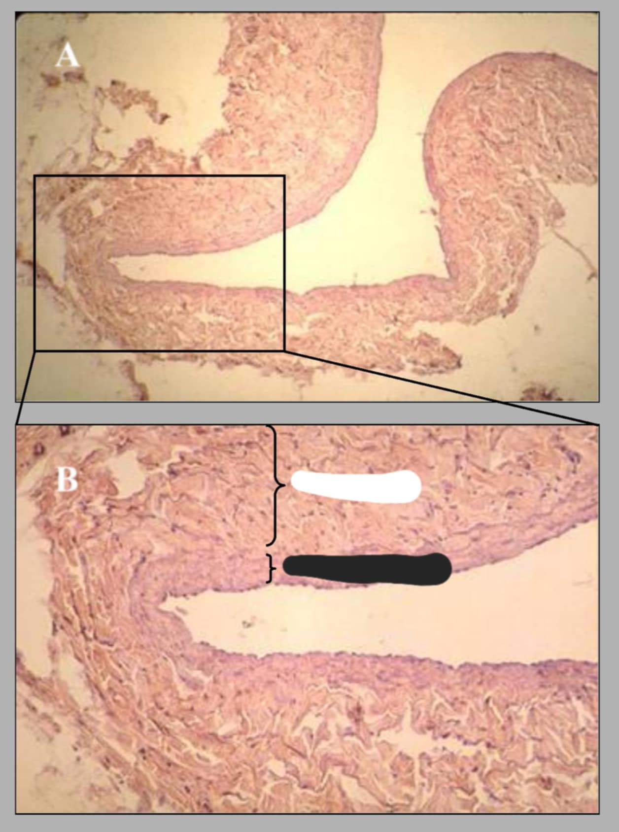
name and parts of this slide.
cross section of external jugular vein of canine.
white-t. adventitia, black-t. intima/media
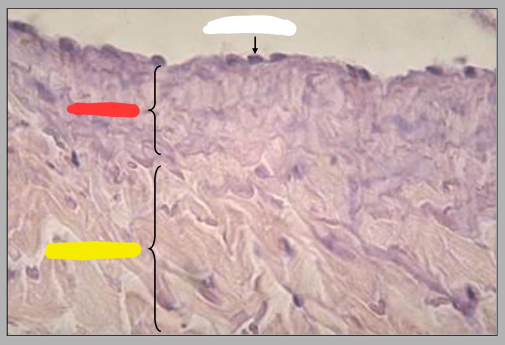
name of this slide and its parts.
inner wall of jugular vein.
white-endothelium, red-t. media, yellow-t. adventitia.
the tunica media is the thin lightly basophilic layer, and the acidophilic layer is the tunica adventitia
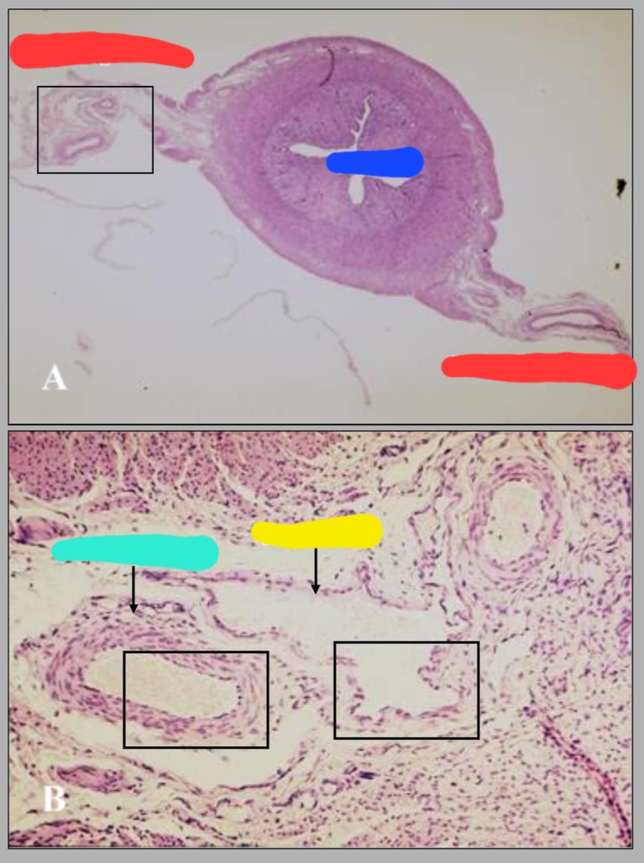
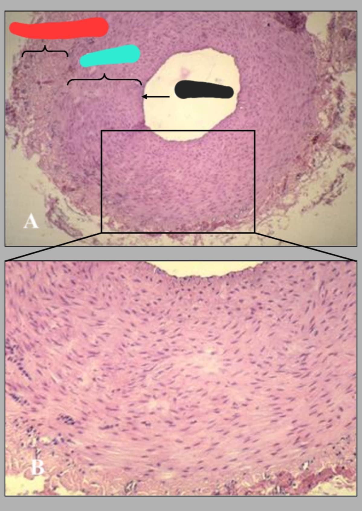
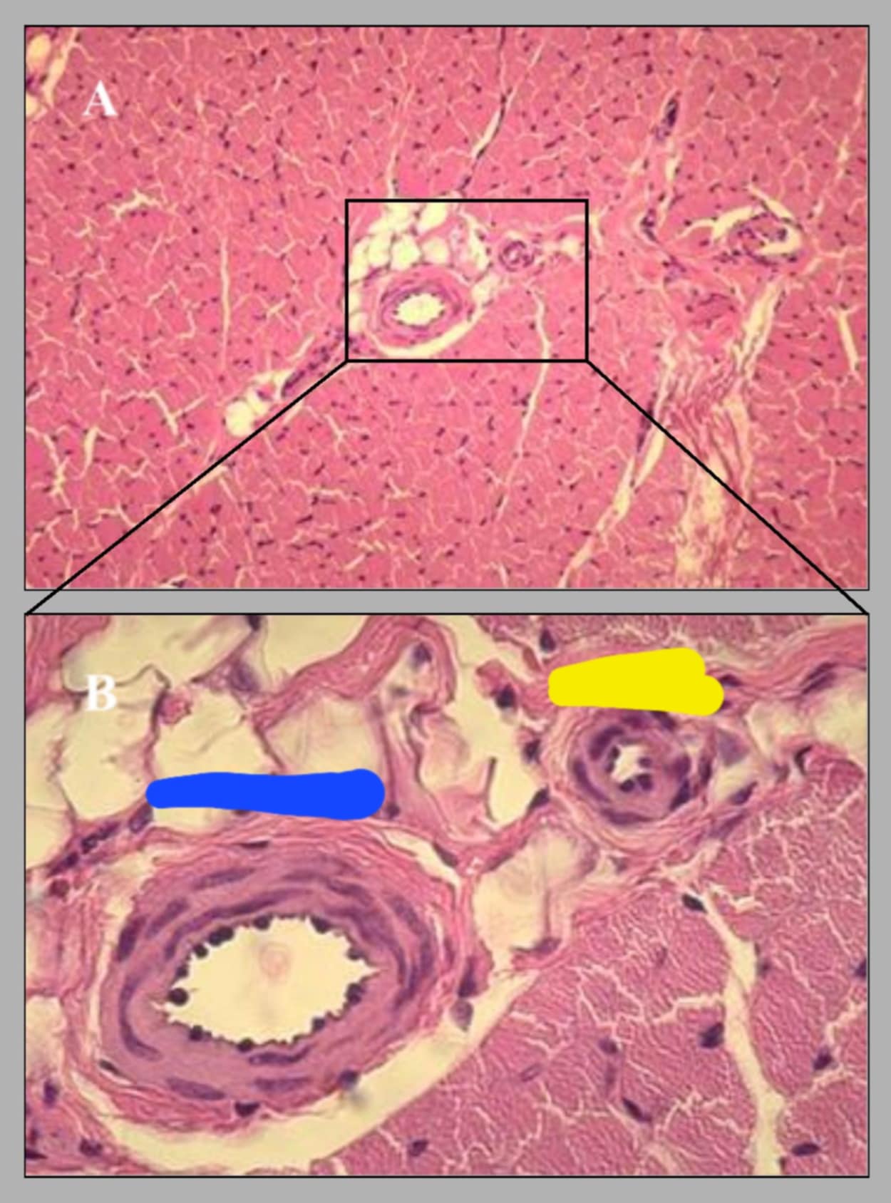
name this slide and state its parts
cross section of canine uterus and accompanying blood supply.
red-broad ligament, blue-uterus, yellow-small vein, cyan-small artery.
Located in the broad ligament are the uterine artery and vein and their branches. Arteries have thicker wall and more regular lumen than veins and veins have thinner wall and irregularly shaped lumen.
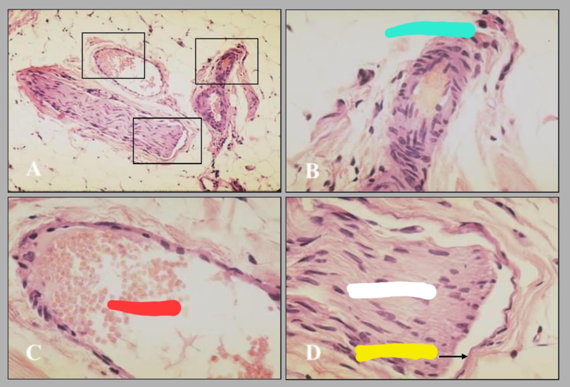
name of this slide?
walls of small artery(a) and small vein(b)
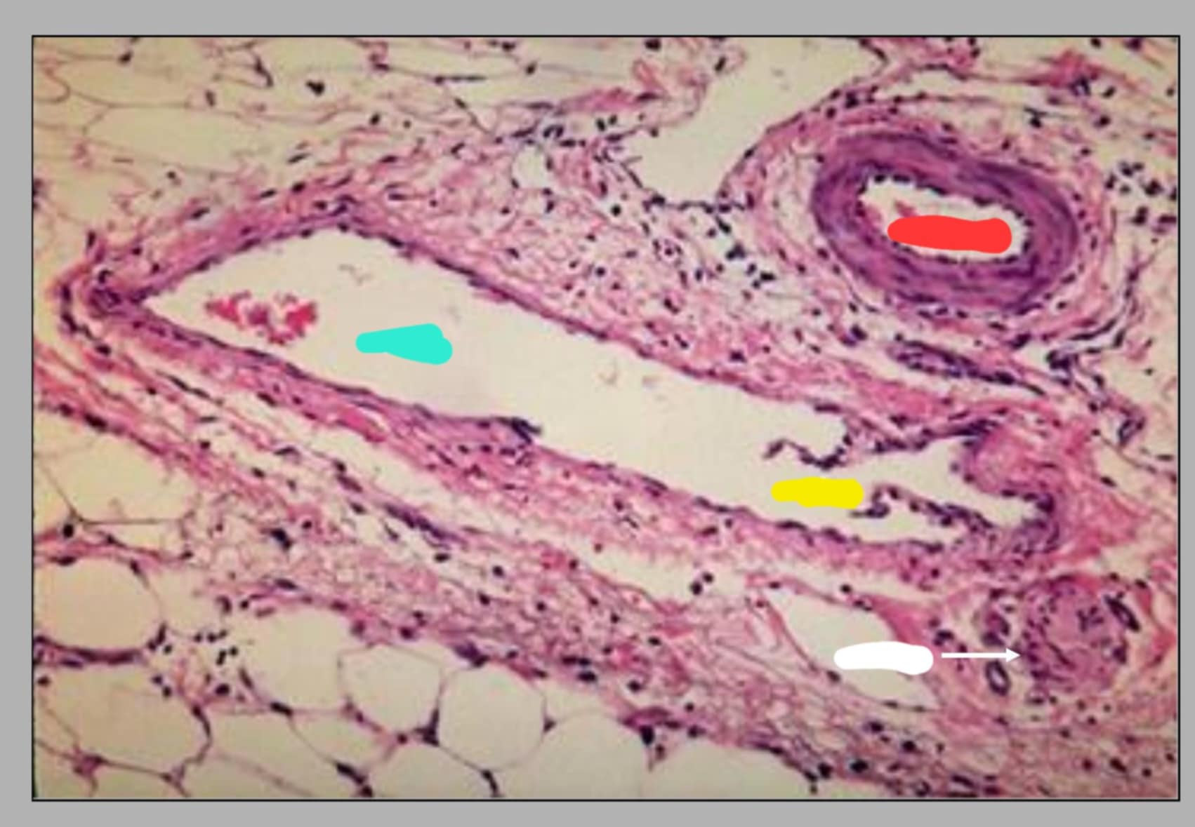
name this slide and state its parts
small artery and vein.
red-artery, cyan-vein, yellow-valve, white-nerve
the artery, vein, and nerve constitutes what is sometimes called ____?
neurovascular triad
name of this slide. and state its parts.
neurovascular triad. cyan-small artery, red-small vein, white-nerve fibers, yellow-perineurium
name this slide and state its parts
cross section of small artery and arteriole.
blue-small artery, yellow-arteriole
name of these 2 slides and state the parts of the 2nd slide
1st slide shows the arteriole and venule, the 2nd slide shows an arteriole sectioned longitudinally. the parts are: red-smooth muscle of t. media and yellow-endothelium
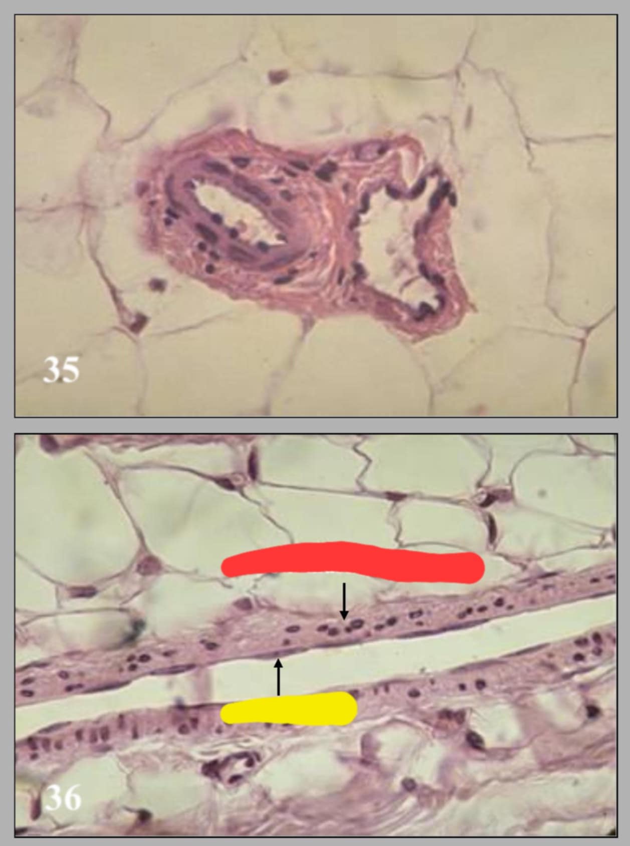
name and parts.
small blood vessel in teased loose connective tissue.
blue-small vein, white-small artery, red-endothelual cell nucleus, cyan-smooth muscle cell nuclei, and yellow-arteriole
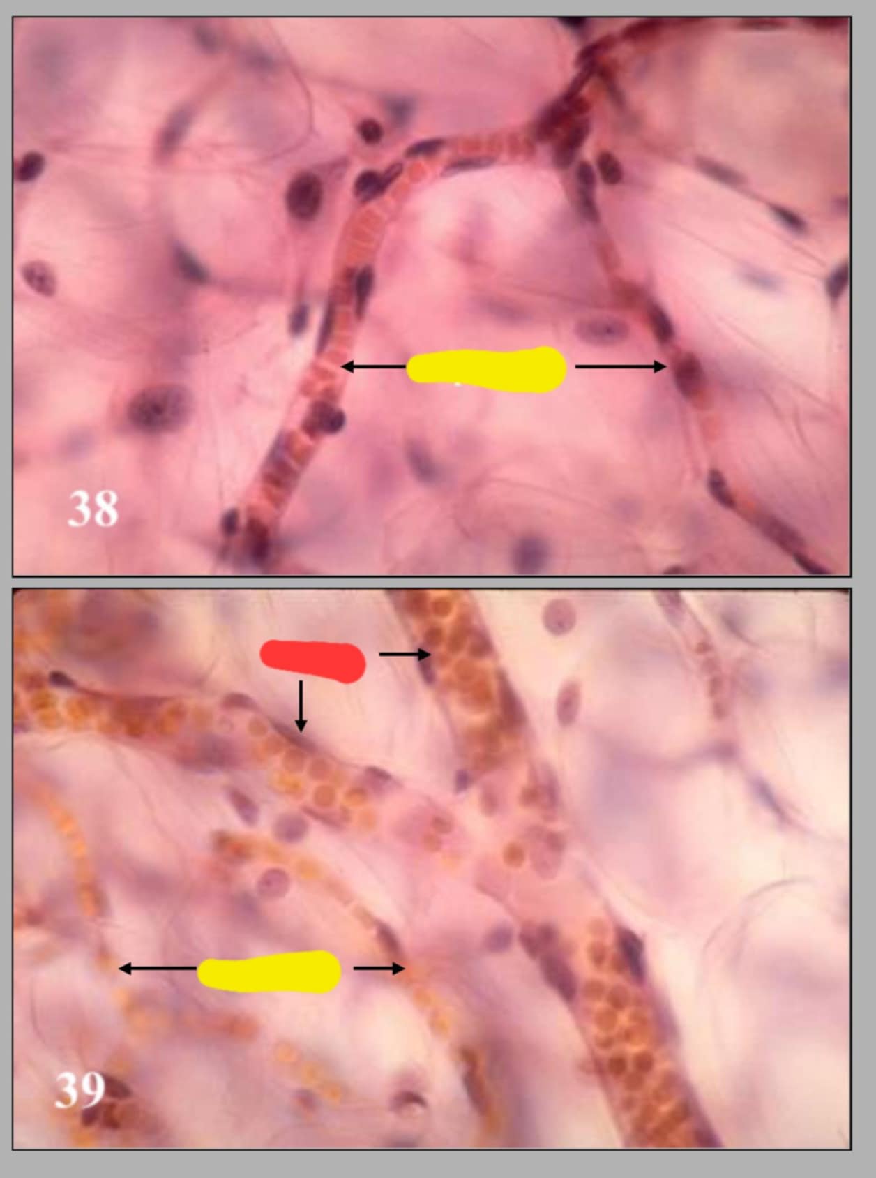
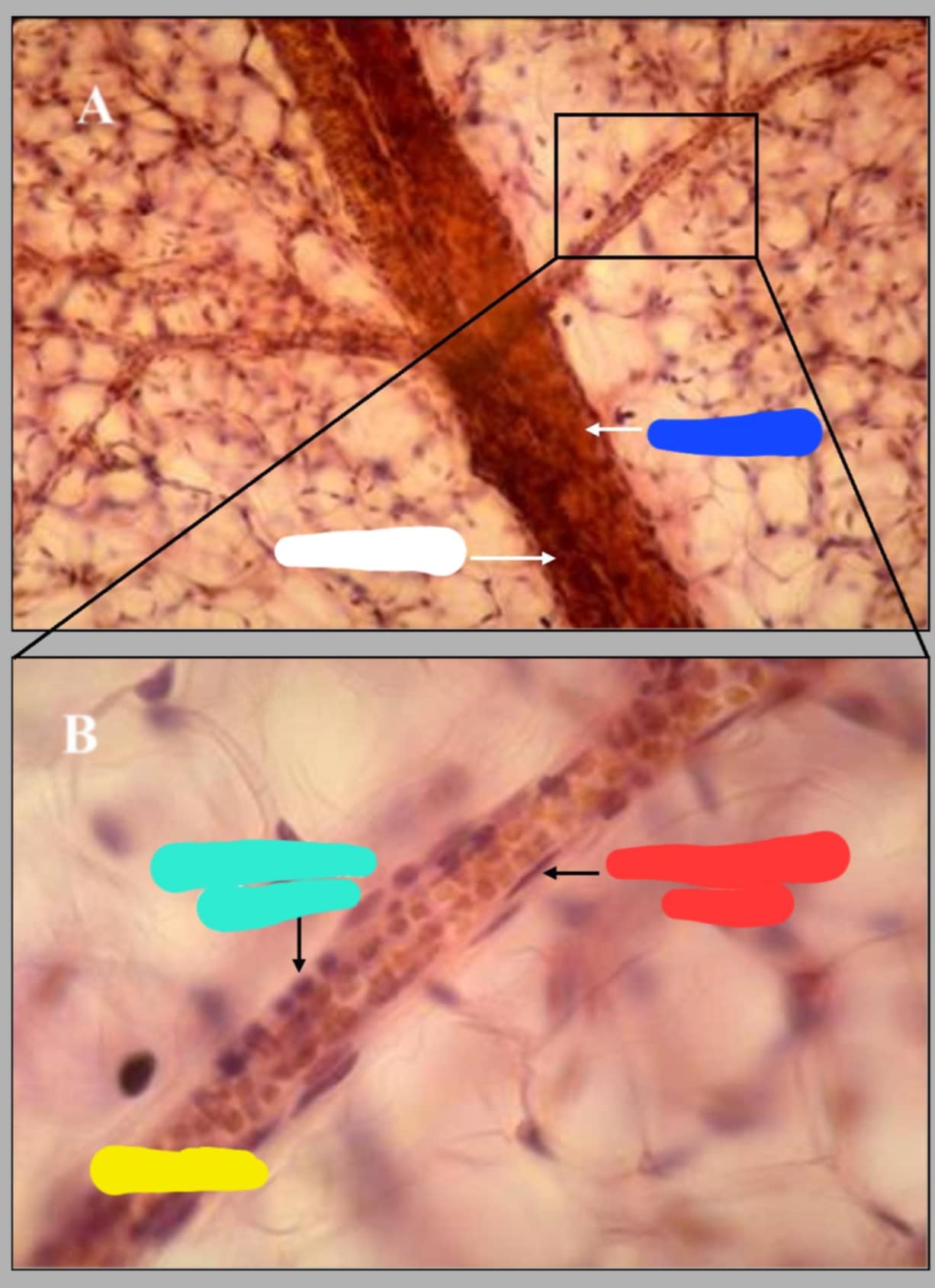
name of the 2 diff slides and their parts
38-capillary in teased loose ct. and the 39-venule in tease loose ct.
yellow-capillaries, and red-venules
is tiny tube lined by a single layer of endothelial cells; 1-2 endothelial cells comprise the wall of capillary. its luminal diameter is large enough to allow the passage of a single row of rbc.
capillary
capillaries empty into _______.
Venules. (they look similar to capillaries but have wider caliber)
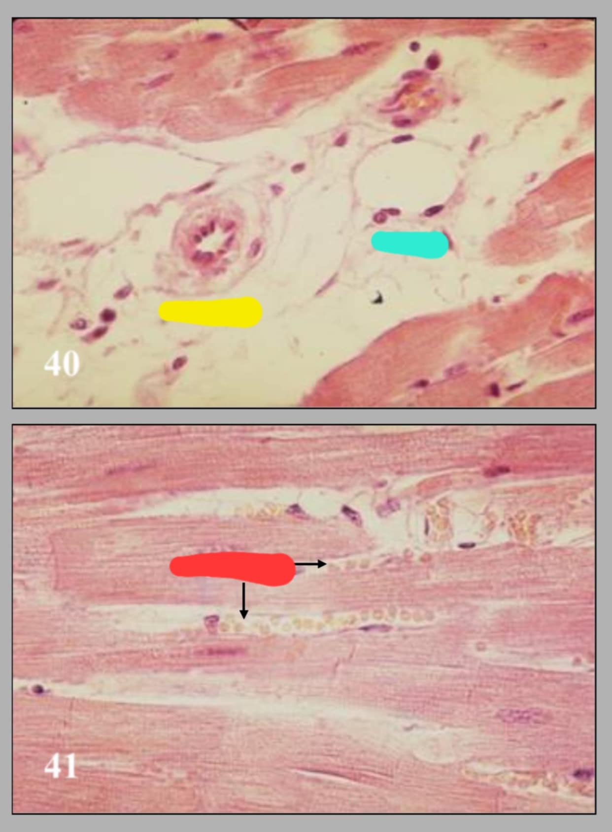
name of these 2 slides and their parts.
40-cross section of arteriole and venule in cardia muscle.
41-capillaries in cardiac muscle.
parts in the 1st slide: yellow-arteriole,cyan-venule. in the 2nd slide: red-capillaries
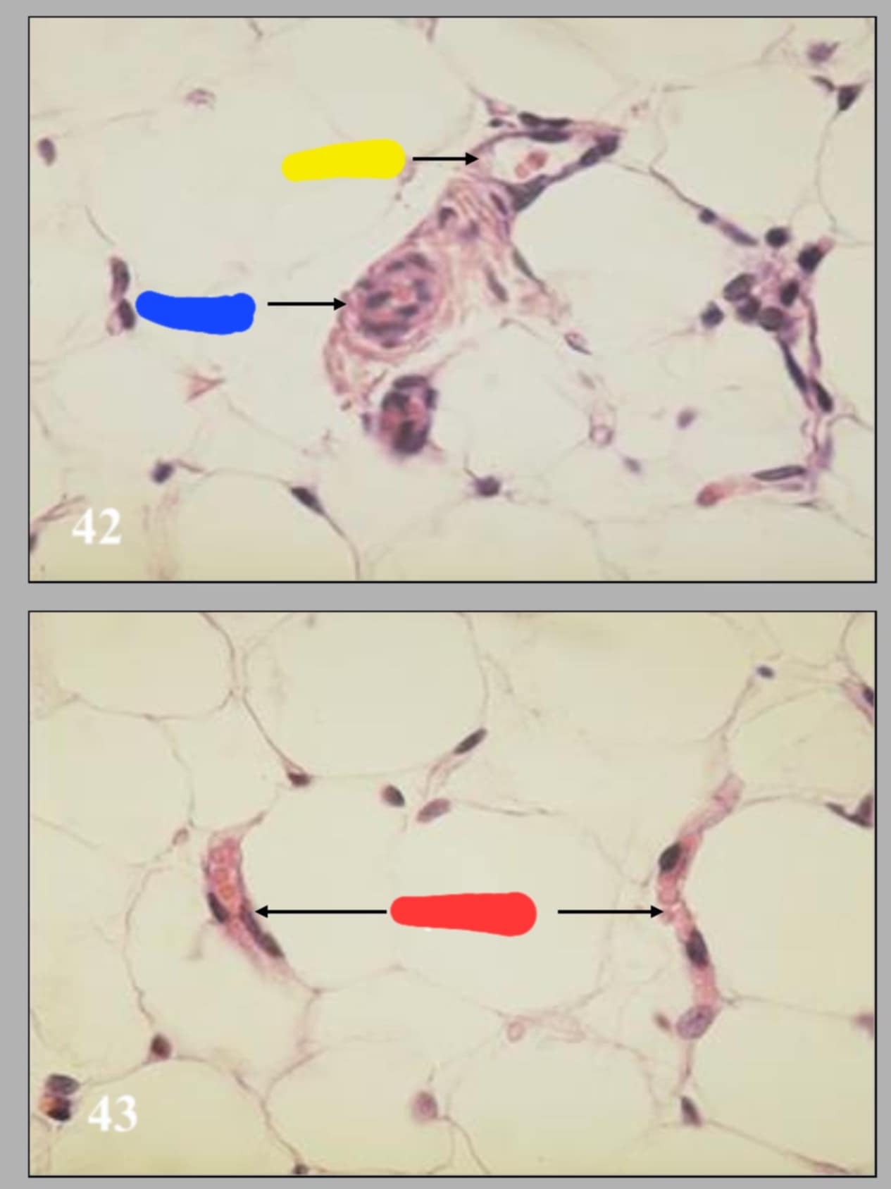
name the 2 diff slides and their parts
42-arteriole and venule in adipose tissue: yellow-venule, blue-arteriole
43-capillaries in adipose tissue: red-capillaries
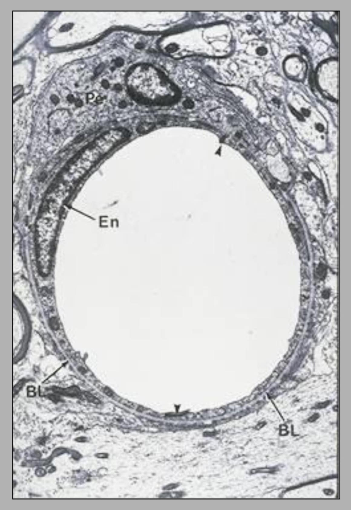
name this slide and state its parts
electron micrograph of capillary in the spinal cord. The entire circumference of this capillary is made up of 2 endothelial cells.
parts: arrowhead-junctions, En-endothelial cell nucleus, BL-basal lamina sureounding the endothelium, Pe-pericyte located just outside the endothelial cells