B2.3 Cell Specialisation
1/29
There's no tags or description
Looks like no tags are added yet.
Name | Mastery | Learn | Test | Matching | Spaced | Call with Kai |
|---|
No analytics yet
Send a link to your students to track their progress
30 Terms
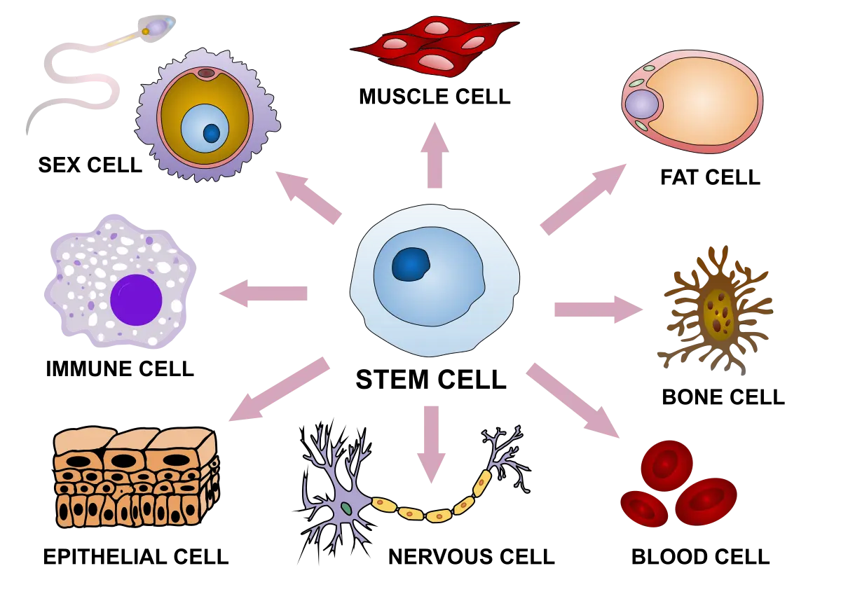
Describe the process of unspecialised cells becoming specialised
After fertilisation, fertilised egg (zygote) divides into many unspecialised cells (called embryonic stem cells)
These cells start off w/ the same DNA & genes
But due to chemical gradients, cells can turn on/off genes to make proteins for specific functions (differentiation)
These gradients cause cells in different parts of embryo to receive different signals → guides cells to specialise in correct location
Define cell differentiation
The process where unspecialised cells become specialised to carry out distinct functions
Describe two properties of stem cells
1) Unlimited division - Has capacity to keep dividing & making cells
2) Differentiation - Depending on type, can develop into different specialised cells
Define a stem cell niche
A specialised microenvironment in tissues where adult stem cells live and are controlled
Describe the functions of stem cell niches (5)
It sends signals that:
1) Support self-renewal (including proliferation = rapid increase of cells)
2) Maintain quiescence (resting, inactive state)
3) Trigger differentiation (for tissue repair/function)
4) Protect stem cells from stress/damage
Describe examples of stem cell niches (2)
1) Bone marrow - Maintains a balance between stem cell storage & production of blood cells
2) Hair follicle - Stores stem cells that control hair growth, rest, & regrowth cycles
Define potency
The ability of a stem cell to develop into different types of specialised cells
Describe the stem cell types (location, what they can become, & example)
Stem Cell Type (by 📉 potency) | Location | Can become | Example |
Totipotent | Very early embryo (zygote & first cell divisions) | Any body cell & placenta | A fertilised egg (zygote) |
Pluripotent | Early-stage embryo (blastocyst) | Any body cell (not placenta) | Embryonic stem cells |
Multipotent | Adult tissues (e.g. bone marrow, skin) | Related cells within one system/tissue | Blood stem cells (RBCs, WBCs, platelets) |
Describe sperm cell’s size/structure & the link to its function (2)
Size: ~5-10 μm in diameter (very small)
Structure → Function:
Small size (minimal cytoplasm) + flagellum → quick movement through fluids & towards egg
Describe egg cell’s size/structure & the link to its function (2)
Size: ~100 μm (1 of largest human cells)
Structure → Function:
Large size + round → nutrient storage for early development
Describe RBC’s size/structure & the link to its function (4)
Size: ~6-8 μm in diameter
Structure → Function:
Small, biconcave disc shape + no nucleus →
Maximises surface area for oxygen binding & gas exchange
Can fit through small capillaries
Describe WBC’s size/structure & the link to its function (2)
Size: ~12-17 μm in diameter
Structure → Function:
Irregular & variable (enlarges during protein synthesis) + nucleus → recognising pathogens & produce immune responses
Describe granule cell’s size/structure & the link to its function (2)
Size: ~4-5 μm in diameter (very small)
Structure → Function:
Small size → allows packing of millions of cells
Describe motor cell’s size/structure & the link to its function (2)
Size: Axon up to 1 m long
Structure → Function:
Long length → transmit signals to muscles over long distances
Describe striated muscle cell’s size/structure & the link to its function (3)
Size: Diameter of 30-40 μm, up to 40 mm in length
Structure → Function:
Long, thick size → generate powerful contractions to move bones
Multinucleated→ greater protein synthesis capacity
What happens in surface area vs volume in cells? (2)
Surface area = where exchange occurs
Volume = where materials are used/produced
What happens to SA:V ratio as cell size grows? (3)
As cells grow, volume increases faster than surface area
This decreases the SA:V ratio
This affects how well substances move in/out of cells
What are the common cell adaptations to increase SA:V ratio?
1) Flattening - Reduces thickness, spreading cell to expose more surface
2) Microvilli - Finger-like projections multiplying cell’s surface
3) Invaginations (infoldings) - Folds on the cell membrane that expand surface inward
4) Biconcave shape - Increases external surface area, while keeping low volume
How do Erythrocytes (type of RBC) adapt to increase SA:V ratio? (2)
Adaptation: Biconcave disc-shape
Function: Maximizes SA & provides a high SA:V ratio for rapid oxygen exchange
How do Proximal Convoluted Tubule Cells (in Kidney Nephron) adapt to increase SA:V ratio? (5)
Adaptations:
Microvilli (on apical surface) - Increase SA facing filtrate
Allows efficient reabsorption into cell
Basal infoldings (on underside) - Increase SA for exchange w/ blood capillaries
Provide space for mitochondria to fuel active transport
→ they both maximise efficiency in reabsorbing valuable substances back into bloodstream
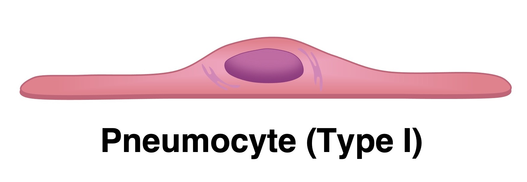
Define pneumocyte
Specialised epithelial cells lining the alveoli of the lungs
Describe the structure & function of type I pneumocytes (2)
Structure: Extremely thin & flat, making up most of the alveolar surface
Function: Minimises diffusion distance for oxygen & carbon dioxide into bloodstream
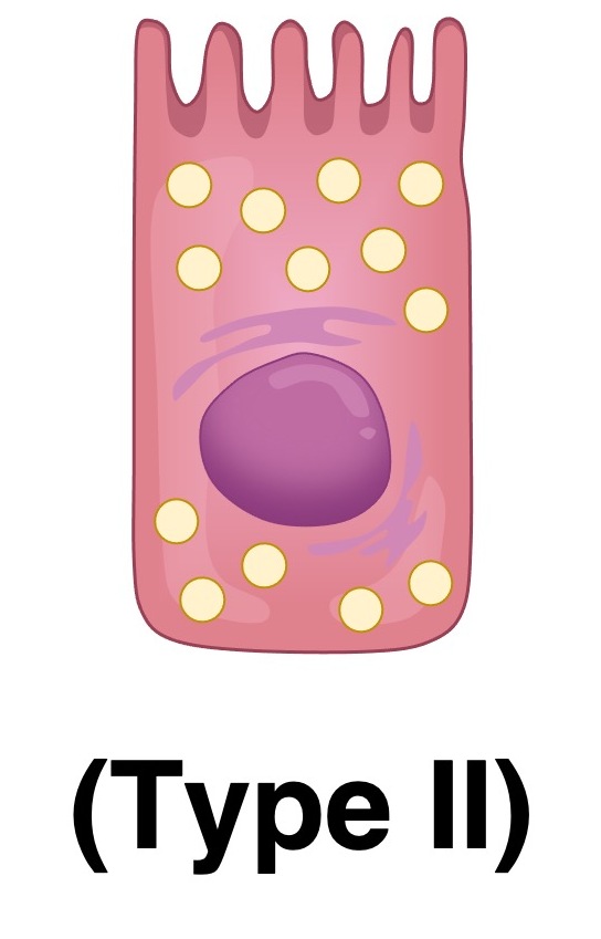
Describe the structure & function of type II pneumocytes (6)
Structure: Cuboidal cells w/ many secretory vesicles
Function:
1) Secretes pulmonary surfactant to support lung function:
Reduces surface tension in alveoli
Prevents alveolar collapse during exhalation
Keeps alveoli open & functional
2) Can divide & replace damaged Type I cell
Describe the adaptations of skeletal muscle cells (3)
Branching - Unbranched
Length - Very long
Nuclei - Multiple nuclei per cell (at edges)
Describe the adaptations of cardiac muscle cells (3)
Branching - Branched
Length - Short
Nuclei - 1-2 nuclei per cell (at centre)
What is the shared adaptation of cardiac & striated muscle cells? (2)
Myofibrils -
Contractile units within both cardiac & striated muscle cells that are composed of actin & myosin
These proteins form sarcomeres (repeating structures responsible for muscle contraction) → giving them striated appearances
Are skeletal muscle fibres cells? (3)
Skeletal muscle fibres are cells, but its features make it atypical:
It is multinucleated
Very long → formed by fusion of multiple myoblasts (embryonic cells)
Define gametes
Specialised reproductive cells that fuse during fertilisation to form a zygote
Describe adaptations of sperm cells, in relation to their functions (6)
Adaptation | Function |
Small size | Enables fast movement over long distances |
Streamlined shape | Reduces water resistance, aids fast swimming |
Flagellum (tail) | Propels the sperm using whip-like motion |
Mitochondria (midpiece) | Provides ATP for tail movement |
Acrosome | Contains enzymes to penetrate egg’s outer layers |
Haploid nucleus | Carries 23 chromosomes |
Describe adaptations of egg cells, in relation to their functions (6)
Adaptation | Function |
Large size | Contains nutrients for embryo development |
Zona pellucida | Protects egg & blocks multiple sperm entry |
Cortical granules | Harden zona after fertilisation to prevent polyspermy |
Cytoplasm rich in organelles | Supports early embryo growth |
Haploid nucleus | Carries 23 chromosomes |