Nervous System - Neurons and Communication
1/44
There's no tags or description
Looks like no tags are added yet.
Name | Mastery | Learn | Test | Matching | Spaced | Call with Kai |
|---|
No analytics yet
Send a link to your students to track their progress
45 Terms
What are the cellular and network properties of neurons?
Organization of the nervous system
Cells of the nervous system
Electrical signals in neurons → lead to action potentials
Cell-to-cell communication in the nervous system
Integration of neural info transfer
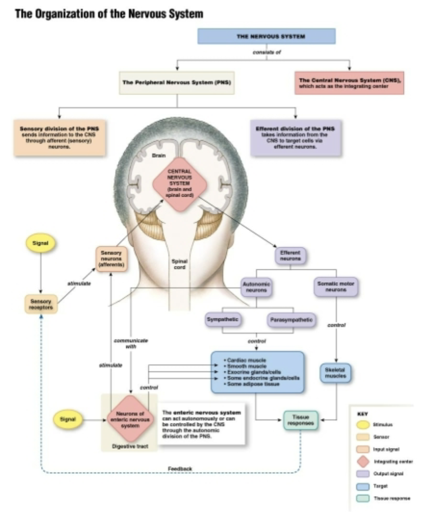
What are the components of the nervous system?
Central nervous system (CNS)
Brain
Spinal cord:
Transmits signals between brain and body
Controls reflexes
📌 Think: CNS = decision-making HQ
Peripheral Nervous System (PNS)
Sensory (afferent neurons)
Carry information FROM the body TO the CNS
Detect: Touch, pain, temp, pressure, body position (proprioception)
📌 Mnemonic: Afferent = Arriving at the CNS
Efferent neurons
Carry commands FROM the CNS TO the body
Control muscles and glands
Somatic motor neurons: Voluntary, controls skeletal muscles
Autonomic nervous systems
Involuntary
Controls smooth muscle, cardiac muscle, and glands
Maintains homeostasis
Sympathetic:
“Fight or Flight”
Activated during stress or emergencies
Parasympathetic
“Rest and Digest”
Active during relaxation
📌 Mnemonic: Efferent = Exiting the CNS
Enteric Nervous System
Controls the gastrointestinal (GI) tract
Can function independently of the CNS
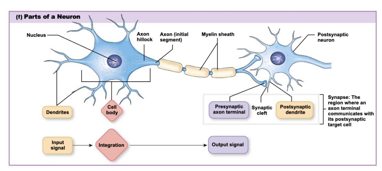
What are the components of a neuron?
Cell body: Contains nucleus and most of the cellular machinery
Dendrites: Receive information
Short, branched extensions
Carry signals toward the cell body
Axon: Sends information
Nerves: PNS
Tracts: CNS
Axon Hillock:
Junction between cell body and axon
Trigger zone for action potentials
If summed input reaches threshold → neuron fires
Axon Terminal:
End of axon
Releases neurotransmitters
Converts electrical signal → chemical signal
Myelin sheath: Insulator (makes signal go faster)
Allows saltatory conduction (jumping between nodes → signaling faster)
Synapse: Site of communication between neurons (or where an axon terminal communicates with its postsynaptic target cells)
Synaptic cleft: Small gap between neurons. Neurotransmitters diffuse across this space
Postsynaptic Dendrite: Receives neurotransmitters
Input signal: Dendrites → Cell body (integration) → Axon hillock (decision point - whether action potential should be released) → Axon → Axon terminals → Postsynaptic dendrite → Output signal
1) What are sensory neurons?
2) How are sensory neurons classified structurally?
1) Carry information from receptors → CNS
2) Pseudopolar: Single process (axon). During development, the dendrite fused with the axon
Bipolar: Two relatively equal fibers extending off the central cell body
1) What are interneurons neurons?
2) How are interneurons neurons classified structurally?
1) Located entirely within the CNS. Integrate and process information. Connect sensory → motor pathways
2) Anaxonic: Have no apparent axon
Multipolar: Highly branched but lacks long extensions
1) What are efferent neurons?
2) How are efferent neurons classified structurally?
1) Carry commands from CNS → muscles or glands. Control responses
2) Multipolar: 5-7 dendrites, each branching 4-6 times. A single long axon may branch several times and end at enlarged axon terminals
What is the function of the synapse?
Presynaptic: The sending cell
Contains the axon terminal
Releases neurotransmitter (chemical synapse)
or passes current directly (electrical synapse)
Postsynaptic: The receiving celll; contains receptors (chemical synapse)
Electric synapses:
Cells are connected by gap junctions
Ions flow directly from cell to cell
Very fast
Bidirectional
Chemical synapse:
Most common type in the nervous system
Cells separated by a synaptic cleft → communication via neurotransmitters
Unidirectional
What’s the difference between slow and fast axonal transport?
Slow axonal transport: Moves soluble material by axoplasmic (cytoplasmic) flow at 0.2-2.5 mm/day
Fast axonal transport:
Moves organelles at rates up to 400 mm/day
Forward (or anterograde) transport: From cell body → axon terminal
Backward (retrograde) transport: From axon terminal → cell body
Local protein synthesis → helps with fast transport
How does fast axonal transport work?
Peptides are synthesized on rough ER and packaged by the Golgi apparatus
Fast axonal transport walks vesicles and mitochondria along microtubule network
Vesicle contents are released by exocytosis
Synaptic vesicles recycling
Retrograde fast axonal transport
Old membrane components digested in lysosomes (which is in the cell body)
What are the function of glial cells?
Glial cells ≠ neurons
Support, protect, insulate, and nourish neurons
Are essential for normal neural function
Organize glial cells by location: CNS and PNS
What are the glial cells of the CNS?
Ependymal cells:
Create barriers between compartments
Form the lining of the ventricles
Source of neural stem cells
Astrocytes:
Take up K+, water, neurotransmitters
Source of neural stem cells
Secrete neurotrophic factors
Helps form a blood-brain barrier
Provide substrates for ATP production
Oligodendrocytes:
Form myelin sheaths
What are the glial cells of the PNS?
Schwann cells:
Form myelin sheaths
Secrete neurotrophic factors
Satellite cells:
Support cell bodies by forming supportive capsules around a ganglion (plural ganglia)
Ganglia: Collections of neuronal cell bodies in the PNS
Nuclei: Collections of neuronal cell bodies in CNS
What is an action potential?
Conduction is the high-speed movement of an action potential along an axon
AP: Wave of electrical signal at constant amplitude
Action potentials are all-or-none
AP’s Go in one direction
AP’s Do not change ion concentration gradients
1) Explain how the signal travels and induces action potential.
2) Explain the steps of action potential.
1)
The action potential travels in one direction down the axon
Pos charges (Na+ influx) move forward
The region behind is refractory, preventing backward movement (explain why signals don’t go backward - like the seps
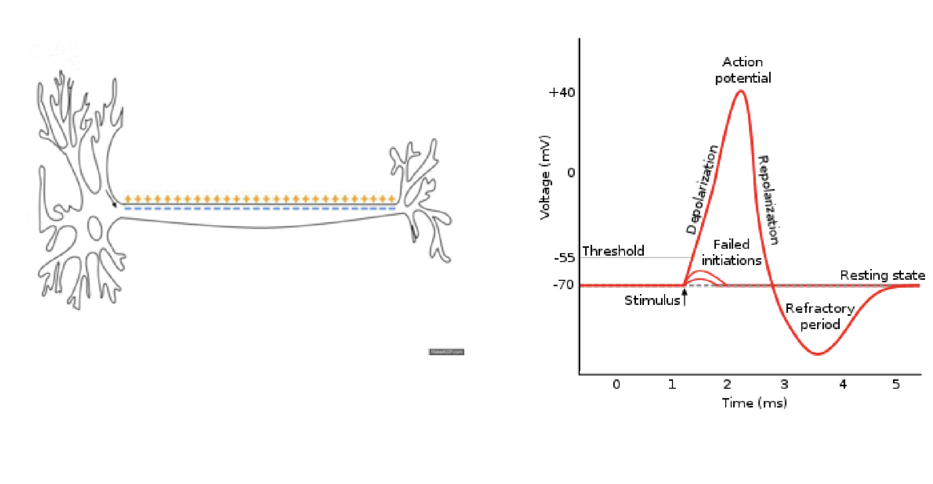
2)
Resting Membrane Potential (~ -70 mV)
Neuron is at rest
Maintained by: Na+/K+ pump, leak channels
Stimulus:
Causes local depolarization
If small → failed initiation
If strong enough → reaches threshold
Threshold (~ - 55 mV)
Point of no return
Voltage-gated Na+ channels open
Depolarization
Rapid Na+ influx
Membrane potential rises to ~ + 40 mV
Repolarization
Na+ channels inactivate
Voltage-gated K+ channels open
K+ exits the cell
Hyperpolarization
Membrane becomes more negative than rest
K+ channels close slowly
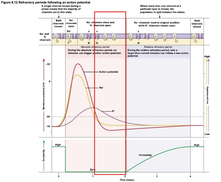
Refractory period:
Absolute: Can’t fire another AP
Relative: Stronger stimulus needed
Return to Resting State:
Na+/K+ restores ion gradients
Neuron ready to fire again
Why can’t an action potential go backward?
Domino analogy: A domino that already fell can’t fall again immediately. Only the next upright domino can fall
Action potential moves forward
The region behind is refractory b/c during depolarization occurs, the Na+ channel becomes inactivated, and it can’t reopen regardless of how strong the stimulus is (basically the steps can’t work backwards - what’s done is done)
What happens when the membrane is at rest?
The membrane potential is influenced by:
Concentration gradient of ions
Membrane permeability to those ions
ECF contains a lot of Na+, Cl-, Ca2+
ICF contains a lot of K+
How are electric signals created?
Through ion movement!
Resting membrane potential determined primarily by:
K+ concentration gradient
Resting permeability to K+, Na+, and Cl-
Voltage-gated channels control ion permeability
Different channels open at different threshold voltages
Kinetics of channel opening and closing varies from one channel type to another
Explain how ions move across the membrane during action potentials.
The neuron is at its resting membrane potential of -70 mV
Depolarization stimulus enters the trigger zone
Membrane depolarizes to threshold and voltage-gated Na+ and K+ channels open
Na+ channels open first leading to rapid Na+ influx that depolarizes the cell (becomes more +)
At peak, Na+ channels close and slow K+ channels open
K+ moves out of the cell
K+ channels stay open and more K+ leaves the cell, hyperpolarizing it
Voltage-gated K+ channels close, less K+ leaks out of the cell
Cell returns to resting ion permeability and resting membrane potential
Why is action potential propagation a “one-way street”?
Potential delay of 1-2 msec between action potentials independent of intensity of trigger
The refractory period always prevents backward conduction
Due to Na+ gases resetting
Relative refractory period follows an absolute refractory period
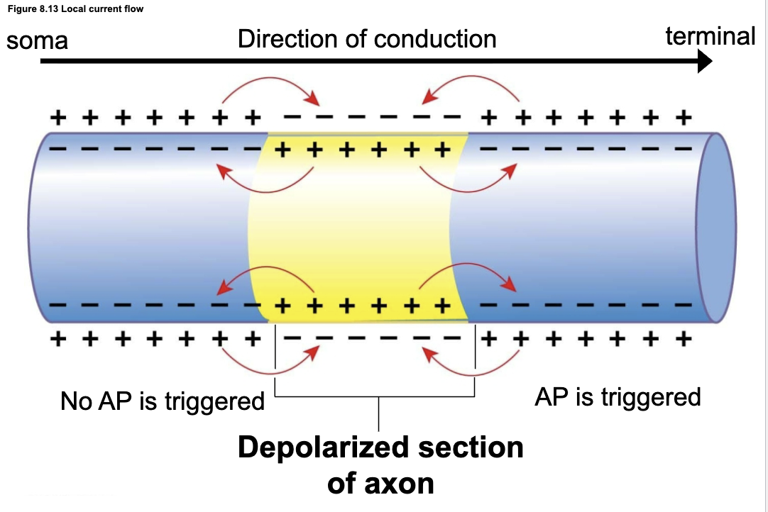
Positive charge spreads along adjacent sections of axon by local current flow
Local current flow causes a new section of the membrane to depolarize
How does an action potential move forward along the axon?
A segment of the axon (yellow) is depolarized → lots of + charge (Na+) inside
That + charge spreads locally to adjacent axon segments
The forward segment (toward the terminal):
Reaches threshold (bc the next segment is still at its resting membrane potential)
Triggers a new action potential
Backward segment (toward soma)
Refractory period (the segment behind just fired so it won’t fire anytime soon/again & Na+ channels are inactivated)
Can’t fire another action potential
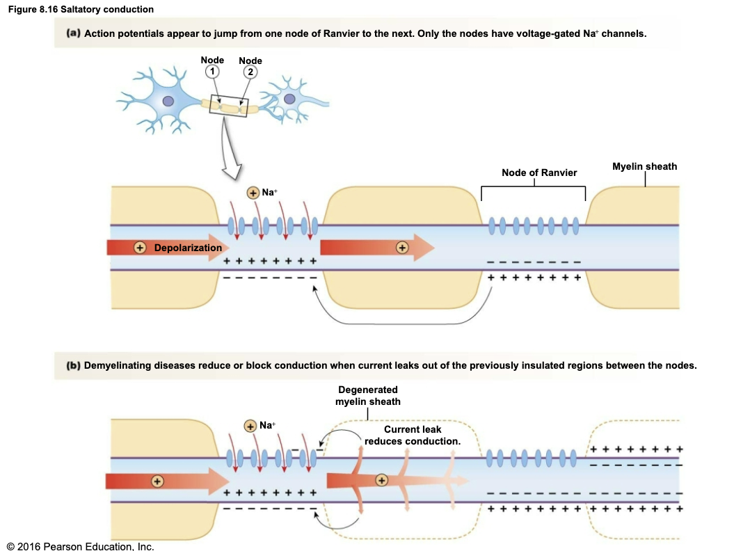
1) How do action potentials travel fast in myelinated axons?
2) What happens when myelin is damaged?
1) Saltatory conduction
The axon is wrapped in myelin (acts as an insulator)
Voltage-gated Na+ channels are only at the Nodes of Ranvier (small gaps in the myelin sheath along a myelinated axon)
When an action potential occurs at one node:
Na+ enters
+ charge spreads rapidly under the myelin
The next node reaches the threshold
The action potential appears to jump from node to node
2) Loss of myelin
The myelin sheath is damaged
Current leaks out of the axon between nodes
Less charge reaches the next node
This leads to slowed conduction or complete conduction block
1) What is the speed of action potentials influenced by?
2) What diseases are associated with demyelinating disease?
1)
Diameter of axon: Larger axons are faster
Resistance of axon membrane to ion leakage out of the cell → myelinated axons are faster
Saltatory conduction between nodes of Ranvier
2)
Multiple sclerosis
Guillain-Barre syndrome
What is the difference between a graded potential and an action potential?
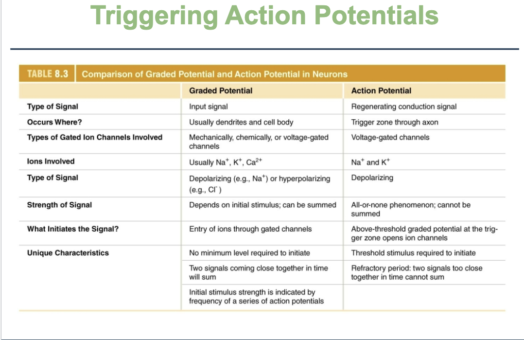
What are the characteristics of graded potentials?
[INCOMPLETE + insert pic + review]
1) Graded potentials decrease in strength as they spread out from the point of origin
A stimulus opens ion channels → local depolarization
The signal spreads out in all directions
Amplitude decreases as it moves away from the stimuli
Explain the process of action potentials
[INCOMPLETE + insert pic + review']
A graded potential above threshold reaches the trigger zone
Voltage-gated Na+ channels open, and Na+ enters the axon
Positive charge flows into adjacent sections of the axon by local current flow
Low current flow from the active region causes new sections of the membrane to depolarize
The refractory period prevents backward conduction. Loss of K+ from the cytoplasm repolarizes the membrane
How can sushi be dangerous?
Tetrodoxin: Blocks voltage-gated Na+ channels
Found in pufferfish, octopuses/octupi/octopodes, some bacteria, some newts
Paralysis throughout the body
Death occurs from 20 mins to 8 hrs after digestion
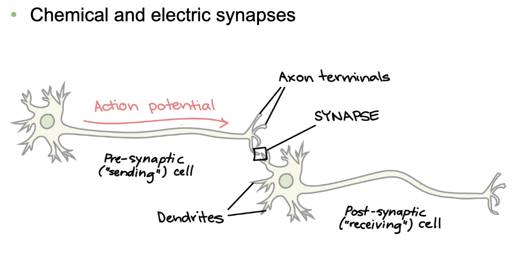
1) How do neurons communicate at synapses?
2) What is the difference between chemical and electrical synapses?
1)
An AP travels down the axon of the presynaptic (sending) neuron
It reaches the axon terminals
The synapse is the area between the presynaptic axon terminal and the postsynaptic dendrite (or soma) → where cell-to-cell communication happens
The postsynaptic (receiving) neuron gets the signal at its dendrites
2)
Electrical synapse:
Pass electrical signals through gap junctions
Ions flow directly between cells
Very fast
Bidirectional
Synchronizes the activity of a network of cells
Chemical synapse:
Neurotransmitters released into the synaptic clef
Bind receptors on postsynaptic cell
Target cell must have matching receptor
Slower
Unidirectional
Highly modifiable
What are neurotransmitters, neuromodulators, and neurohormones?
Neurotransmitters and neuromodulators: Paracrine signals that act at short distances (neurocrines)
Neurotransmitters are fast acting at synpases
Neuromodulators are slow acting at synaptic and non-synaptic sites
Autocrine signals can act on the neurons that release them
Neurohormones act over long distances
Secreted into the blood and distributed throughout the body
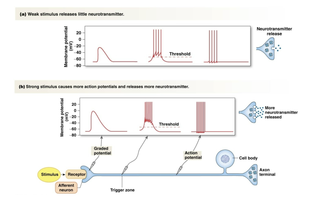
List the different type of neurocrine receptors
All neurotransmitters bind to specific receptors
Ionotropic receptors: Ligand-gated ion channels
Mediate rapid responses
Alter ion flow across membranes
Metabotropic receptors:
G protein-coupled receptors (GPCRs)
Mediate slower responses
Some open or close ion channels
Note:
Agonist and antagonist molecules either mimic or inhibit activity by binding to receptors
What are the 7 kinds of neurotransmitters/neurocrines?
Acetylcholine, amines, amino acids, peptides, purines, gases, lipids
What is acetylcholine?
Synthesized from choline and acetyl CoA
Cholinergic receptors
Nicotinic:
Skeletal muscle, autonomic division of PNS, and CNS
Monovalent cation channels → Na+ and K+
Muscarinic:
CNS and autonomic parasympathetic division of PNS
G protein-coupled receptors
What are amines?
Active in the CNS
Each is derived from single amino acid
Tryptophan → Serotonin
Histidine → Histamine
Tyrosine → Dopamine → Norephinephrine/Noradrenaline → Epinephrine/Adrenaline
What is norepinephrine (amines)?
Secreted by noradrenergic neurons
Major neurotransmitter of the autonomic sympathetic division of the PNS
Adrenergic receptors
Alpha and beta
G protein-coupled receptors
What are amino acids?
Excitatory:
Depolarize target cells by opening ion channels to allow the flow of positive ions into the cell
Glutamate: Primary excitatory neurotransmitter in the CNS, also acts as a neuromodulator
Aspartate: Excitatory neurotransmitter in the brain
Inhibitory:
GABA: Primary inhibitory neurotransmitter in the brain
Hyperpolarizes target cells by opening Cl- channels
What are glutamate receptors?
Glutamate can act as a neurotransmitter or a neuromodulator
AMPA (alpha-amino-3-hydroxy-5-methylisoxazole-4-propionic acid)
Ligand-gated monovalent cation channel (Na+)
NMDA (N-methyl-D-aspartate)
Non-selective cation channels (Na+, K+, Ca2+)
Opening of channel requires glutamate binding and a change in membrane potential
What are peptides?
Substance P and opioid peptides
What are purines?
Adenosine, AMP, ATP
What are some gaseous neurotransmitters that diffuse in the cells?
NO, CO, H2S
What is an example of a lipid?
Eicosanoids: Some are endogenous ligands for cannabinoid receptors
How are neurotransmitters released?
Classic exocytosis:
Vessicles fuse with membrane
Neurotransmitters spill into synaptic cleft
Vesicle membrane is incorporated into axon terminal membrane
Vesicles are recycled by endocytosis and refilled with neurotransmitters
Kiss-and-run:
Vesicles fuse with membrane at the fusion pore
Neurotransmitters pass through a channel
Vesicles pull back from fusion pore
Vesicles are refilled
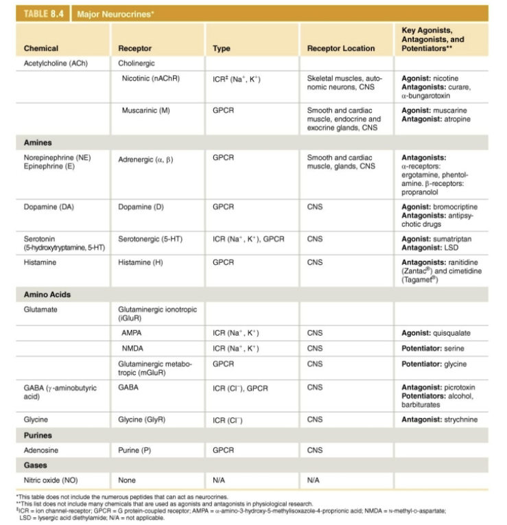
How does a chemical synapse release neurotransmitters?
An AP depolarizes the axon terminal
The depolarization opens voltage-gated Ca2+ channels and Ca2+ enters the cell
Calcium entry triggers exocytosis of synaptic vesicle contents
Neurotransmitter diffuses across the synpatic cleft and binds with receptors on the postsynaptic cell
Neurotransmitter binding initiates a response in the postsynaptic cell
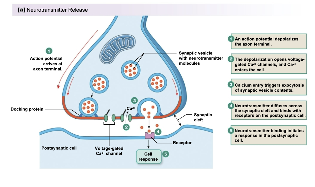
How does neurotransmitter termination occur in a chemical synapse?
Neurotransmitters can be returned to axon terminals for reuse or transported into glial cells
Enzymes inactivate neurotransmitters
Neurotransmitters can diffuse out of the synaptic cleft
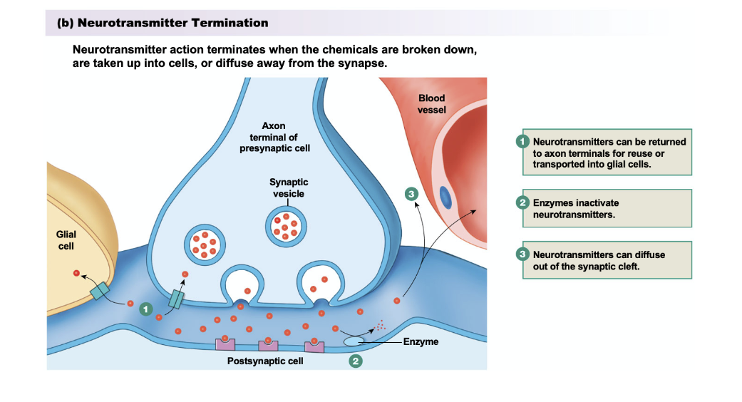
How does a stronger stimulus affect the release of neurotransmitters?
A single action potential releases a set amount of neurotransmitter
A stronger stimulus produces more frequent action potentials leading to a more neurotransmitter release
CNS neurons have different patterns of firing, in addition to frequency
Bursts
Pacemakers
How is neural info integrated?
Divergent and convergent pathways at synapses
Postsynaptic responses may be slow or fast
Synaptic plasticity is a change in activity at the synapses based on past activity