Sub-cellular structures
structures found in cells that include the nucleus, mitochondria, chloroplasts and ribosomes
Nucleus
the control centre of the cell
Cytoplasm
the substance found in living cells (outside the nucleus), where chemical reactions take place
Cell membrane
a layer that holds the cytoplasm in a cell and controls what enters and leaves the cell
Mitochondria
the structures in the cytoplasm where energy is produced from chemical reactions
Ribosomes
small structures found in the cytoplasm of living cells where protein synthesis takes place
Cell wall
a protective layer, found outside the cell membrane of plant, fungal and bacterial cells, that helps to support the cell
Cellulose
a carbohydrate that makes up the cell wall of plants
Vacuole
a fluid-filled cavity in a cell that is used for storage and support
Chloroplast
a sub-cellular structure containing chlorophyll, which is found in plants and algae that carry out photosynthesis
Prokaryotic
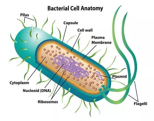
organisms, such as bacteria, that do not have a nucleus or organelles such as mitochondria
Eukaryotic
cells that have a nucleus and sub-cellular organelles such as mitochondria
flagellum
an organelle that enables movement
slime capsule
slimy layer covering a bacterial cell that prevents cell from drying out and helps cell stick to surfaces
Plasmid
a small circle of bacterial DNA that is independent of the main bacterial chromosome
Resolution
the ability to distinguish two objects from each other
Electron microscope
a device that fires electrons at a specimen to obtain a high resolution image
Chromosomes
long molecules found in the nucleus of all cells; made from DNA
DNA
nucleic acid molecules that contain genetic information and make up chromosomes
Gene
A region of DNA that codes for a specific sequence of amino acids (which chain up to make up a polymer; protien)
Double helix
two linked strands that wound around each other to resemble a twisted ladder in a helix-like shape.
Magnification
The ability to make a small object look bigger
What is the formula for image size?
Imagesize = actual image x magnification
Pili
short, hair-like structures on the cell surface of prokaryotic cells. They can have a role in movement, but are more often involved in adherence to surfaces
protein synthesis
process of making proteins, it occurs in 2 main steps, transcription and translation.
mrna
Messenger rna, is a single strand copy of a dna, its is made because transporting the whole dna would be unnecessary.
Trna
transfer rna is an adaptor molecule that serves as the physical link between the mRNA and the amino acid sequence of proteins. It floats outside of the ribosomes and contains 3 sets of nucleotides called anticodons and each carry an amino acid.
stage 1 (transcription)
the process of when the mrna is created and transported out of the nucleus into the ribosomes. This is carried out by an enzyme which unzips the dna and makes a copy of the relevant strand. This copy is the mrna. The mrna is the transported out of the nucleic pores and into the ribosome.
splicing
the process of when introns are removed and exons are added the the mrna
introns
a region of a gene that does not encode for anything (irrelevant)
exons
a region of a gene that encodes for proteins
stage 2 (translating)
The mrna is is used as a guide to sort the trna into the correct order by reading the mrna 3 nucleotides (triplets) at a time and attaching a trna complementary set of nucleotides (anticodons).In this process, the amino acids that the trna carry are also ordered which makes them join together with hydrogen bonds, making them form a polypeptide. The polypeptide will go on to make a protein.
polypeptide
a chain of amino acids
monomer
a molecule that can be bonded with identical molecules to form a polymer
polymer
a substance made from monomers
order of magnitude
Differences in size are often described as differences in order of magnitude. (example) If you increase a number by one order of magnitude, you are multiplying the number by 10. If you decrease a number by one order of magnitude, you are dividing the number by 10, which is equivalent to multiplying by 0.1
Scanning electron microscope
A type of electron microscope that uses a beam of electron across the surface of a specimen where reflected electrons are collected to produce an image
Transmission electron microscope
A type of electron microscope that uses a beam of electron to pass through a thin sample. The electrons are detected and creates an image.
Electron microscope
A microscope that uses a beam of electrons.
.images created are in black and white and specimen need to be dead as electrons pass through a vacuum
.expensive and not portable
.difficult preparation (thin slices especially tem)
Max magnification of electron microscope
x500,000
Max magnification of light microscope
x600
Max resolution of light microscope
0.2 um (micrometers)
Max resolution of electron microscope
0.1 nm (nanometers)
Light microscope
A type of microscope that passes light through an object placed on a slide. The object is observed through the objective and eyepiece lens that magnify the object.
.it can view image in natural colours
.it is cheap and portable
.easy to prepare ( slide w specimen and specific stain)
Methylene blue
A stain that highlights the nucleus of an animal cell
Iodine
A stain that highlights plant cell nuclei
crystal violet
A stain that highlight bacterial cell walls
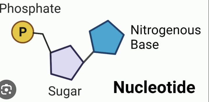
Nucleotide
A phosphate group, ribose sugar and nitrogenous base
Nitrogenous bases
Adenine, thymine
Cytosine, guanine