NEUR200 - Auditory System, Parietal Lobe, Motor System, Movement, Frontal Lobes, Sex
1/117
Earn XP
Description and Tags
Exam 3
Name | Mastery | Learn | Test | Matching | Spaced |
|---|
No study sessions yet.
118 Terms
What is the name of the auditory receptor in transduction?
Cochlea
Cochlea
an auditory nerve containing hair cell receptors vibrates and opens up K+ channels
K+ ions moving into the cell causes an electrical signal → release of neurotransmitters
(T/F) Outer part of ear is more receptive to lower pitches and lower part of ear is more receptive to higher pitches
False— should be the other way around
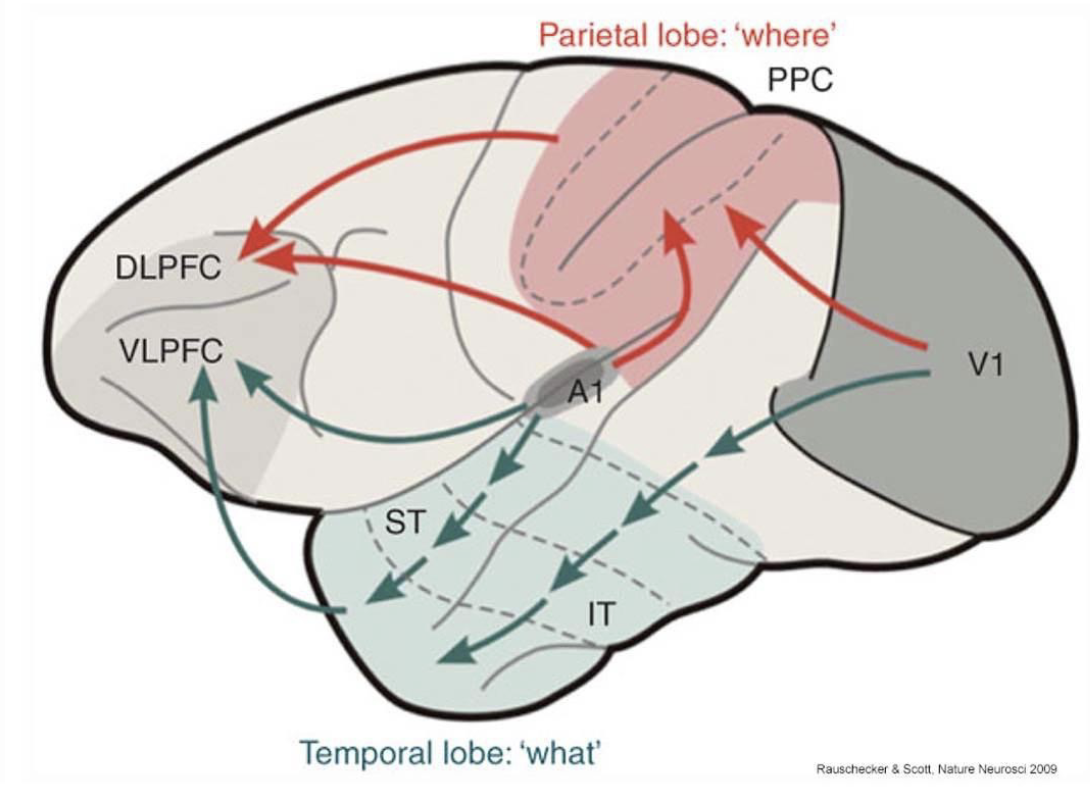
What Pathway (Auditory)
Starts in A1, ends in superior temporal sulcus
involved in object recognition

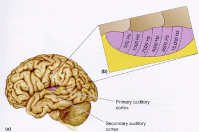
(T/F) In primary auditory cortex (A1), neurons located to left respond to lower frequencies and neurons located to the right respond to higher freqs
True
Where pathway (audition)
Starts in A1, ends in posterior temporal cortex and parietal cortex
Involved in object location
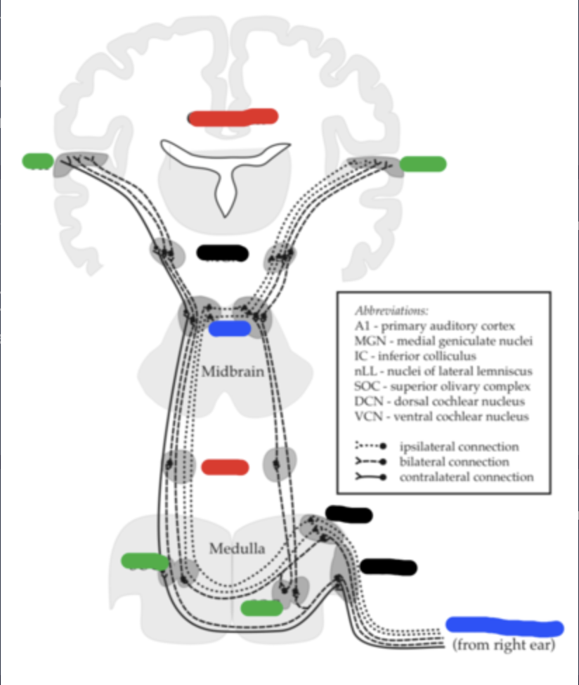
Primary auditory pathway
1) medulla
2) inferior colliculus (in midbrain)
3) Medial geniculate nucleus (of thalamus)
(4) Primary Auditory cortex (A1)
(T/F) Anterior temporal parts are higher than posterior temporal parts
False; Posterior -> higher, anterior -> lower
(T/F) A1 is organized with columns
True
Robert Knight & Pasley
used intracranial recordings of brainwaves from non-primary auditory cortex in the human superior temporal gyrus to determine what words a person has heard
What chronic brain disorder is associated with temporal lobes?
Epilepsy
Insula
(drawn in)
Part of the insula is in temporal lobe
In front of the STG
Connects the mind w/ body
Reacts to changes in the body
Contributes to hunger cravings
Activates when the autonomic nervous system activates
Studied in relation to emotions

Middle temporal gyrus
(in orange)
Auditory and visual processes
Contains ITS
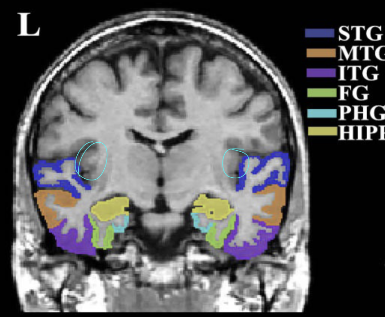
Superior temporal gyrus
(in dark blue)
auditory ventral stream
Contains STS

Inferior temporal gyrus
(in purple)
Visual ventral stream (tgt w/ fusiform gyrus)

Which regions in temporal lobe is amygdala located in?
Medial (middle) temporal regions
Amygdala
emotions related to memories
Activates during fight-or-flight (ex: goosebumps)
Fear emotions
Hippocampus
(in yellow)
location / spatial memory
consolidating memory

Ventral pathway corresponds to what function?
Object recognition
Which temporal lobe is associated with crossmodal matching b/w auditory and visual info and is involved in biological motion perception & social cognition (developing hypothesis abt others’ intentions)
Superior temporal sulcus (STS)
Amusia
impairment to make pitch discriminations
Area is larger in musicians
heschl’s gyrus is affected
Wernicke’s aphasia
Can recognize words but they are meaningless
“Word deafness”
Anterograde amnesia
Difficulty remembering new events
Bilateral removal of the medial temporal lobes
What occurs in Inferotemporal cortex?
Problems of conscious recall (left: verbal; right: visual)
Temporal-lobe personality
Pedantic speech
Egocentrism
Persisting to talk about personal problems
Proneness to anger outbursts
Caused from epilepsy / damage to temporal lobe (limbic system)
What’s another term for temporal-lobe personality?
Geschwind syndrome
Temporal lobes anatomy
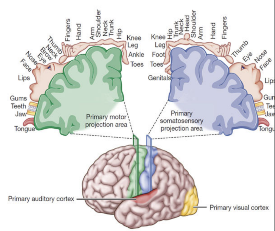
What is the term for map of the body?
Homunculus
What does Dr. Ramachandran’s study on phantom limb sensation show?
After amputation, the map of the body is restructured
Neurons don’t die because they still keep getting input from other parts of the body even though the limb is gone → sensations are still being felt in the phantom limb
Somatosensory cortex map
Posterior parietal cortex
PE, PF, & PG
Spatial information outside your body
Anosognosia
Unawareness of the illness
Balint’s syndrome
Simultagnosia and optic ataxia (problems with guided reach; inability to see where objects are in space)
Neglect syndrome
Not aware of the right visual field when left parietal lobe is affected and vice versa
What is the difference between hemianopia & neglect syndrome?
Not a vision problem!
Different from hemianopia (moving their heads bc they know they’re missing something)
Contrasts with patients with neglect who don’t move their head in awareness
Gerstmann Syndrome
Difficulty grasping spatial info: Dysgraphia (reading), dyscalculia (math), problems w right-left discrimination, etc.
Posterior parietal lobe issue
Which part of parietal lobe is good with identifying spatial info?
Right part of parietal lobe
Apraxia
Loss of skill movement not caused by weakness or motor problems
Ex: problems riding a bike, etc.
Where do problems with sensorimotor transformation occur?
Posterior parietal cortex
What is the correlation between tickling yourself & sensorimotor transformation?
When you tickle yourself, it doesn’t work because the frontal lobe is sending the command to tickle and the spinal cord is directly receiving it and is sending an efference copy of discharge signal to the sensory areas (parietal lobe in this instance) and thus doesn’t get as active as it would if the information wasn’t already fresh in our brains
What is corollary discharge?
efference copy of an action command that inhibits sensory responses so that they don’t interfere with the execution of the motor task
What communication does sensorimotor transformation involve?
communication between motor areas and sensory areas (output in cortex-frontal lobe and input areas-auditory lobes, etc.)
(T/F) Issue with sensorimotor transformation can occur in parietal lobes
False — Issue with frontal lobe and sensory areas NOT parietal lobes!!
Why does our brain send an efference copy to sensory areas?
Our brain sends an efference copy to sensory areas because the brain cares about survival and is always prone to alertness
Who discovered homunculus?
Penfield
What were Penfield’s other discoveries in relation to motor areas?
Primary motor cortex / M1 elicits movements
M1 and somatosensory cortex align nicely
What were Penfield’s experiments about?
He was using cats and stimulated different parts of the brain which did different things → discovered homunculus
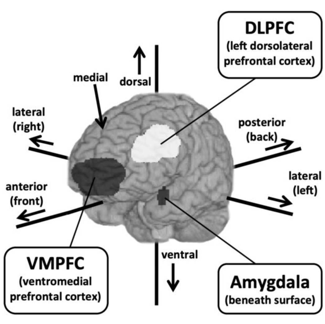
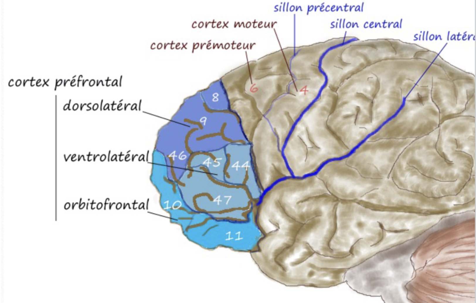
Prefrontal cortex
Movement organization (into sequences) and motor inhibition

What is motor inhibition?
involves higher cognition/decision-making when executing movements such as when putting down your phone to do work
Which motor area includes mirror neurons?
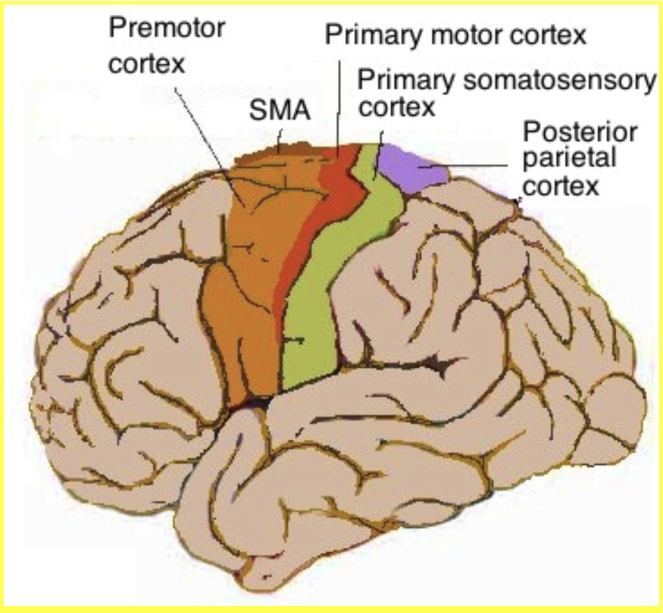
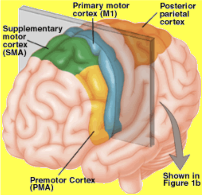
Premotor cortex
Premotor cortex
Part of the brain that observes people’s actions
Selects which part of the body to move and what movements to execute

Which 3 motor areas are involved in motor planning?
Posterior parietal cortex, prefrontal cortex, and SMA
Mirror neurons
active when we observe when someone is doing something and when we automatically mimic that behavior (ex: yawning)
Understanding intentions
Empathy- individuals with Autism spectrum disorder have less activation in MNS
What is supplementary motor area (SMA) involved in?
moving your whole body
(T/F) Cortex is involved in complex, skilled actions (playing viola) and is less important for more involuntary actions such as sneezing, laughing, or crying
True
What are the 2 subcortical motor areas?
Basal ganglia & cerebellum
Basal ganglia
Contains Striatum (caudate nucleus & putamen), Substantia nigra (in dopaminergic pathway), and Globus pallidus
Critical for learning new habits and automatic responses (ex: driving)
Spontaneous movement (ex: twitching, blinking)
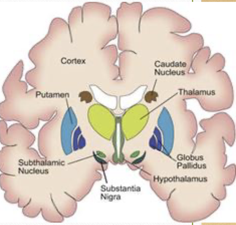
What are the 5 symptoms/disorders caused from damage to basal ganglia?
Dyskinesia: unwanted movements
Huntington’s disease: caudate and putamen cells are destroyed
Tourette’s syndrome (putamen cell damage): involuntary movements
Parkinson’s: loss of dopamine cells in substantia nigra
OCD: can be caused from strep throat
Which brain region has the most number of neurons?
Cerebellum
How many neurons does cerebellum have?
billion. neurons.
What are the functions of the cerebellum?
Involved in rapid movements that require aim, timing, and alterations of movements:
Ex: rhythm
Judging which visual stimulus/thing moved faster
What is one symptom/disorder resulting from damage to the cerebellum?
Ataxia
What is ataxia?
Problem with motor coordination—> similar to effects of alcohol intoxication
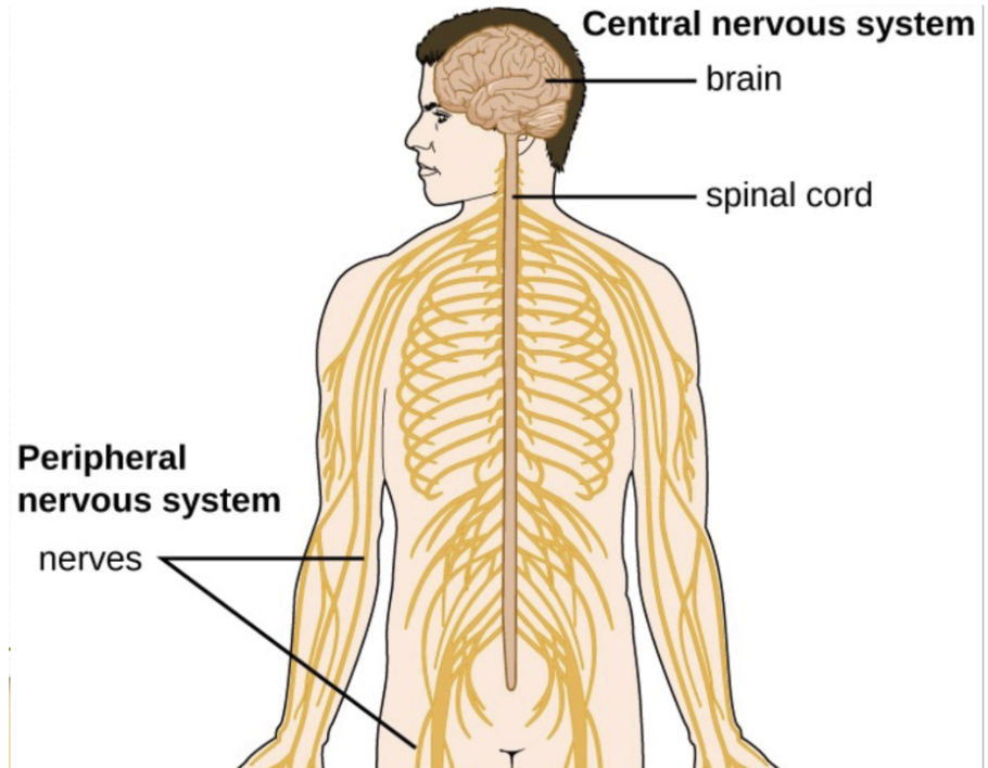
Somatic Nervous System
Where the brain controls movement and receives sensory info from body (spinal nerves) and head & neck (cranial nerves)
What does movement depend on?
muscle contraction
Neuromuscular junction
Synapse b/w a motor neuron axon and muscle fiber
What neurotransmitter is released from the neuromuscular junction?
Axons release aCh at this junction, which excites the muscle to contract
What is the difference between fast-twitch and slow-twitch fibers?
Fast-twitch fibers:
Fast contractions and rapid exhaustion (ex: anaerobic – think oxygen!)
Slow-twitch fibers:
Slow contractions and no fatigue (ex: lips when talking, aerobic)
(T/F) Everyone has the same amount of fast-twitch and slow-twitch fibers.
False — people develop varying amounts due to specialization in aerobic and anaerobic activities
Where do neurons in Autonomic Nervous System send and receive information from?
heart, intestines, etc.
(T/F) PNS and SNS are independent.
True!
Sympathetic Nervous System (PNS)
Increase heart rate, breathing, and decreases digestive activity (fight-or-flight)
Synthesized by adrenal cortex (upper part of kidneys)
Which neurotransmitter is related to SNS?
Relates to norepinephrine (synthesizer: locus coeruleus)
Parasympathetic NS (PNS)
Decreases heart rate and raises digestive activity; conserves energy
Vegetative
Vagus nerve
carries signals between brain, heart, and digestive system
found in the parasympathetic nervous system
help with return to calm state
Which 2 neurotransmitters are associated with PNS?
aCh & serotonin (for sleep-wake cycles)
(T/F) Premotor areas include Broca’s area
True — includes broca’s area, SMA, and the dorsal and ventral premotor cortex

Which areas do premotor areas receive spatial info from to navigate how to go to places?
Receive connections from parietal and other frontal areas
(T/F) SMA is in the middle of the two hemispheres of the brain.
True
What’s another term for spindle neurons?
von economo
Where are spindle neurons located?
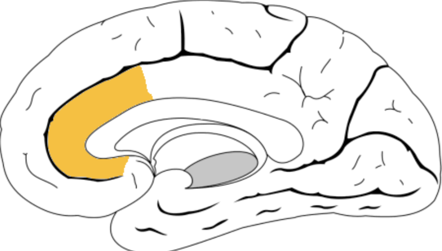
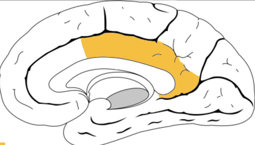
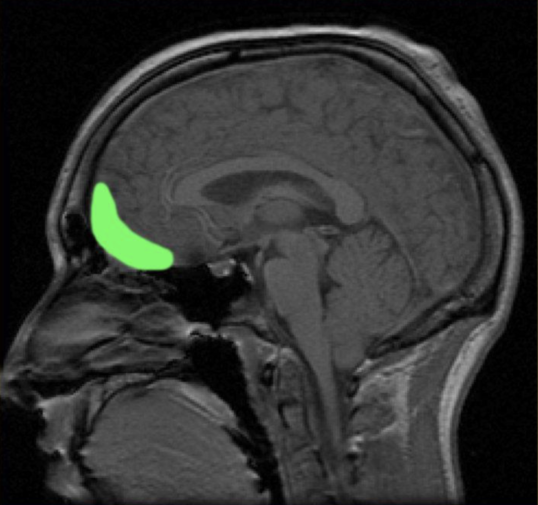
Anterior Cingulate Cortex (ACC)
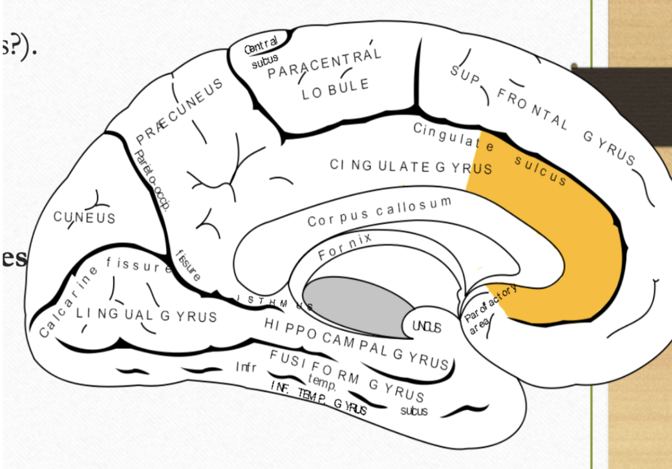
Ventral Anterior Cingulate Cortex / Ventral ACC
Working in the present w the outside
Connections w/ default-mode network and memory areas
Important for self-referential thinking(individualism/self-centered thinking), thinking, and regulating emotions/moods
Ex: dogs are more selfless due to less activity in this part of the brain
Which psychological disorder(s) is associated with Ventral ACC
Many! But schizophrenia (paranoia) would be a key disorder
Which areas is the ventral ACC connected to?
Connections from DLPFC, posterior cingulate cortex, and medial temporal cortex (hippo & amygdala)
What are the four parts of the prefrontal cortex?
Dorsal (posterior) ACC
Orbitofrontal cortex
Ventrolateral
Ventromedial ACC
What are the functions of dorsal (posterior) ACC?
Important for attention, cognitive control, consciousness
Connects motor areas and insula (hint: connects mind w body)
Orbitofrontal cortex
Connections from the insula, amygdala, and other areas of temporal lobe (superior temporal & inferotemporal cortex)
Connections sent to hippocampus and amygdala to modulate physiological responses of the body
(T/F) Spontaneous speech is caused from damage to the prefrontal cortex
True
Which area(s) of the brain result in rule breaking & risk taking when it is damaged?
Orbito FC and ACC
What are the three symptoms resulting from damage to Dorsolateral PFC?
Perseveration & response inhibition and diminished automatic responses (detecting patterns) (DLPFC)
Ex: shape game: boy made mistakes and kept making them bc he didn’t learn from them
Associative learning (DLPFC): problems learning from experience
Temporal memory issues (DLPFC) (learning in which order things happened)
(T/F) OFC lesions result in reduced sex drive.
False: they result in abnormal sexual behavior (masturbating in public)
What do DLPFC lesions result in?
Reduced sex drive
Which area(s) of the prefrontal cortex is associated with pseudodepression & pseudopsychopathy?
OFC / orbitofrontal cortex
What is the difference between pseudodepression & pseudopsychopathy?
depression- apathy, indifference, reduced sexual interest
pseudopsychopathy- coarse language, promiscuous sexual behavior
Voluntary gaze/eye movement is a result from damage to which region?
Frontal eye fields (FEF) in Premotor cortex—associated with attention & eye movement
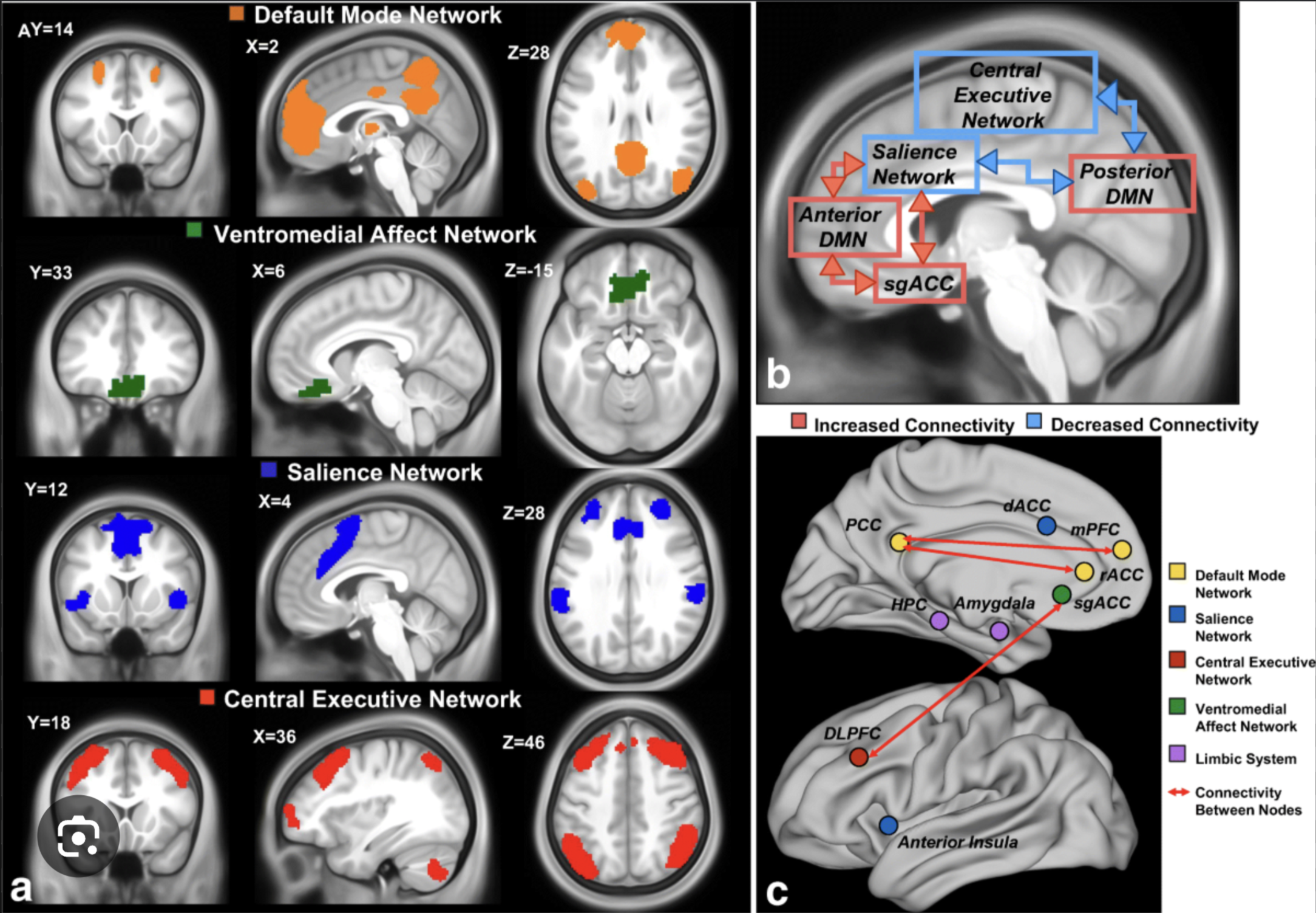
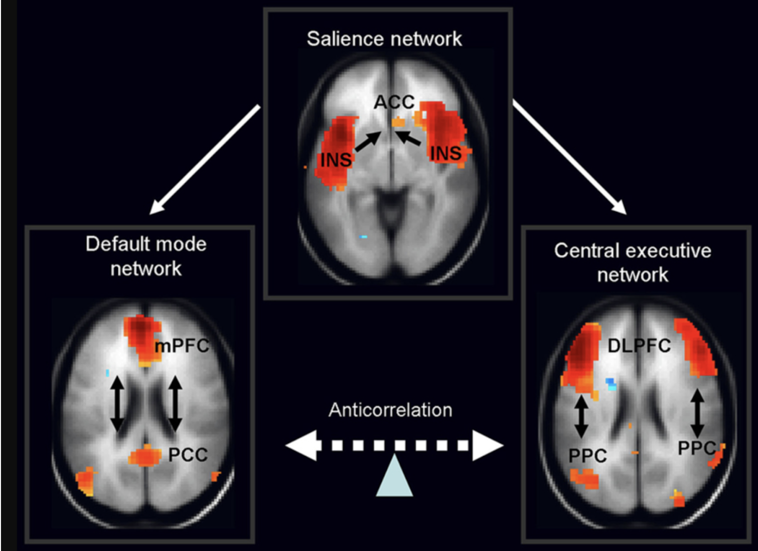
(T/F) Default mode network is independent from the salience network.
False— they are both correlated
What is the function of default mode network?
mind-wandering, thinking about one’s past or future
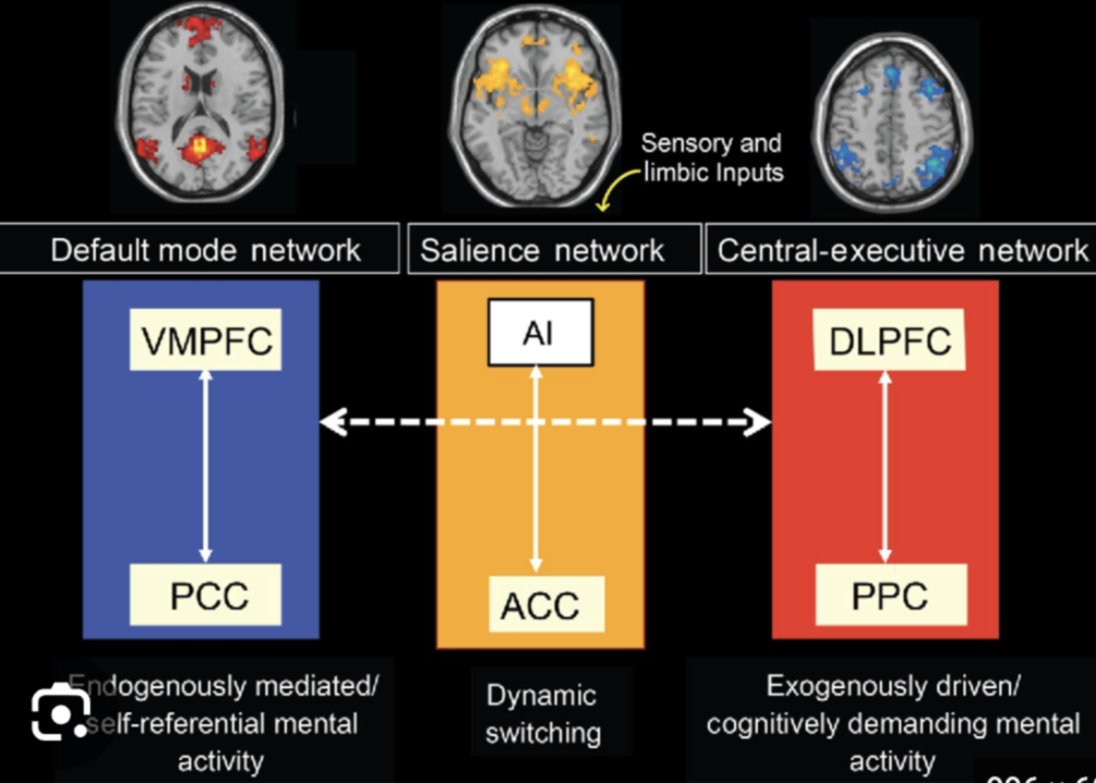
Which areas are in the default mode network?
medial prefrontal cortex/mPFC, posterior cingulate cortex / PCC, medial temporal lobes
What does the dorsal attention system focus on?
Top-down visuospatial
Network locations & activities