Dental Radiology Exam 2-Analysis of errors and artifacts
1/58
There's no tags or description
Looks like no tags are added yet.
Name | Mastery | Learn | Test | Matching | Spaced | Call with Kai |
|---|
No analytics yet
Send a link to your students to track their progress
59 Terms
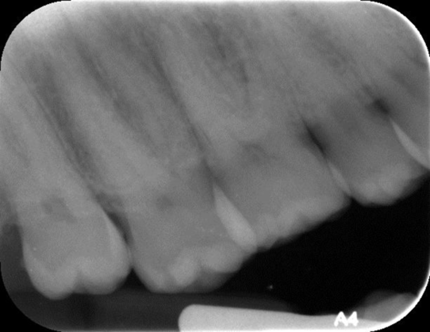
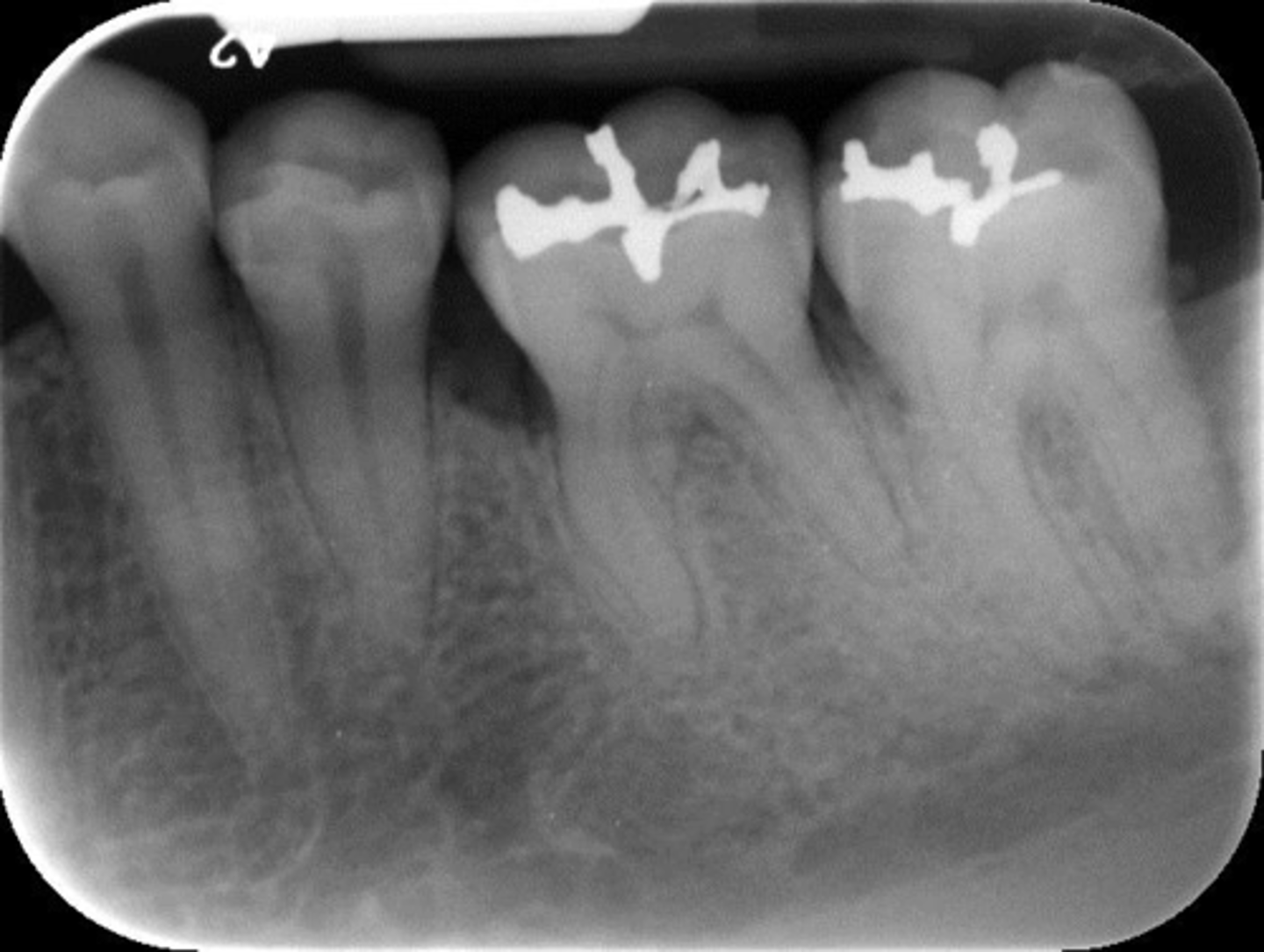
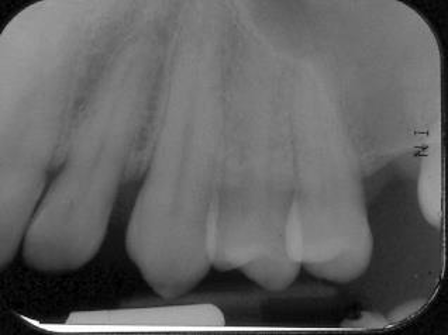
What is this projection of?
Premolars projection
Projection (technique) errors include:
Apical areas "cut off"
Horizontal Angulation
Vertical Angulation
Partial images -"Cone Cuts"
Plate Bending
Phalangioma
Double Exposure
Movement
Exposed Backwards
Plate Placed Incorrectly
Appliances & glasses
Thyroid collar
Exposure, scanning and handling errors include:
Scanning Errors
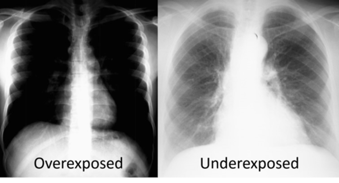
Underexposed
Overexposed
Moisture Contamination
Scratched or damaged plate
List the three basic film quality evaluation criteria and the factors that control them.
1. All rads must have acceptable image characteristics of detail, definition, density and contrast
2. All crowns and roots, including apices, are fully depicted together with inter proximal alveolar crests, tooth contact areas, and surrounding apical bone regions
3. Images of all teeth and other structures are shown in proper relative size and contour with minimal distortion and without overlapping images, where anatomically possible
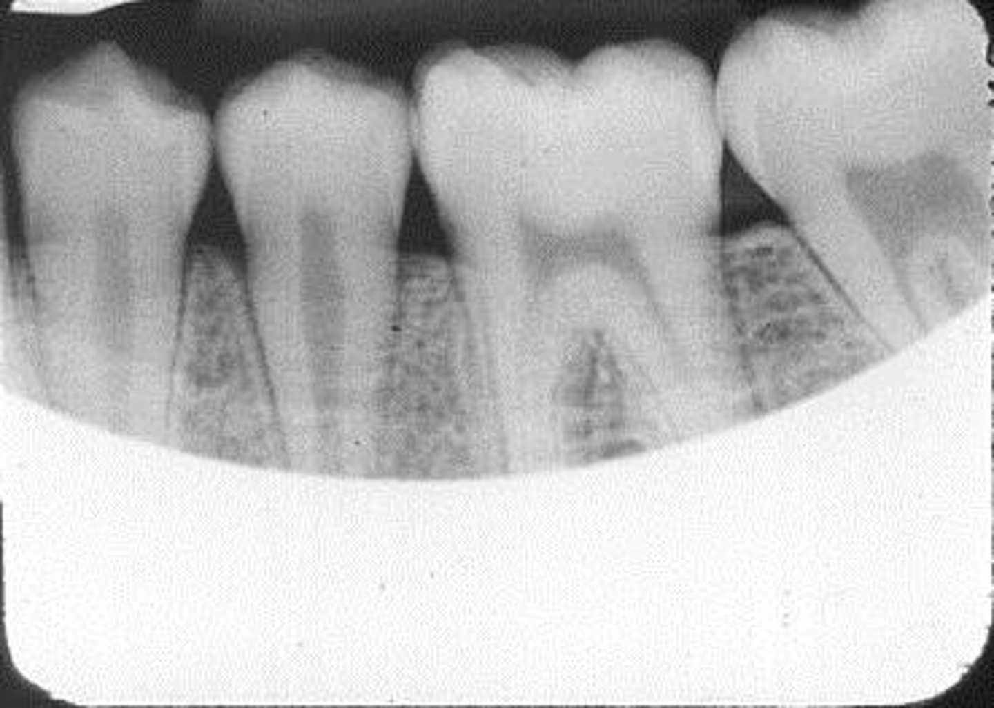
Bitewing radiograph evaluation criteria
1. No overlapping of interproximal contacts
2. Crowns centered
3. Crest of the alveolar bone should be visible
4. The occlusal plane should be horizontal
5. Buccal and lingual cusps shouldn't be separated excessively
State the three groups wherein the majority of errors are produced
1. Technique errors
2. Exposure and processing errors
3. Film-handling errors
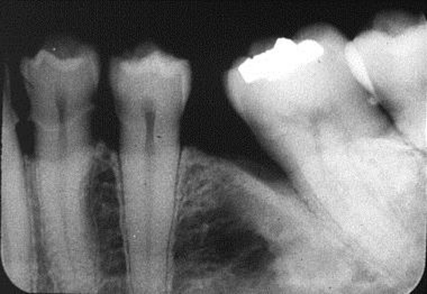
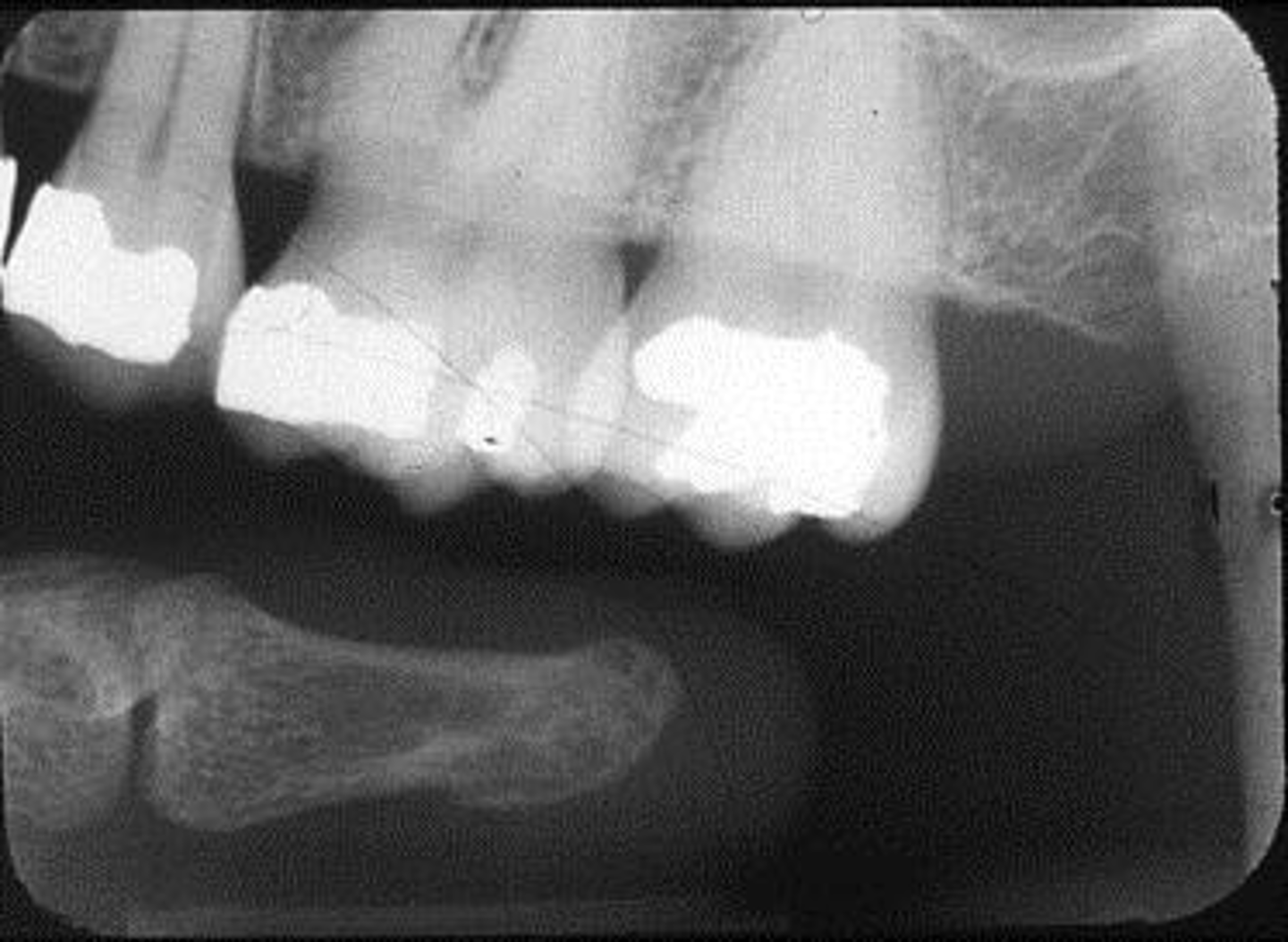
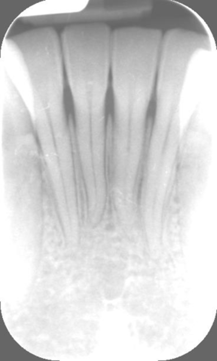
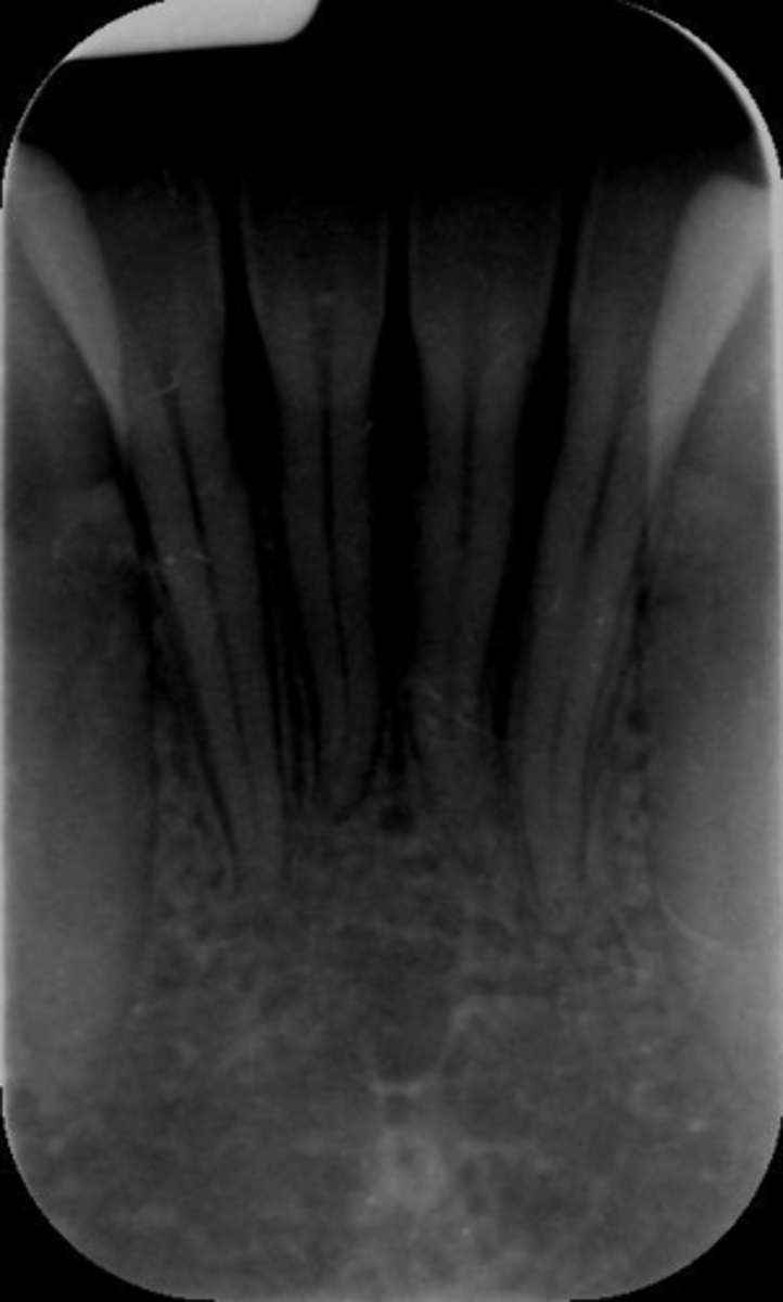
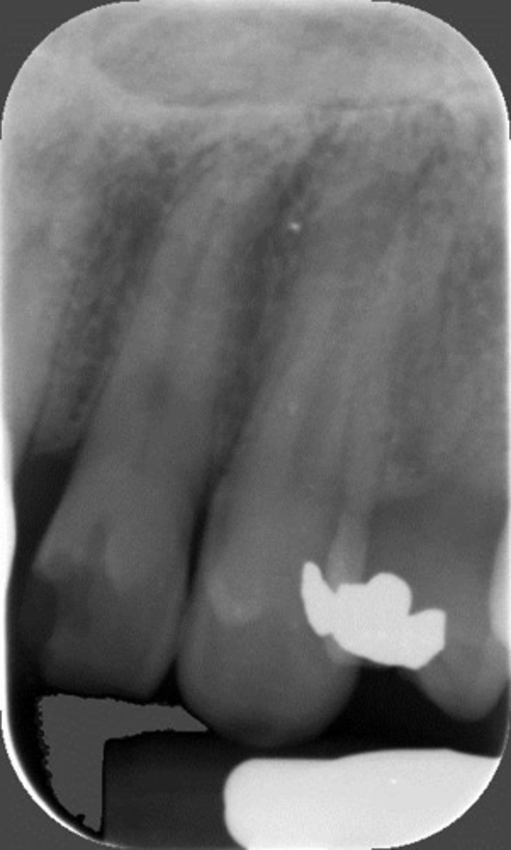
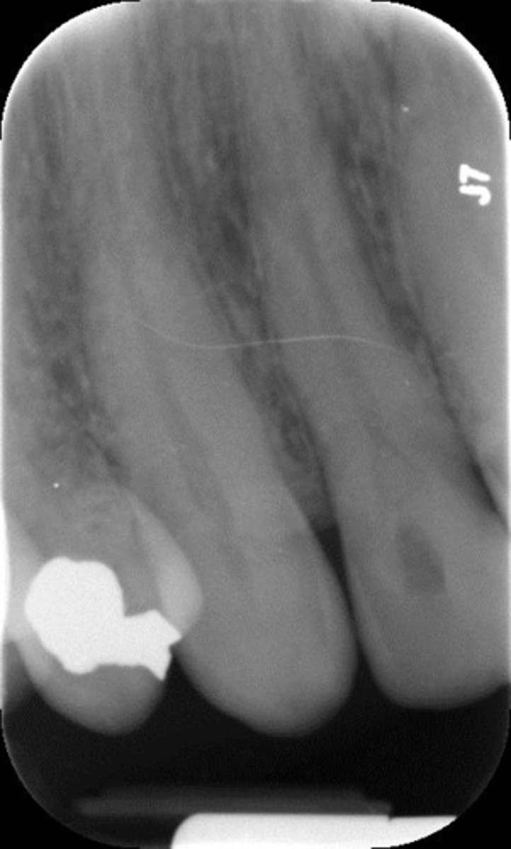
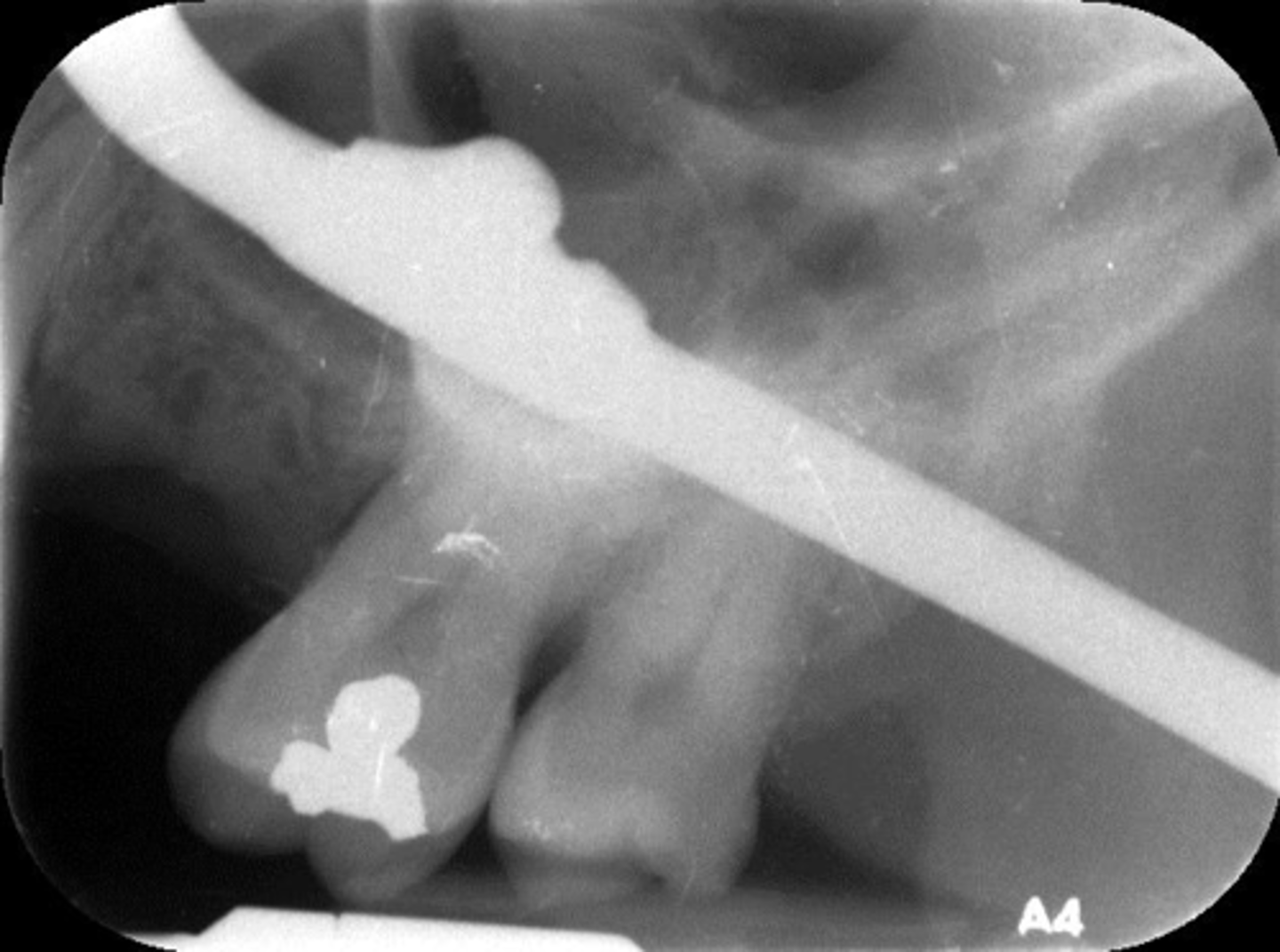
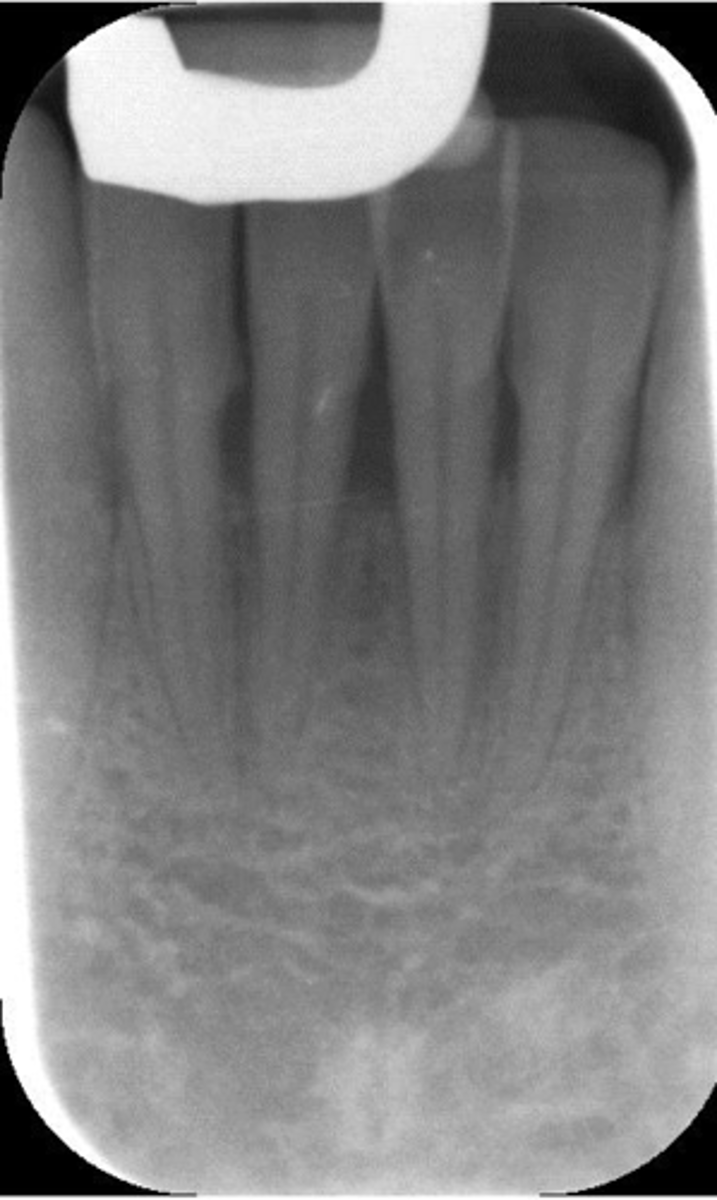
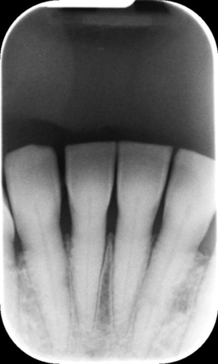
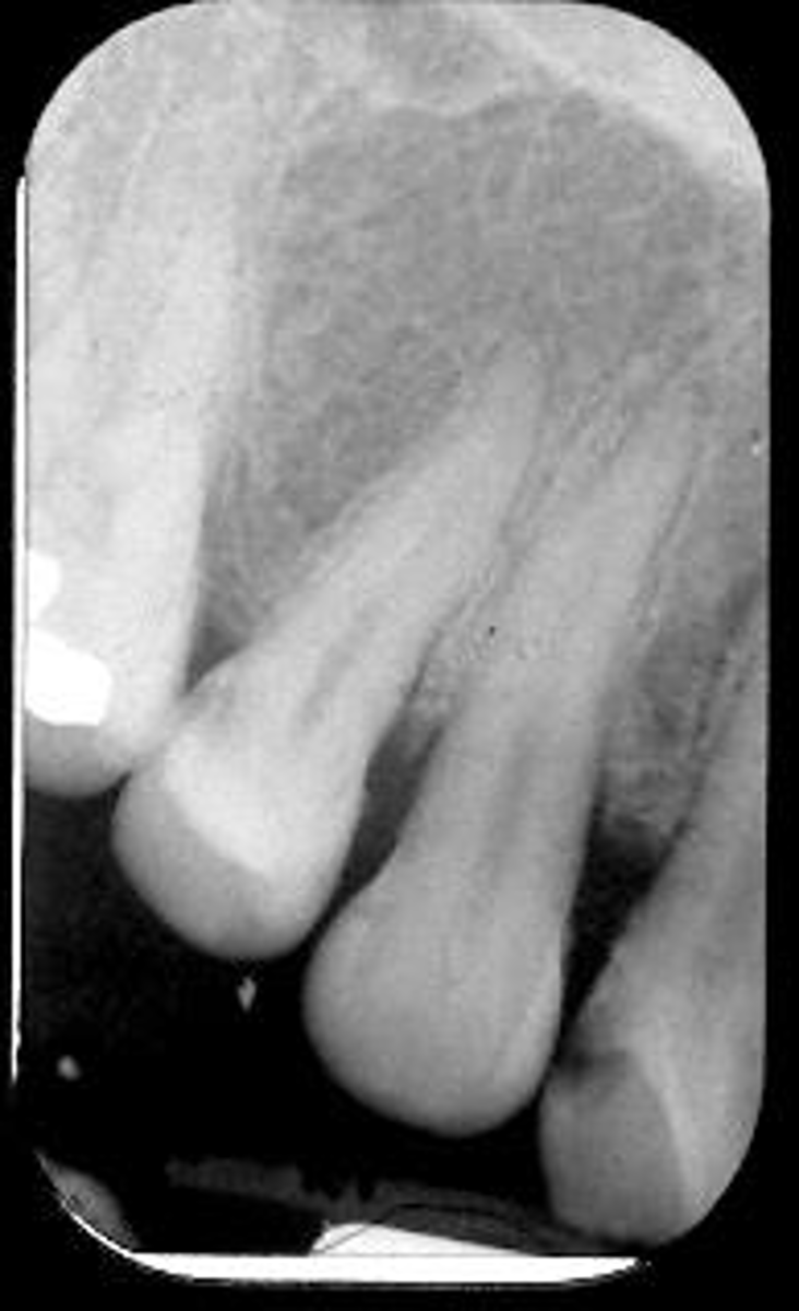
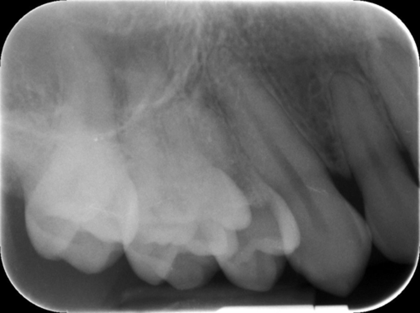
Absence of apical structures
Appearance-Apices "cut off"
Cause-plate too close to the teeth
Correction-Make certain the incisal-occlusal surfaces of the teeth touch the bite-block. Position plate in the middle of the oral cavity when working in the maxillary arch
The X-ray beam must travel ____ the embrasures of the teeth.
Through
Apices "cut off"-maxillary teeth
Usually caused by the plate being placed too close to the teeth in the paralleling technique
Technique for taking images on patients with shallow palates
Increase vertical angulation slightly
Use bisecting angle technique
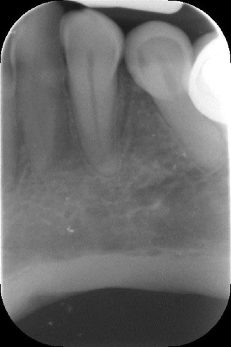
Apices "cut off"-mandibular teeth
Usually caused by the plate not being pushed into the lingual vestibule, or positioned on top of the tongue
Dropped plate corner
Appearance-Occlusal plane appears tipped/tilted
Cause-Plate was not parallel to the incisal-occlusal surfaces of teeth
Correction-Make sure incisal-occlusal surfaces of teeth touch the bite-block (cotton roll under biteblock, not on the side of teeth to be radiographed)
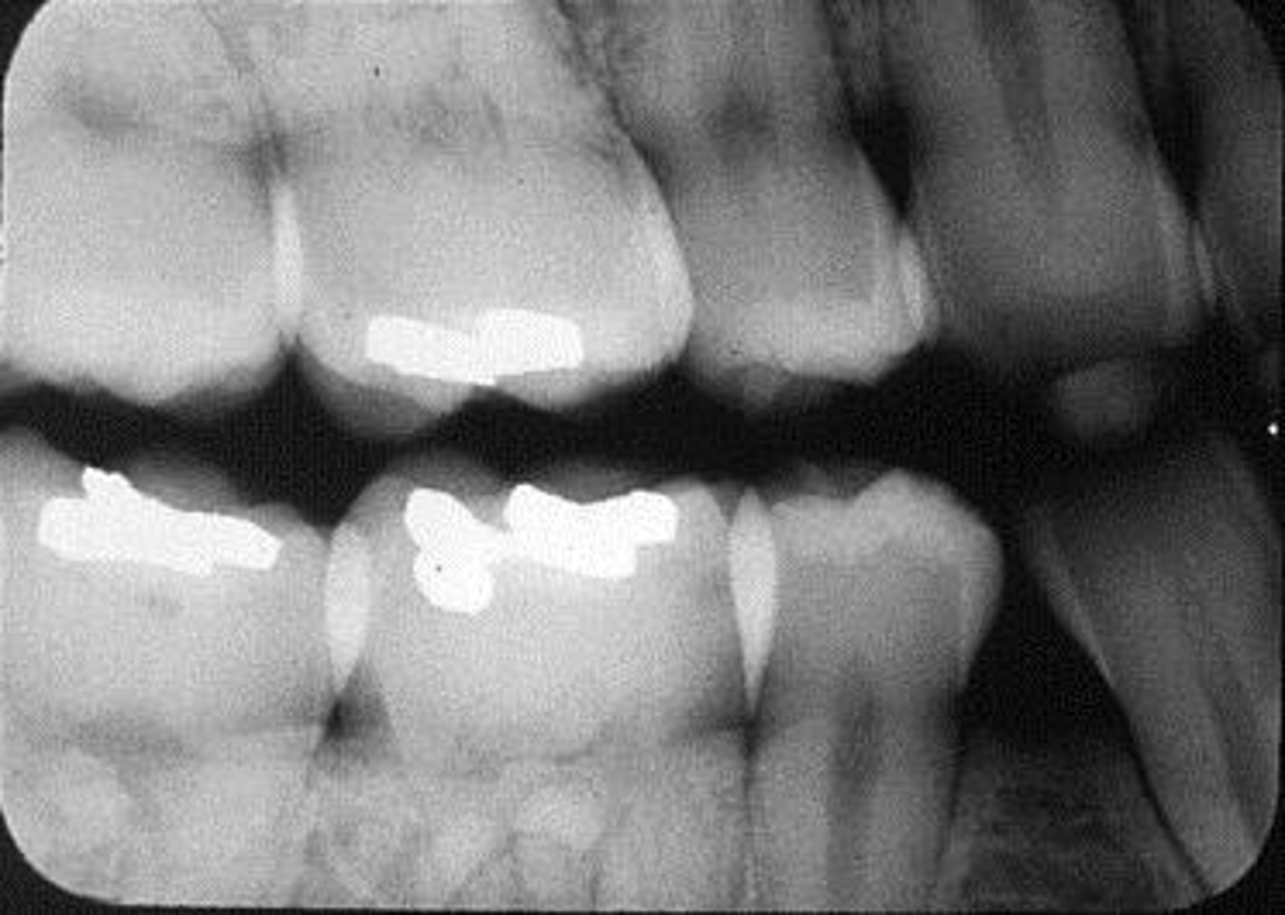
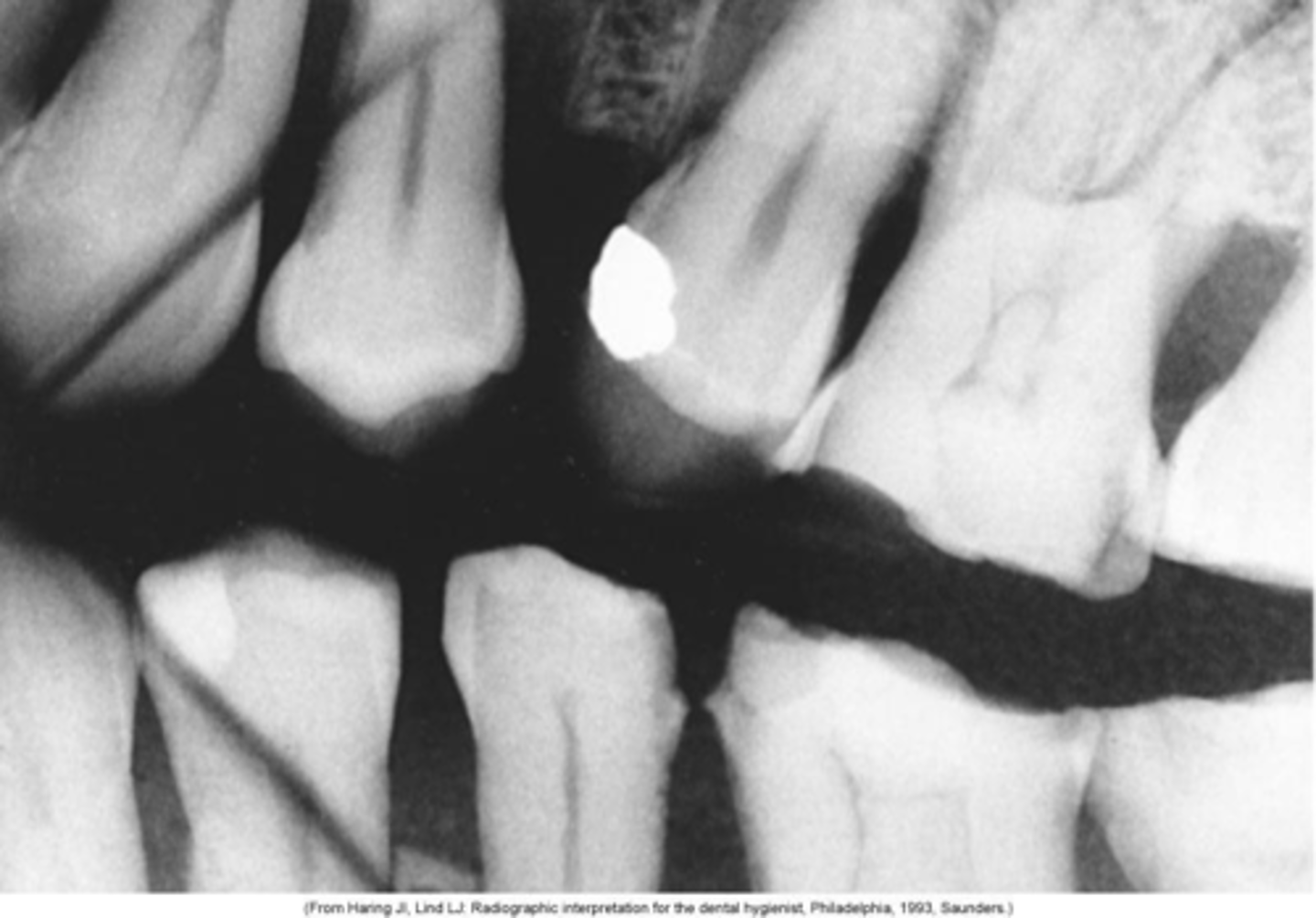
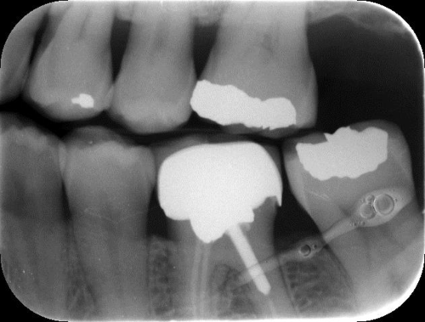
Incorrect horizontal angulation
Appearance-Overlapped contacts or roots
Cause-Plane of plate not parallel to long axis of teeth. Central ray was not directed through inter proximal spaces.
Correction-Position plate parallel to the teeth. Direct x-ray beam through interproximal regions
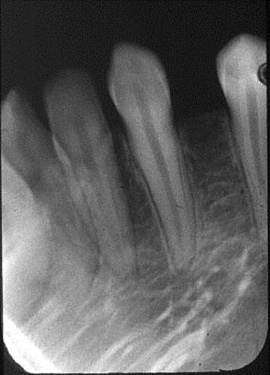
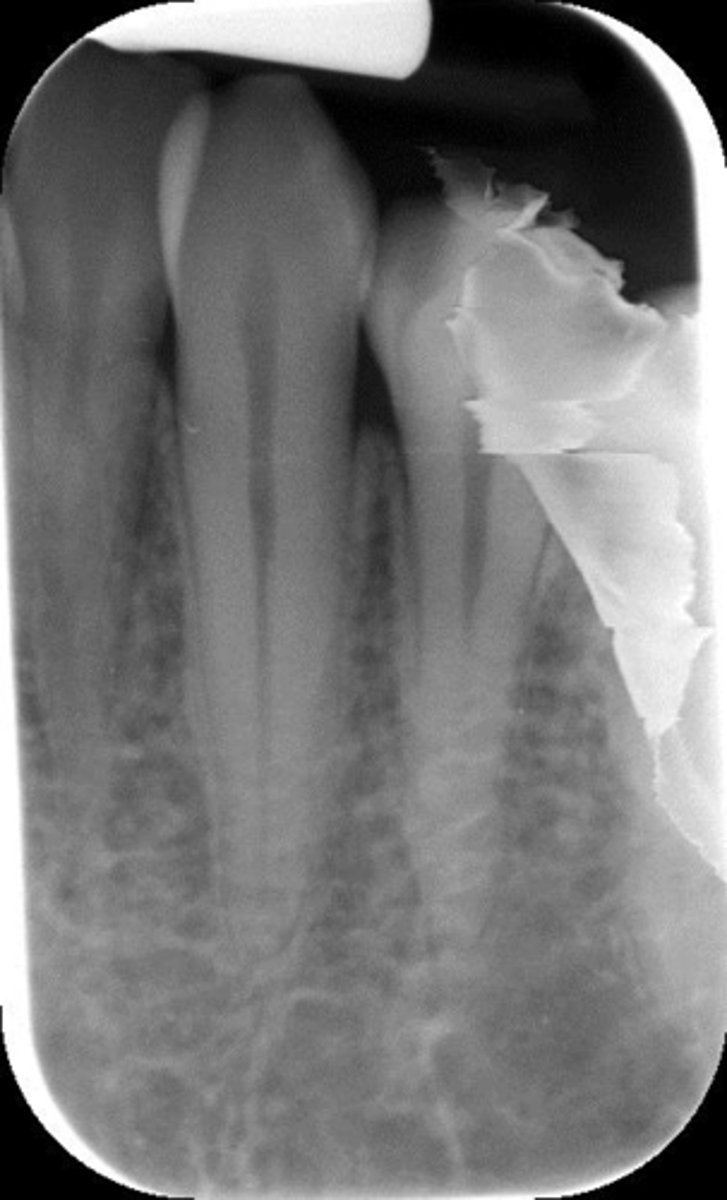
Incorrect vertical angulation-foreshortened images
Appearance-Short teeth with blunted roots
Cause-Excessive vertical angulation
Correction-Align BID with Rinn instrument bar
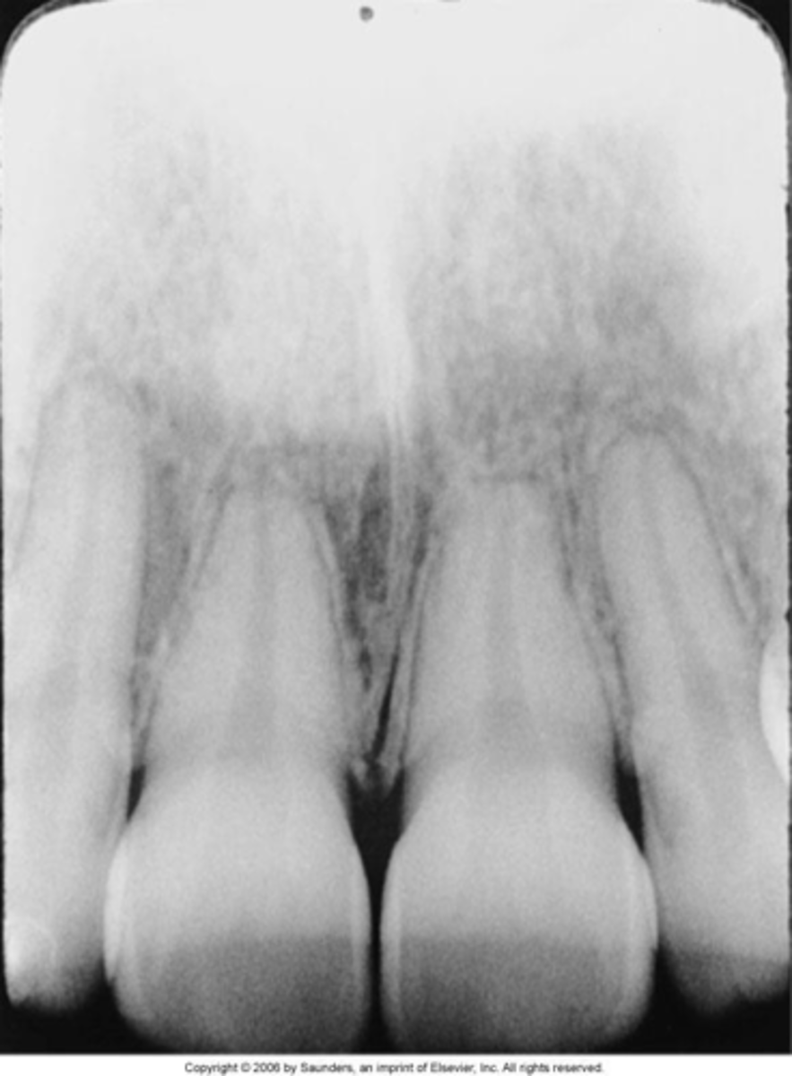
Incorrect vertical angulation-elongated images
Appearance-Long, distorted teeth
Cause-Insufficient vertical angulation
Correction-Use adequate vertical angulation
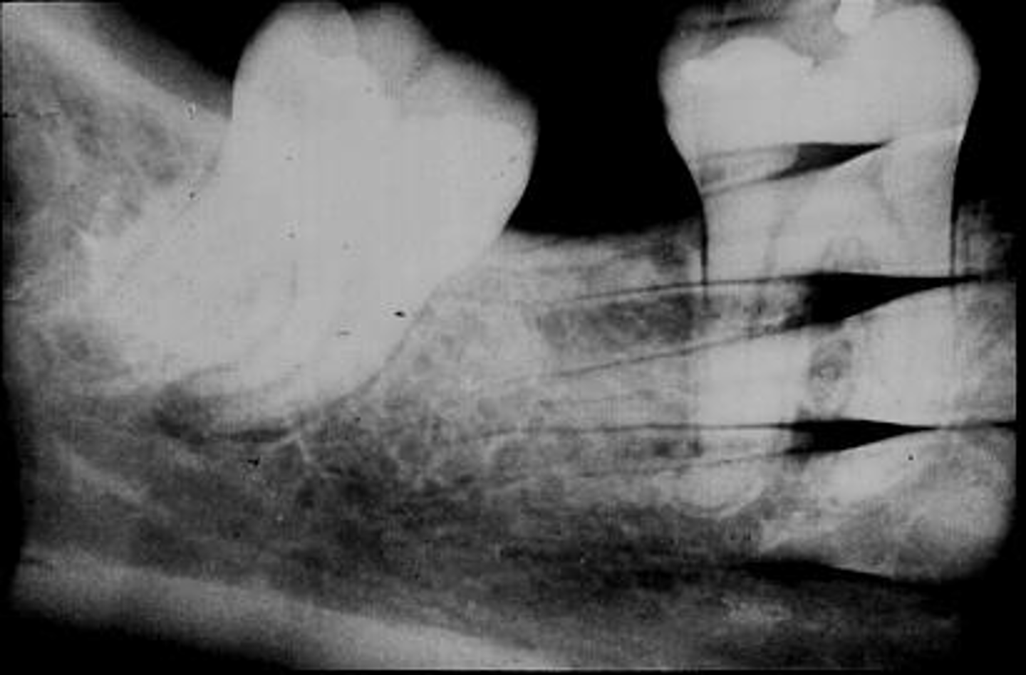
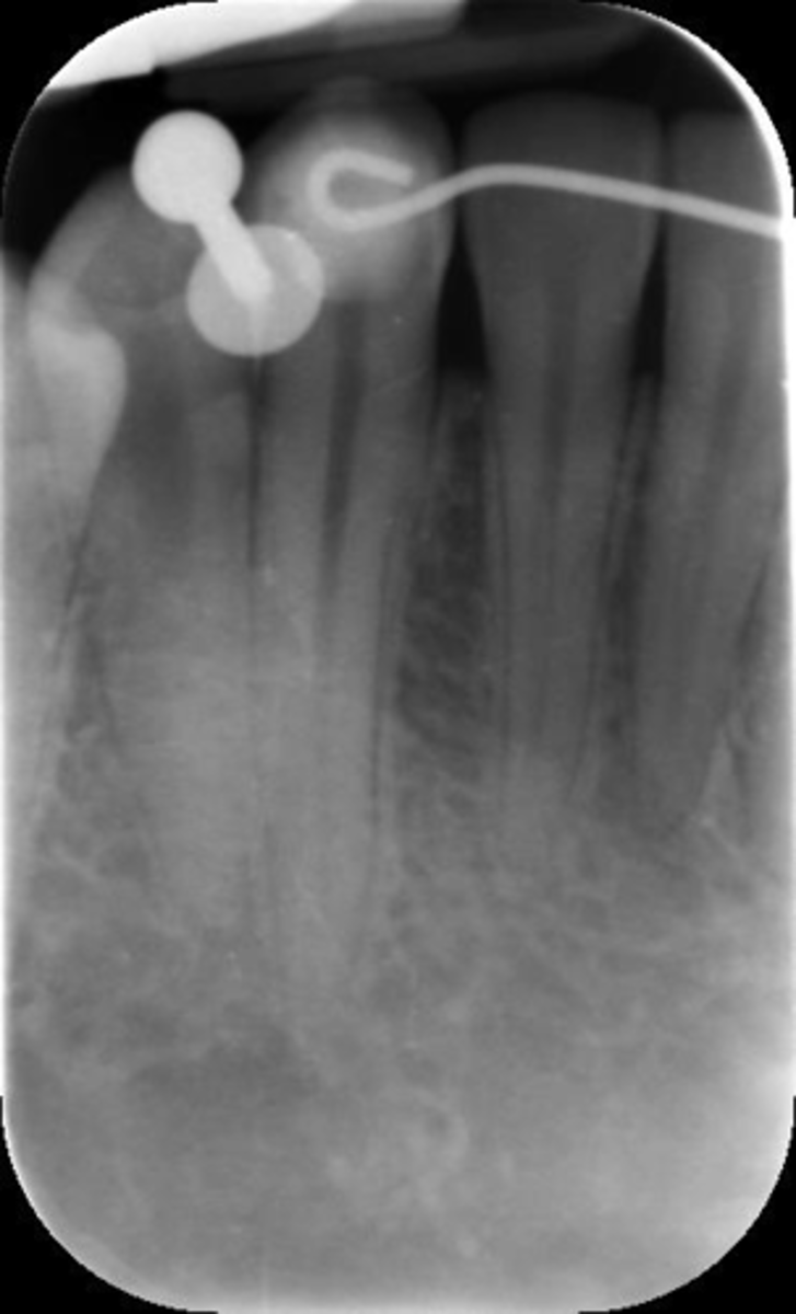
BID alignment problems cone-cuts
Appearance-A clear area appears on the plate
Cause-BID was not properly aligned with the periapical plate holder or, incorrect setting of the RINN instrument
Correction-Make certain the x-ray beam is centered over the plate
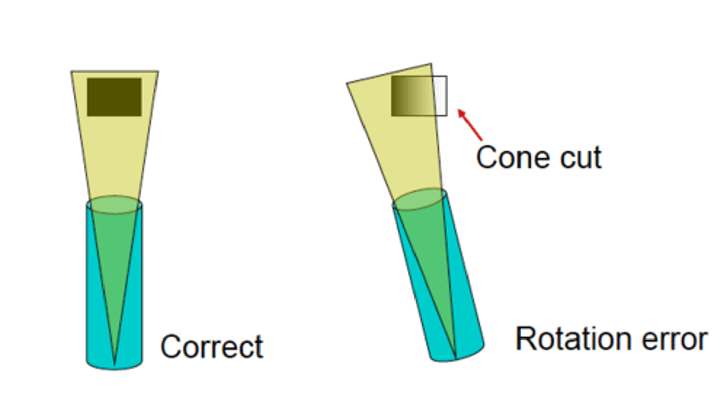
Rotation error
Can cause cone cut
Improper alignment of the BID*
Overlapping inter proximal contact ares*
This is a Rotation error, because just by rotating the BID toward the center of the plate, it can be avoided
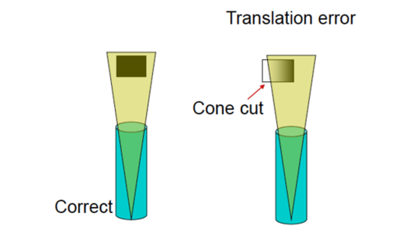
Translation error
A cone-cut can also result from a Translation error, when the BID is directed to the plate with the correct angulation*, but too far mesially or distally.
Therefore, radiation failed to hit the X ray plate in its entirety.
*Open inter proximal contacts
Film/plate bending
Appearance-Images appear stretched and distorted
Cause-The plate was bent excessively
Correction-Check plate placement before exposure
Film/plate creasing
Appearance-A thin radiolucent line appears on the plate
Cause-The plate was creased
Correction-Do not bend or crease the plate
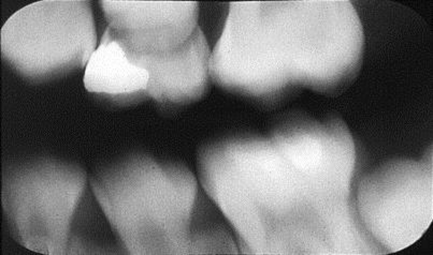
Phalangioma
Appearance-The patient's finger appears on the plate
Cause-The patient's finger was positioned in front of the plate
Correction-Make certain the patient's finger is placed behind the plate (if necessary)
Double exposure
Appearance-A double image appears on the plate
Cause-The plate was exposed twice in the patient's mouth
Correction-Always separate exposed and unexposed plates
Movement
Appearance-Blurred images appear on the plate
Cause-The patient moved during exposure of the plate
Correction-Instruct the patient to remain still while the plate is being exposed
Reversed plate (exposed backwards)
Appearance-Light images with a lighter circle appear on the radiograph
Cause-The plate was placed backwards in the mouth and then exposed
Correction-Always place the front side of the plate adjacent to the teeth

How do you prevent positioning errors?
Avoid faulty plate placement -center the plate over the area to be radiographed
Distal surface of the canine on radiographs
Not routinely seen on the canine projection due to overlap of the lingual cusp of the first premolar
Underexposed or overexposed rads are affected by
kVp, mAs, source to plate distance
Backwards plate is caused by:
•Improper placement of wrapped plate in the mouth
•Improper wrapping of the plate in dispensary
It is your responsibility to verify that the plate has been correctly wrapped
X rays must hit the plate on the blue side
You should avoid taking radiographs that lack
Diagnostic quality
Retakes cause
Expose patients to unnecessary radiation
Waste time
Faulty radiographs interfere with interpretation
Wrong insertion of the plate in the scanner window
Scanning error caused by
Remove appliances
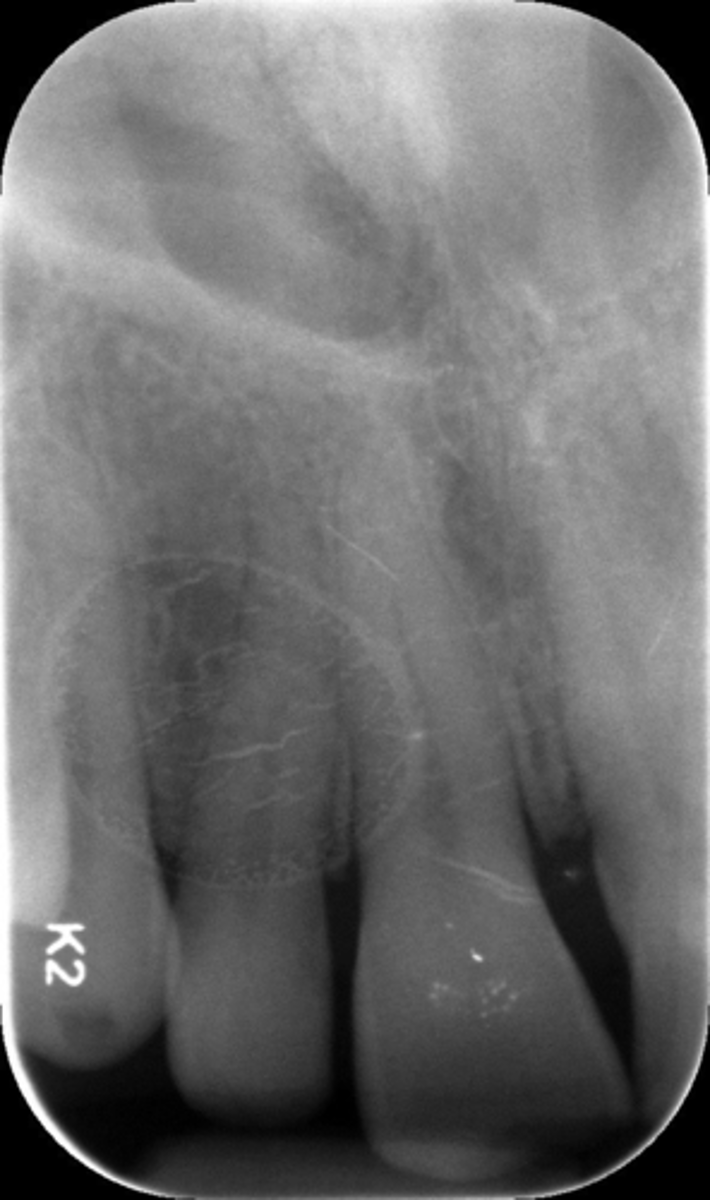
What is this error?
Remove glasses
What is his error?
Thyroid collar
What is this error?
Underexposed
What is this error?
Overexposed
What is this error?
Moisture contamination
What is this error?
Moisture contamination
What is this error?
Damaged plate
What is this error?

Damaged plate
What is this error?
Wrong film direction, backwards, positive angulation, wrong film size, no open contacts
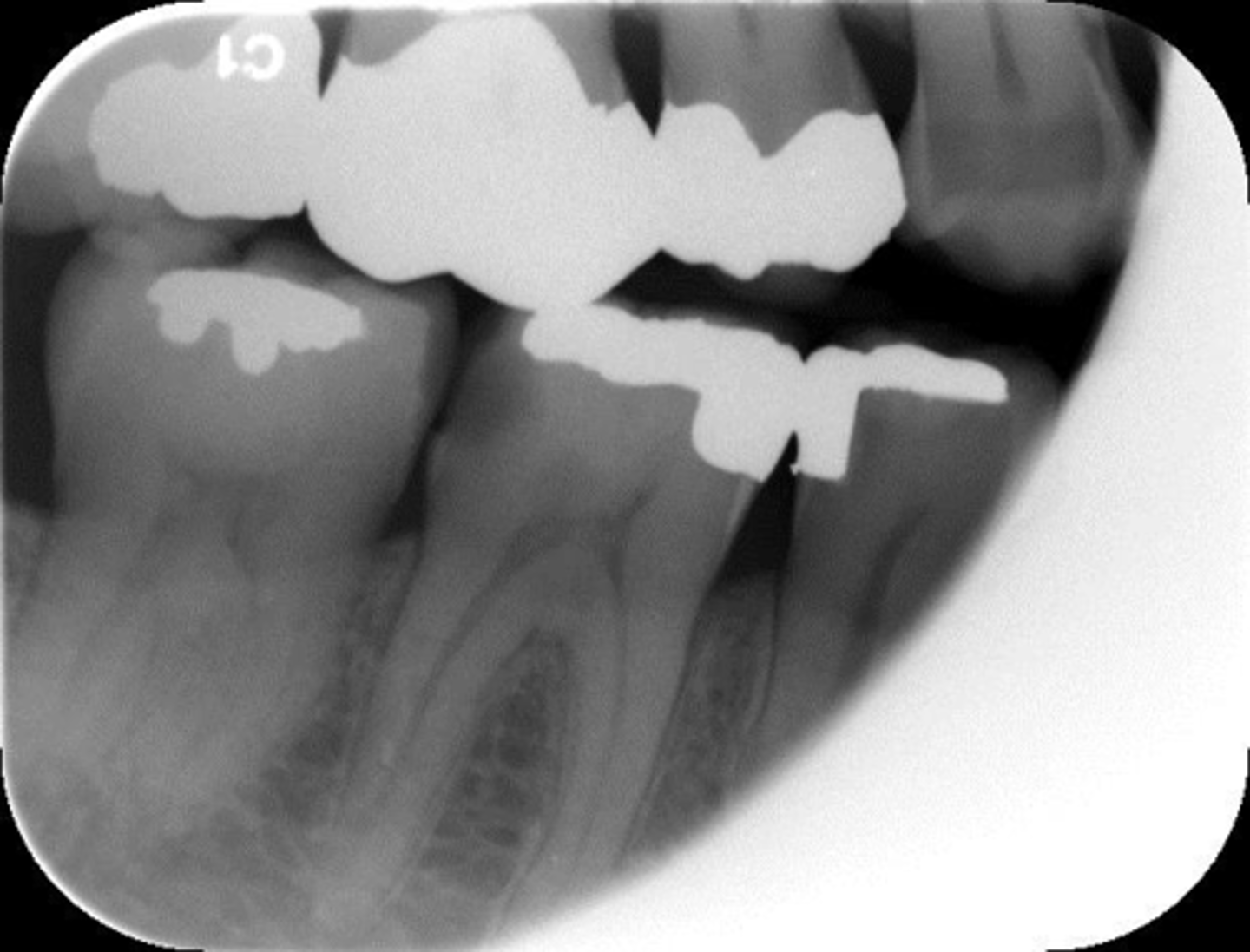
What is this error?
Double exposed, damage to plate
What is this error?
Moisture contamination
What is this error?
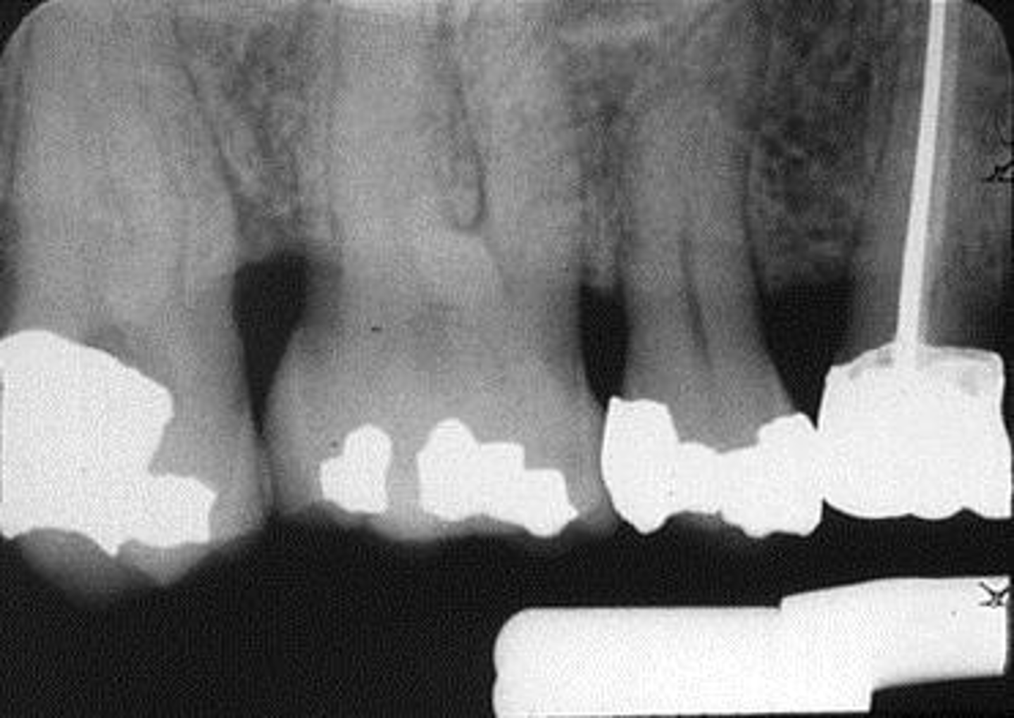
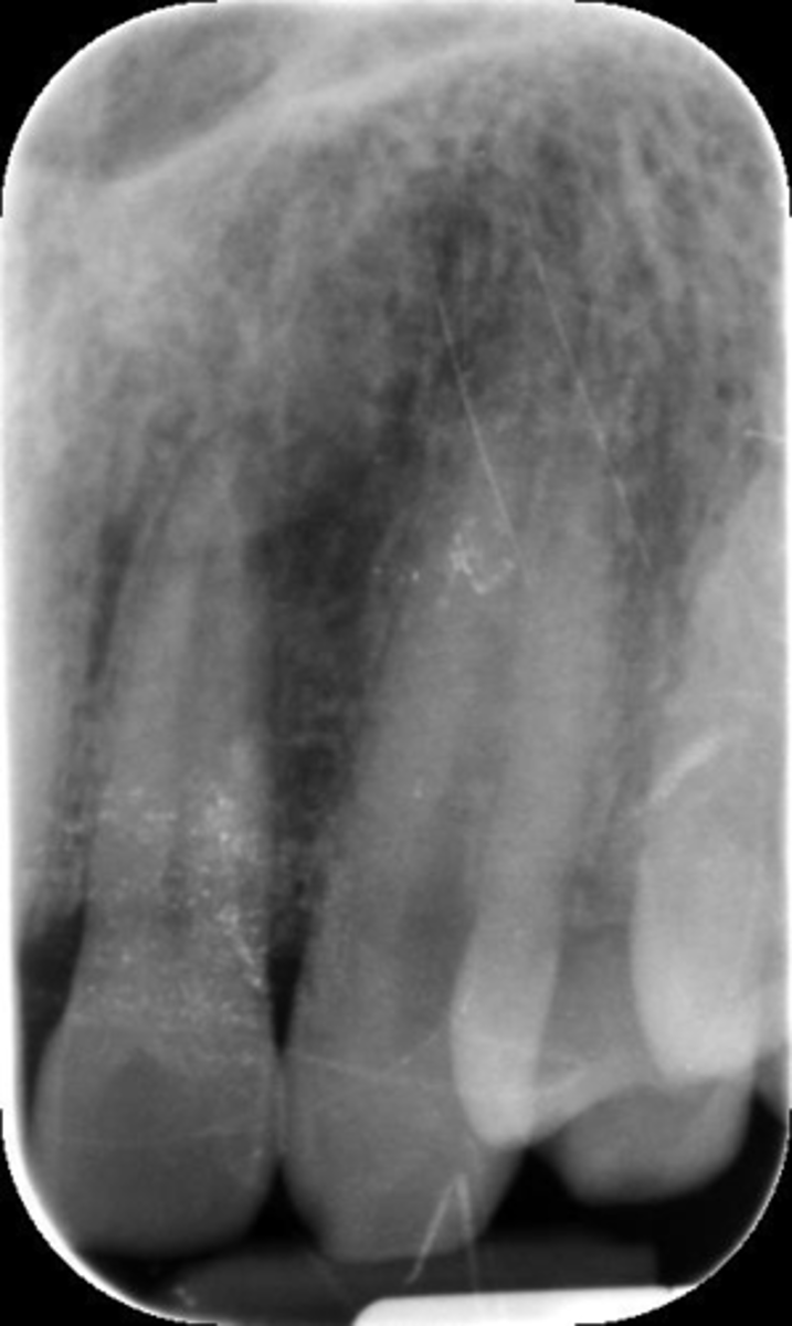
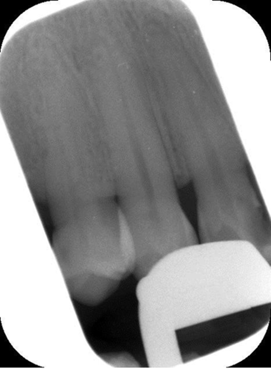
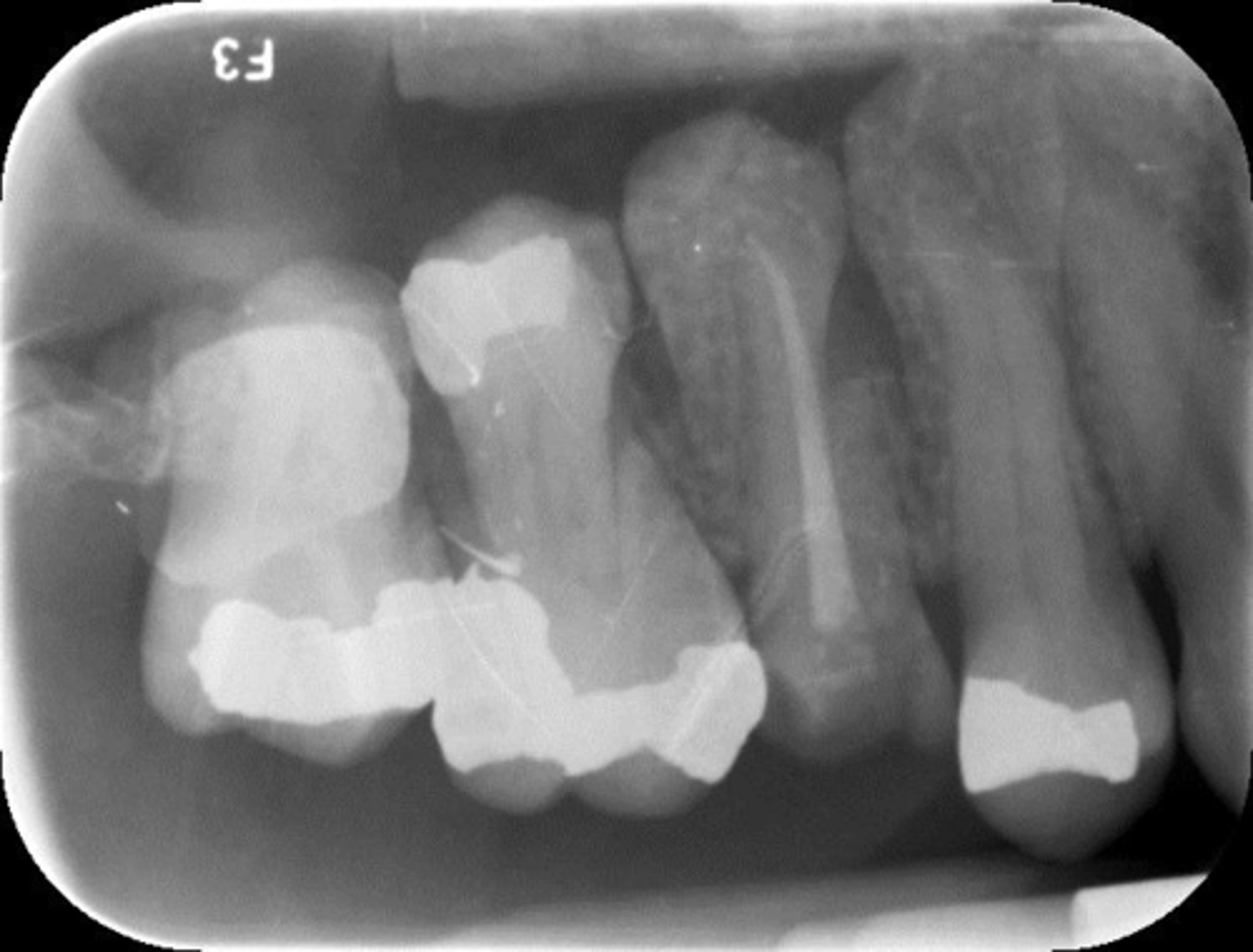
Elongated, vertical angulation, film holder (metal bar)
What is this error?
Damage, elongation, can't see apices
What is this error?
Water damage
What is this error?

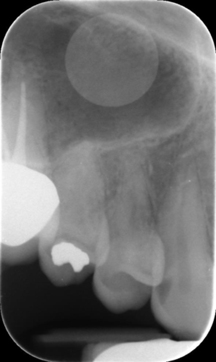
Artifact
What is this error?
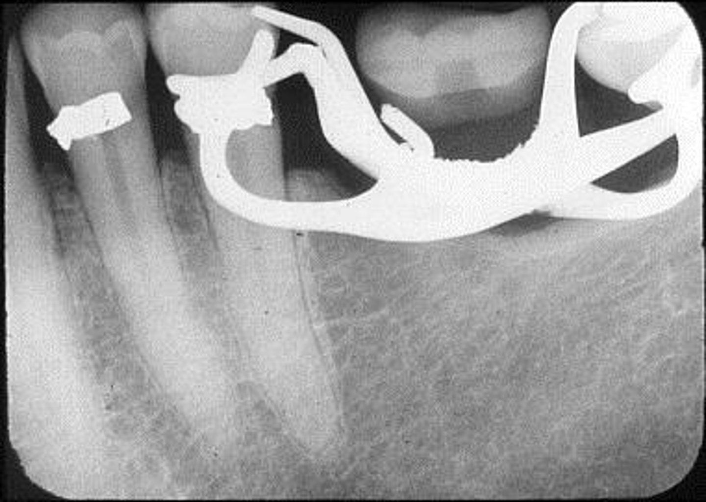
Rinn instrument bar, vertical angulation
What is this error?
Damage
What is this error?

Moisture contamination
What is this error?

Backwards film
What is this error?

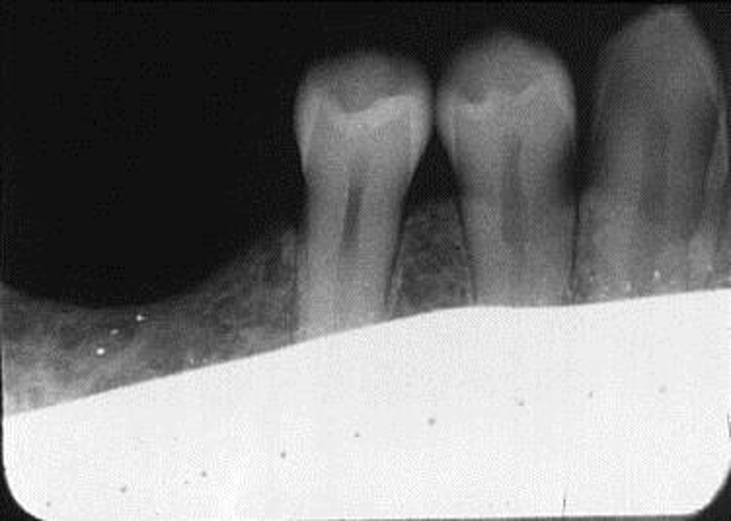
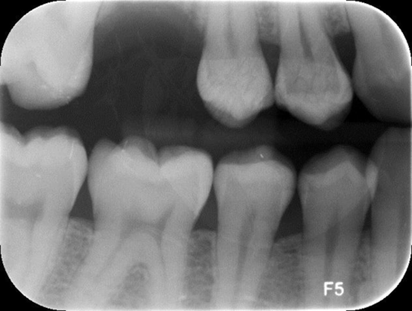
Patient is not biting down
What is this error?
Positive vertical angulation
What is this error?
Positive vertical angulation
What is this error?
Lip piercing
What is this error?
Film fall, drop corner
What is this error?
Double image
What is this error?
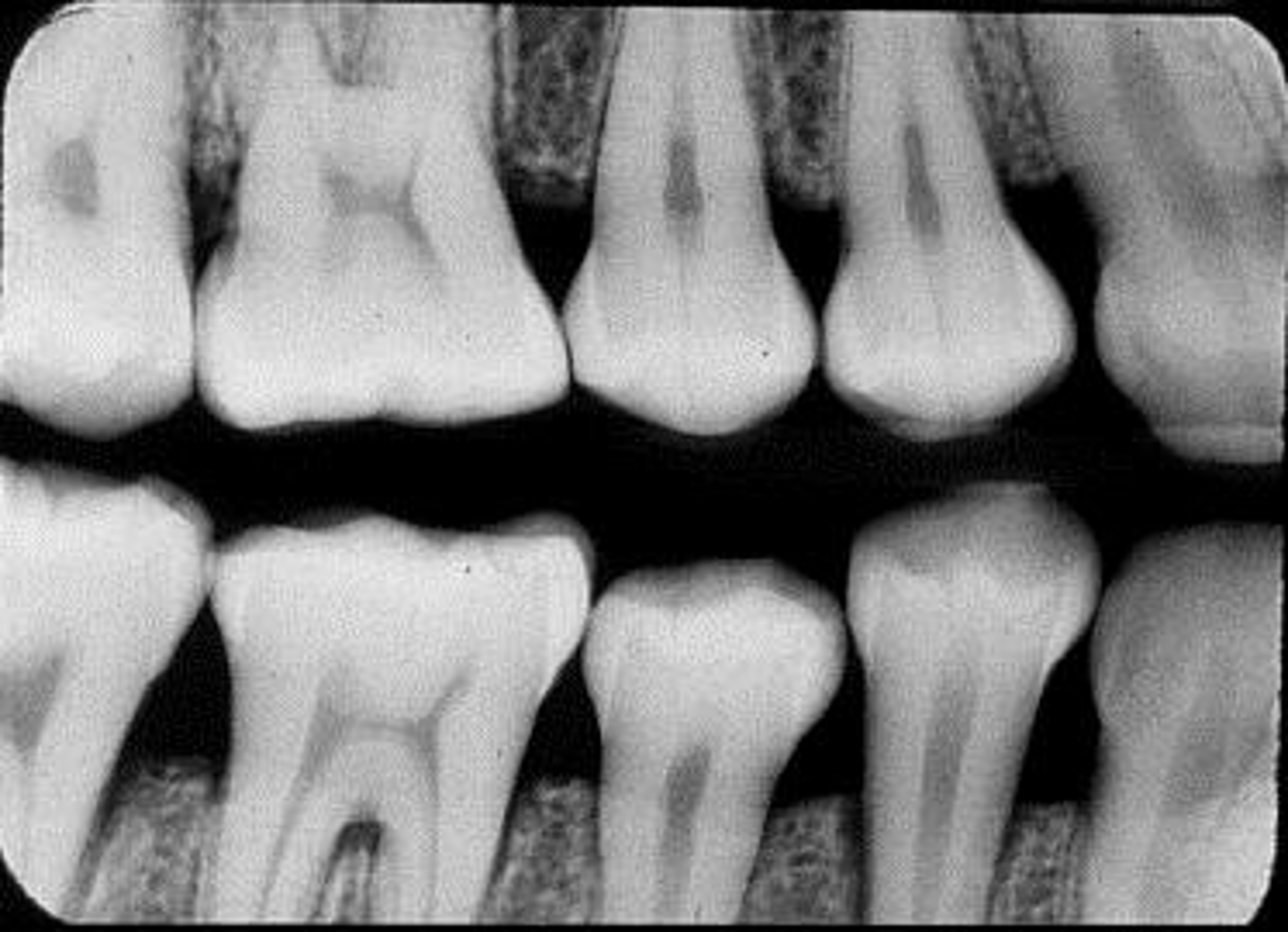
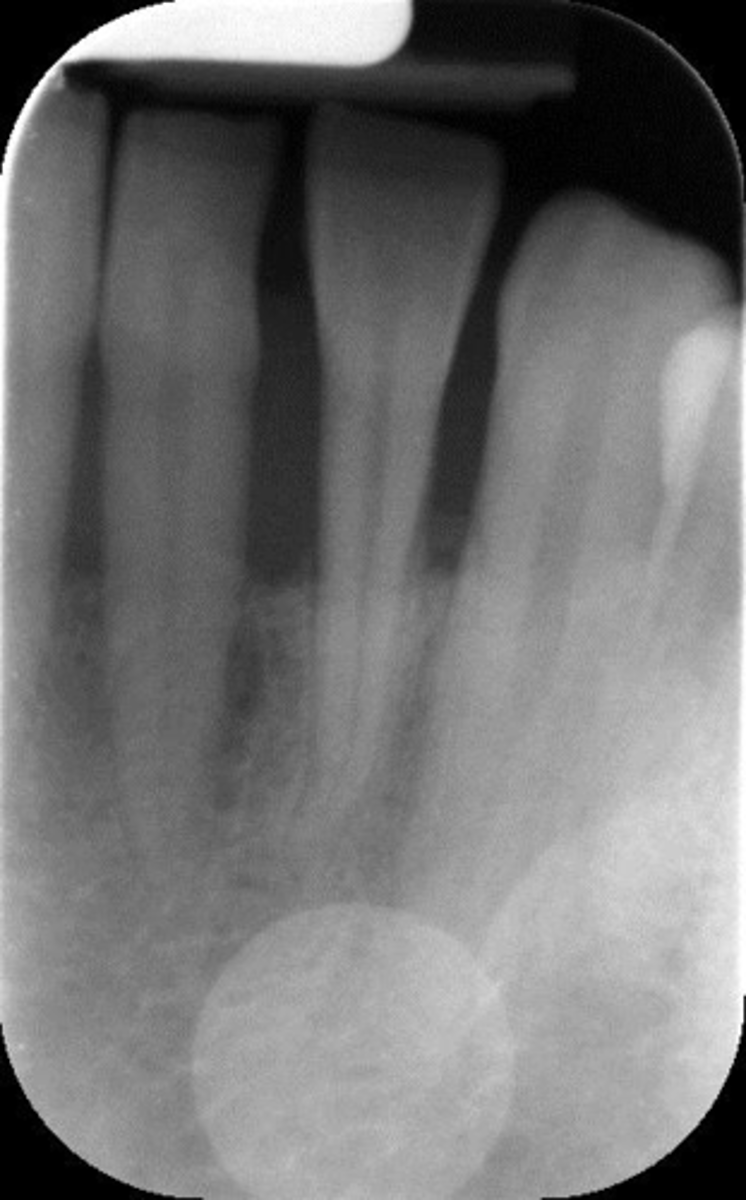
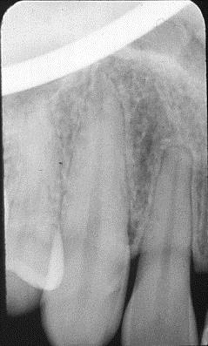
Horizontal angulation, no apices show, vertical angulation
What is this error?
Cone cut, rotation
What is this error?