Human Biology
1/196
There's no tags or description
Looks like no tags are added yet.
Name | Mastery | Learn | Test | Matching | Spaced |
|---|
No study sessions yet.
197 Terms
What is the hierarchical structural organisation within the body from smallest to largest?
Cells, Tissues, Organs, Systems
What are cells?
Cells are the structural and functional units that carry out the life processes (respiration, ingestion, excretion, response, growth, reproduction & movement).
What is the cell theory?
The cell theory states that all organisms are made of cells, are the basic unit of life and arise from pre-existing cells.
Name all the main organelles in the cell
Cell membrane
Cytoplasm
Centrioles
Nucleus
Nucleolus
Endoplasmic reticulum (rough & smooth)
Ribosomes
Mitochondria
Golgi body
Vesicles
Lysosomes
Cytoskeleton
What is the structure and function of the cell membrane?
Structure: The outer lining of a cell.
Function: Determines which substances get into or out of the cell. It allows regulation of cells internal environment.
What is the structure and function of the cytoplasm?
Structure: It is the jelly-like or watery material inside the cell that fills all the space between the nucleus and the cell membrane.
Function: Suspends organelles in place.
What is the structure and function of the centrioles?
Structure: Small structure in the cytoplasm, consisting of microtubules
Function: Involved in cell division and the formation of cell structures such as flagella and cilia.
What is the structure and function of the nucleus?
Structure: Membrane bound: double membrane – contains Deoxyribonucleic Acid (DNA)
Function: Contains DNA/ hereditary information. It controls the structure of the cell and the way it functions.
What is the structure and function of the nucleolus?
Structure: Spherical structure found in the cell's nucleus
Function: Plays a role in ribosome manufacturing.
What is the structure and function of the endoplasmic reticulum (smooth and rough)?
Structure:
(Rough) Membrane bound – network of cisternae which ribosomes bind to membrane
(Smooth) Membrane bound: network of cisternae, does NOT have ribosomes on its surface
Function:
(Rough) Protein synthesis, modification and folding.
(Smooth) Synthesises lipids.
What is the structure and function of the ribosomes?
Structure: Made up of proteins and rRNA
Function: Makes proteins.
What is the structure and function of the mitochondria?
Structure: Membrane bound: double membrane – inner membrane highly folded, contains DNA
Function: Obtains energy from organic compounds (cellular respiration).
What is the structure and function of the golgi body?
Structure: Membrane bound: stack of cisternae that are not connected to each other
Function: Processes/modifies and packages proteins and digestive enzymes.
What is the structure and function of the vesicles?
Structure: Membrane-bound: sacs that store and transport materials
Function: Transports materials into, out or within the cell.
What is the structure and function of the lysosomes?
Structure: Membrane bound: vesicle containing digestive enzymes
Function: Digests cellular waste material and foreign matter.
What is the structure and function of the cytoskeleton?
Structure: Framework of protein fibres that give cells shape
Function: Consists of microfilaments and microtubules that give the cell its shape and assist the movement of materials, organelles or the whole cell.
Diagram of the cell

Identify cellular inputs
Oxygen, water and nutrients
Identify cellular outputs
Carbon dioxide, metabolic wastes, water, heat and energy
The chemical and word equation for cellular respiration.

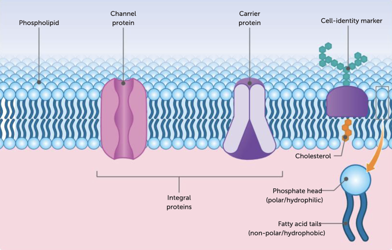
Describe the structure of the cell membrane (fluid mosaic model)
The membrane is said to be fluid because the molecules of which it is made are constantly changing positions, it is said to be mosaic because it is composed of many different kinds of molecules. The main structure of the membrane is composed of phospholipid molecules, which are lipid molecules containing a phosphate group. The phospholipids are arranged in two layers, known as a bilayer. Each phospholipid molecule has a head that is hydrophilic (water loving), and a tail is hydrophobic (water hating). The phospholipids are arranged in the two layers with their heads on the outside and the tails on the inside. Embedded in the phospholipid bilayer are cholesterol and protein molecules. They are important for the function integrity and stability of the membrane. Cell membranes have a variety of protein molecules, including receptor proteins, channel proteins, carrier proteins and cell identity markers. Carrier proteins bind to specific molecules and change shape to move them across the membrane. They can facilitate both passive (no energy) and active transport (required ATP energy), often moving larger molecules like glucose. Channel proteins create open pores in the membrane, allowing ions or small molecules to pass through. They typically facilitate passive transport (No ATP required) and are highly selective for specific ions or molecules. Aquaporins are special proteins that help water and small neutral molecules pass through cell membranes without using energy.
What are the transport methods of the cell membrane?
Simple diffusion
Osmosis
Facilitated diffusion
Active transport
Endocytosis
Exocytosis
SImple diffusion
Molecules can diffuse only if the membrane is permeable to them. They go from an area of high concentration to a low concentrated area (concentration gradient). It is passive transport (no ATP energy required). Examples of substances transported are water, oxygen, alcohol, fatty acids, steroids, ions such as sodium, potassium, calcium, lipids and soluble drugs.
Osmosis
Passive Transport (NO ATP ENERGY REQUIRED). The diffusion of water molecules through a differentially permeable membrane from an area of higher water concentration to an area of lower water concentration. Transports: Water
Facilitated diffusion
Passive Transport (NO ATP ENERGY REQUIRED). The process whereby proteins allow the movement of substances through the cell membrane along the concentration gradient (from high conc. area to low conc. area). Transports: Glucose, amino acids
Active transport
Active Transport (REQUIRES ATP ENERGY). The use of energy to move substances, usually ions, across a cell membrane against the concentration gradient (from low conc. area to high conc. area). Transports: Certain ions, glucose and amino acids
Vesicular transport (endocytosis)
Active Transport (REQUIRES ATP ENERGY). The process by which a cell takes in materials by enfolding and enclosing them; includes phagocytosis (taking solids into the cell) and pinocytosis (when the vesicles take liquids into the particles). Transports: Cholesterol, iron ions
Vesicular transport (exocytosis)
Active Transport (REQUIRES ATP ENERGY). The process whereby the contents of the vesicles of cells are pushed out through the cell membrane. Transports: secretions
Types of transport table
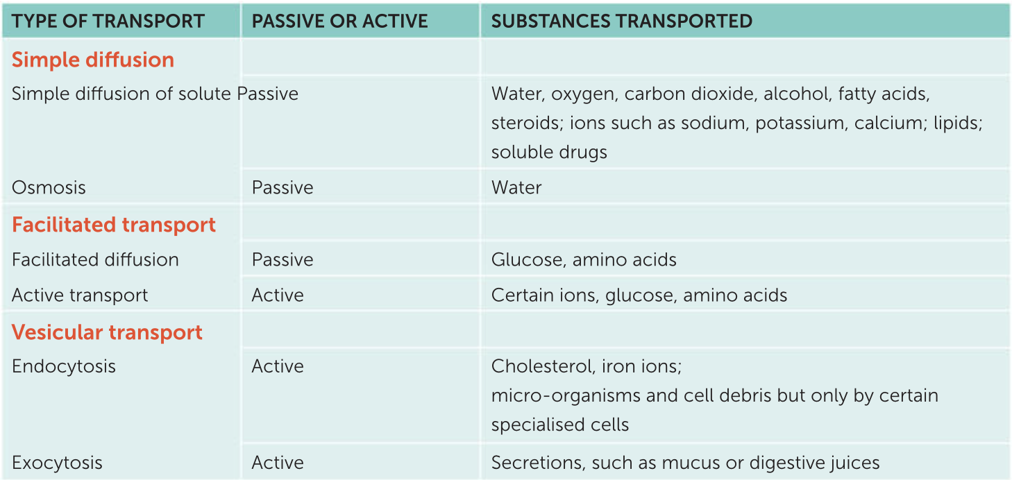
Describe the difference between passive and active transport
Passive transport does NOT require ATP energy to move molecules across the cell membrane as it follows the concentration gradient(from high to low conc.) while active transport DOES require ATP energy to move these molecules as it goes against the concentration gradient(from low to high conc.)
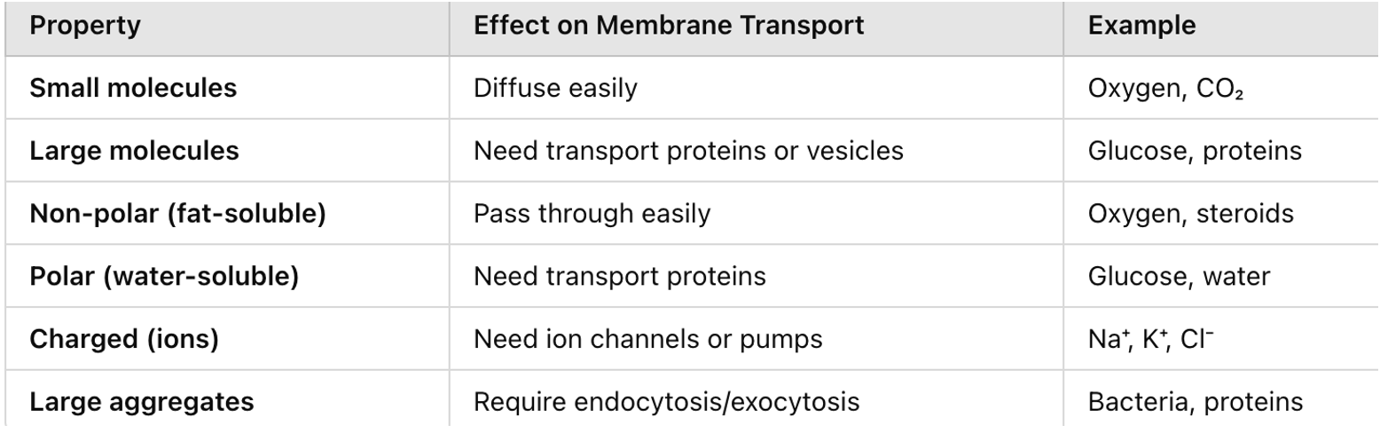
Explain how surface area to volume ratio affects the exchange of materials across the cell membrane for large and small bodies.
Small cells exchange materials more efficiently because they have a high surface area compared to their volume. This allows faster movement of nutrients and waste across the cell membrane. In contrast, large cells have a lower surface area-to-volume ratio, making it harder to exchange materials quickly, which can slow down important processes.
Explain how surface area to volume ratio affects the exchange of materials across the cell membrane for different shapes.
Round shapes, like spheres, have less surface area compared to their volume, limiting the exchange of materials. However, flat or long shapes provide more surface area, allowing faster and more efficient movement of substances across the cell membrane.
Explain how concentration gradients affects the exchange of materials across a cell membrane.
A concentration gradient is the difference in the concentration of a substance between two areas such as inside and outside of the cell. A concentration gradient occurs when there is a difference in the concentration of a substance between two areas such as inside and outside of the cell. Materials naturally move from an area of high concentration to low concentration through a process called diffusion e.g. simple diffusion, facilitated diffusion and osmosis. If the concentration gradient is from low to high its going against the natural gradient meaning it using ATP energy e.g. active transport, endocytosis and exocytosis.This movement continues until concentrations are equal or has reached equilibrium. A steeper concentration gradient allows materials to move quickly while a weaker gradient slows down the process.
Explain how the size and form of particles can affect how materials are exchanged across the cell membrane.
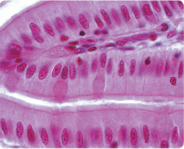
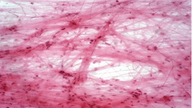
Describe the structure and function of epithelial tissue
It is a covering or lining tissue. The outer layer of the skin is epithelial tissue. Organs including the heart, kidney, intestines, liver and lungs are epithelium. It is also lines the inside of organs, so the inner layer of the heart, stomach, intestines and other hollow organs is made up of epithelium. The cells that make up epithelium are very closely joined together. They vary in shape from thin and flat to column-shaped and cube-shaped, depending on the particular tissue.
Describe the structure and function of connective tissue
The connective tissue provides support for the body and helps to hold all the body parts together. The cells in the tissue are not close together. They are seperate from each other by large amounts of material that is not made of cells. This non cellular material is called the matrix. Connective tissue include bone, cartilage, tendon, ligaments and fat storage tissues. Blood is often classified as a connective tissue. The matrix in blood is the liquid in which blood cells are suspended
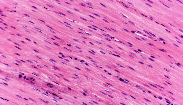
Describe the structure and function of muscular tissue
The cells of muscle cells, called muscle fibres are long and thin and can contract to become shorter. There are three different types of muscular tissue which are skeletal, smooth and cardiac muscle.
Skeletal muscle makes up the muscle that are attached to the bones. We have voluntary control over these muscles so that we can move parts of our bodies when necessary. Thus, they are sometimes called voluntary muscles. Under a microscope, skeletal muscles fibres seem to have stripes or striations, across them so another name of this is striated muscle.
Smooth muscle doesn’t have any striations, and therefore is called non-striated muscle. It is found in the stomach and intestines, in walls of blood vessels, in the iris of the eye, in the uterus and many other organs. We cannot smooth muscles voluntarily so it is also called involuntary muscle.
Cardiac muscle makes up most of the heart. When heart muscles contracts, it pumps blood. Heart muscles cannot be voluntarily contolled.
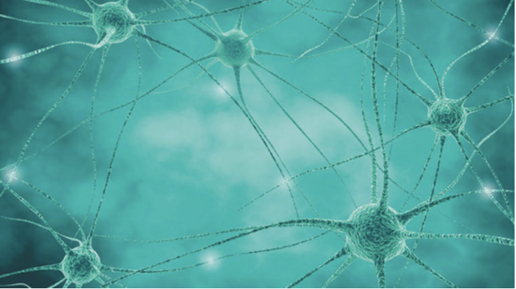
Describe the structure and function of nervous tissue
Nervous tissue is made up of specialised nerve cells called neurons. Neurons have long projections from the body of the cell. When part of a neuron is stimulated, messages can be carried along these projections from one part of the body to another. Nervous tissues are found in the brain, spinal cord and the nerves.
Metabolism
Define metabolism
All the reactions that take place in cells, and therefore in the organism of which the cells are a part.
Differentiate between anabolic and catabolic reactions
Anabolic reactions are when small molecules build up into larger ones. It is where energy is released.
Catabolic reactions are when large molecules are broken into smaller ones. It is where energy is used up.
Provide examples of common anabolic and catabolic reactions
An example of an anabolic reaction is protein synthesis.
An example of catabolic reaction is digestion.
Recall the structure of organic and inorganic compounds and give examples of each
Organic compounds are molecules that have a carbon chain. They also contain a number of hydrogen atoms that may include atoms of oxygen, nitrogen and sulfur. Examples of them are carbohydrates, lipids, proteins and nucleic acids.
Inorganic compounds are not based on a carbon chain. Most don’t contain carbon atoms at all, but those that do, such as carbon dioxide, are small molecules. Examples of them are water, minerals and vitamins.
Describe carbohydrates
Carbohydrates are the main source of energy for cells. They are also used for cellular respiration. Simple sugars, particularly glucose are used in cellular respiration to release energy. Complex carbohydrates, such as starch are broken down into simple sugars. Carbohydrates contain atoms of carbon, hydrogen and oxygen; there are twice as many hydrogen atoms as oxygen atoms. Simple sugars are called monosaccharides. Glucose, fructose and galactose are examples of monosaccharides. Simple sugars are able to join together to form larger molecules. Disaccharides, such as sucrose, maltose and lactose are formed when two simple sugars are joined together. Polysaccharides are larger carbohydrate molecules formed when many simple sugars are joined together. Glycogen, cellulose and starch are examples of it.
Describe lipids
Lipids include fats and oils and are important for building enzymes/proteins and for transport. They are broken down to fatty acids and glycerol. The glycerol can then enter the glycolysis pathway of cellular respiration and is broken down to release energy in similar ways as glucose. Other examples of lipids are phospholipids, which are important in the cell membrane, steroids, including cholesterol and the sex hormones. Each lipid consist of one molecule of glycerol and one, two or three fatty acid molecules. The most common fat which is stored in the body is triglyceride, which is composed of glycerol and three fatty acid molecules.
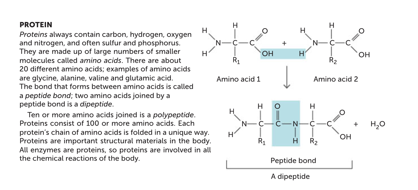
Describe proteins
Proteins are organic compounds that are made up of many amino acids. They are used to maintain the structure of the cell and cell signalling. It is also another source of energy. With regards to metabolism, the most important proteins made are enzymes. Enzymes influence metabolism by controlling the chemical reactions that occur in the body. An amino acid is a molecule that contains both an amino group and a carboxylic acid group. When two amino acids bond together, they form a peptide bond, releasing water. There are 20 different amino acids found in proteins, each differing in the structure of the side chain. Proteins consist of 100 or more amino acids. Each protein has a characteristic shape due to the folding of the chain. Shorter length amino acids include dipeptides, with two amino acids joined and polypeptides, made up of more than 10 amino acids.
Describe nucleic acids
Other organic compounds include nucleic acids such as ribonucleic acid (RNA) and deoxyribonucleic acid (DNA). DNA consists of two chains of nucleotides that contain the sugar deoxyribose. It is generic material in the nucleus that stores inherited information. RNA is made up of single strands of nucleotides that contain the sugar ribose. These molecules carry information from DNA in the nucleus to the ribosomes for protein production.
Describe water
Water is important in metabolism because it is the fluid in which other substances are dissolved. Some of the cell’s chemical reactions occur in water, and in others water molecules actually take part in the reaction.
Describe minerals
Minerals are important for metabolism because they may be part of enzymes, may function as cofactors for enzymes, or may be part of substances such as adenosine triphosphate (ATP) that are involved in metabolism.
Describe vitamins
Vitamins act as coenzymes for many of the chemical reactions for metabolism.
Cellular respiration
It is the process by which organic molecules, taken in as food, are broken down in the cells to release energy for teh cells activities.
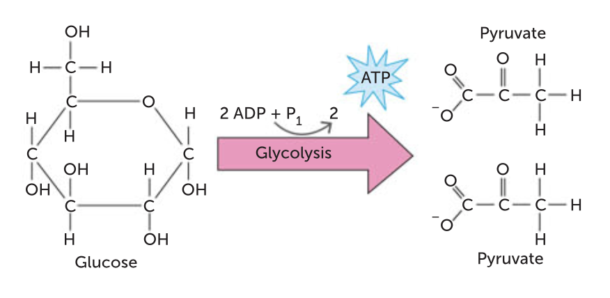
Glycolysis
The first phase in the breakdown of glucose does not require oxygen. A glucose molecule is broken down, in a series of 10 steps, to two molecules of pyruvate.
Describe the process of Aerobic respiration
The complete breakdown of glucose to carbon dioxide and water requires oxygen. The pyruvate produced from glycolysis is completely broken down to carbon dioxide and water. Aerobic respiration occurs in the cytosol (the initial breakdown of glucose) and mitochondria (Krebs cycle and electron transport chain) of the cell. To complete the breakdown of glucose, the two pyruvate molecules produced in glycolysis must enter the mitochondrion, where enzymes are available to allow the next series of reactions to occur. For the pyruvate to enter the next pathway it is first converted to acetyl coenzyme A (acetyl CoA). To do this, a carbon dioxide molecule is removed from the pyruvate and the remaining two- carbon structure joins to CoA. No ATP is produced. The acetyl CoA then enters the krebs cycle. Here carbon atoms in the acetyl CoA are released in carbon dioxide. For every acetyl CoA that enters the krebs cycle, one molecule of ATP is produced. This means that two ATP molecules are produced. The final stage of cellular respiration is the electron transport system, the only stage that uses oxygen. Here electrons are passed between molecules, finally resulting in oxygen molecules forming water. Around 26-34 ATP molecules is produced from this. Aerobic respiration can produce up to 38 ATP molecules.
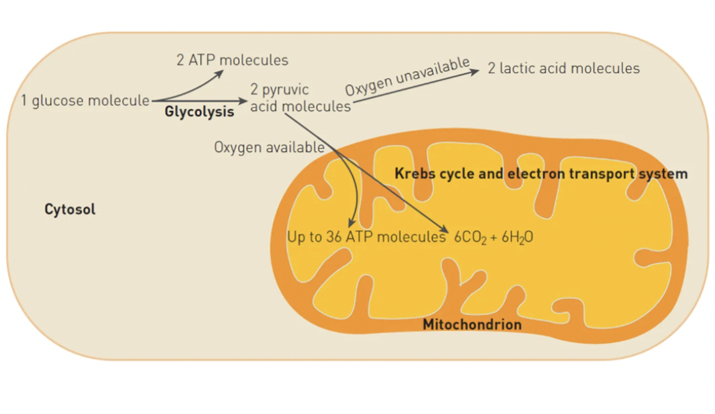
Describe the process of Anaerobic respiration
If no oxygen is available, the pyruvate produced in glycolysis is converted to lactic acid by fermentation. The fermentation stage of anaerobic respiration doesn’t produce any additional ATP, however, the glycolysis of one molecule of glucose releases enough energy to convert two molecules of ADP to ATP. Therefore anaerobic respiration allows cells to produce some energy in the absence of oxygen. The enzymes required for anaerobic respiration are available in the cytosol of the cell. Therefore anaerobic respiration occurs in the cytosol. Lactic acid from anaerobic respiration is taken by the blood to the liver where it can be recombined with oxygen to form glucose and eventually glycogen. Anaerobic respiration is important in during vigorous physical activity.
What do cells require for metabolism to be efficient?
Cells require oxygen, carbohydrates/simple sugars, proteins/amino acids, lipids/fatty acids & glycerol, vitamins and minerals for metabolism to be efficient.
Describe enzyme function
Enzymes: an inorganic substance (usually a protein) that increases the speed of chemical changes without being altered or destroyed in the change
Substrate: a molecule upon which an enzyme acts
Active site: the part of an enzyme molecule that combines with the substrate
Co-enzyme: non-protein organic molecules that are essential for the functioning of an enzyme
Enzyme inhibitors: substances that slow down or stops an enzyme’s activity
Catalysts: A substance that lowers the activation energy of a reaction, increasing the rate of the reaction without being consumed
Cofactors: The ions or inorganic molecules required by enzymes to catalyse a reaction
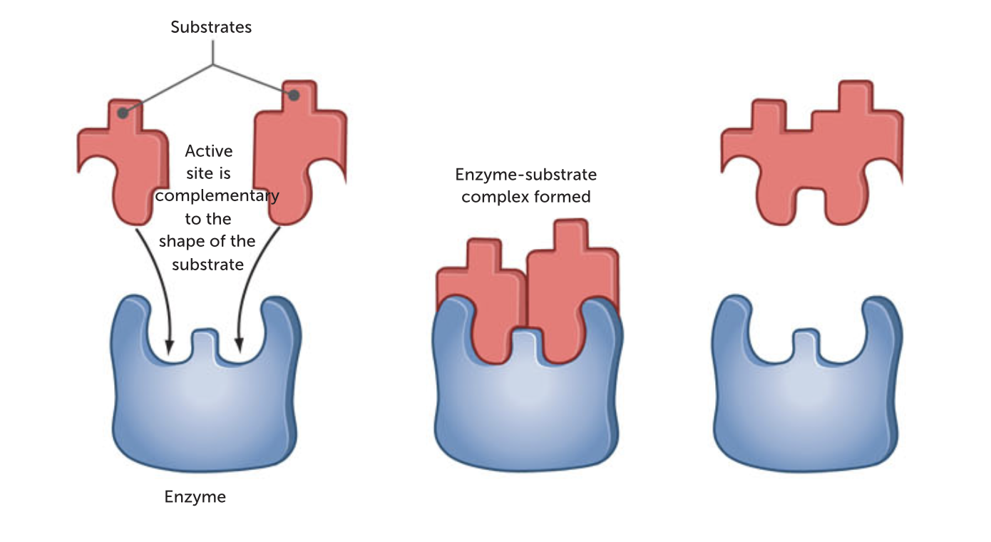
Lock and key model of enzymes
The lock and key model states that the shape of the enzyme (the key) is always complementary to the shape of the substrate (the lock). Therefore, the two will fit exactly to form the enzyme substrate complex.
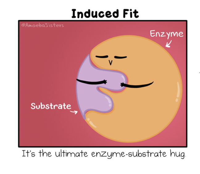
Induced fit model of enzymes
The induced fit model states that when the enzyme and substrate join, they form weak bonds that cause the shape of the enzyme to change, creating complementary shapes.
Discuss the importance of activation energy
Activation energy is the minimum amount of energy required to break the bonds of the reacting particles in a chemical reaction; the energy needed to start a chemical reaction. It plays a vital role in regulating metabolic reactions, ensuring the reactions occur at the right time and under controlled conditions
Explain how the concentration of enzymes can affect enzyme functioning
The higher the concentration of enzymes, the faster the rate of a chemical reaction because there are more enzyme molecules to influence reactants. By regulating the type and number of enzymes present, the body is able to control which reactions occur and the rate at which they are produce.
Explain how Ph can affect enzyme functioning
Enzymes are very sensitive to the pH of the medium in which a reaction is taking place. Each enzyme has an optimum pH at which it will work most effectively
Explain how temperature can affect enzyme functioning
At low temperatures, enzyme activity is slow because molecules move less, leading to fewer enzyme-substrate collisions. As temperature increases, enzyme activity rises until it reaches an optimal temperature (around 30 – 40°C in humans), where the reaction rate is highest. However, if the temperature exceeds this optimal range, the enzyme's structure begins to break down, causing it to denature and lose its function. Denaturation is irreversible and results in the reaction stopping
Explain how Inhibitors, co-factors and co-enzymes can affect enzyme functioning
Explain how the concentration of reactants and products can affect enzyme functioning
They usually speed up the chemical reactions because the reaction can occur quicker, however when the number of reactants exceeds the number of enzymes then it becomes saturated and the reaction rate doesn’t speed up any more.
Respiratory System
Describe the structure and function of the nose
Structure: The lining of the nose and nasal cavity is convoluted and lined by mucous membranes.
Function: It allows air to enter the body.
Describe the structure and function of the pharynx
Structure: A muscular tube from the nasal cavity to the top of the trachea and oesophagus.
Function: Air travels through it.
Describe the structure and function of the larynx
Structure: A cartilage structure joining the pharynx and trachea.
Function: Allows people to make sounds.
Describe the structure and function of the trachea
Structure: It is made up of c shaped cartilage rings that hold the structure open.
Function: It carries the air into and out of the lungs.
Describe the structure and function of the bronchi
Structure: It has c shaped cartilage rings.
Function: It carries air to and from the lung.
Describe the structure and function of the bronchioles
Structure: Very fine tubes with walls of smooth muscle.
Function: Carries air into the alveoli. It can control the flow of air into the lungs.
Describe the structure and function of the alveoli
Structure: They are tiny air sacs that have very thin walls.
Function: It allows a net flow of oxygen to pass from the airways into the blood and carbon dioxide to pass from the blood into the airways.
Describe the structure and function of the lungs
Structure: A spongy air filled organs that take up the whole chest cavity.
Function: To supply oxygen to the body.
Describe the pathway of air into and out of the lungs
Air travels into the nose and mouth, down the pharynx and larynx, through the trachea, into the bronchi, bronchiole and alveoli, where gas exchange occurs and air moves out the opposite way.
What is the function of cartilage in the trachea?
The c shaped cartilage rings in the trachea holds the structure open. This ensures that air can always pass through it.
What is the function of the ciliated epithelium in the trachea
The epithelial lining of the trachea produces mucus.
What is the function of the mucous membranes in nose and trachea.
The mucus traps dust and debris which prevents it from entering the lungs.
Inspiration
Inspiration is the process of taking air into the lungs. For air to flow into the lungs the pressure of air must be less than the atmospheric pressure outside the body. By increasing lung volume the air pressure in the lungs decreases. To increase the volume of the lungs, the diaphragm and intercostal muscles contract. The diaphragm becomes flatter and the rib cage moves upwards and outwards. The pleura attached to the wall of the chest draws lungs to expand. Air flows into the lungs until pressure becomes equal.
Expiration
Expiration is breathing out. The diaphragm and intercostal muscle relax, so the diaphragm bulges more into the chest cavity and the ribcage moves downwards. This reduces the volume of the chest cavity Air pressure in the lungs is now greater than the pressure outside the body. Air flows out through the trachea and nose until air pressure is equal.
Describe the features of a large surface area to volume ratio in the respiratory system
The alveoli give the lungs a huge internal surface area, so that large amounts of gases can be exchanged in a relatively short time.
Describe the features of the thin alveoli membranes
The membrane that forms the wall of the alveolus is very thin, so that gas molecules do not have far to travel when moving into or out of the blood.
Describe the features of the moist fluid lining in the alveoli
It is important that the membrane of the alveolus be covered by a thin layer of moisture because gases can diffuse into and out of the blood only when they are dissolved in fluid.
Describe the features of the extensive network of capillaries
Each alveolus is well supplied with blood vessels (capillaries), so that as much blood as possible is close to the air in the alveolus. The continuous flow of blood helps to maintain a difference in concentrations of oxygen and carbon dioxide between the blood and the air in the lungs.
Explain the process of gas exchange including the rate and direction of the exchange
The blood in the capillaries surrounding the alveoli is brought to the lungs by the pulmonary arteries. This blood has been through the capillaries of the body, where much of the oxygen has been taken up by the body cells. Therefore, the blood that comes into the capillaries around the alveoli has a low concentration of oxygen. Oxygen dissolves in the moisture on the inside of the alveolus and diffuses through the membrane, the walls of the capillaries and into the blood. The blood arriving to the capillaries of the alveoli has come from the body circulation where it has picked up carbon dioxide produced by respiration in the cells. The concentration of carbon dioxide in the alveolar capillaries is higher than the concentration in the air in the alveolus. Therefore, carbon dioxide diffuses out of the blood and into the air in the alveolus.
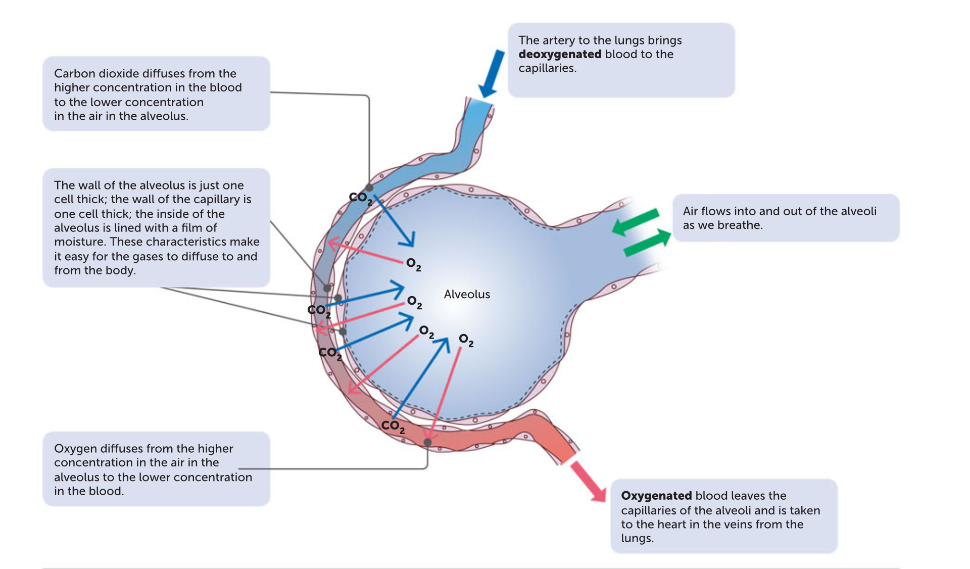
Concentration gradient for diffusion in the alveoli
For diffusion of gases into and out of the blood, there must be a concentration gradient – that is, a difference in gas concentration between theair in the alveoli and the blood in the capillaries. The concentration gradient for oxygen and caarbon dioxide is maintained by:
the constant flow of blood through the capillaries. As the blood flowing through the capillaries around each alveolus picks up oxygen and loses carbon dioxide, it is replaced by more blood being pumped into the capillaries. This ‘new’ blood is low in oxygen and high in carbon dioxide, so the concentration gradient is maintained
the movement of air into and out of the alveoli as we breathe in and out. The air that has picked up carbon dioxide from, and lost oxygen to, the blood is replaced by ‘new’ air with each breath.The ‘new’ air is low in carbon dioxide and high in oxygen.
Emphysema
It is a disease usually caused by long term exposure to irritating particles in the air taken to the lungs. The irritating particles cause damage to the alveolus. They lose their elasticity, are often replaced with fibrous tissue, and may break down, reducing the internal surface area of the lung. Because of the loss of elasticity in the lung tissue, the lungs become more inflated, and breathing no longer occurs passively but requires voluntary effort. This is caused by smoking, dust inhalation and highly polluted cities.
Lung cancer
Lung cancer is similar to most other cancers in that it involves the development of a mass of cells that divides in an uncontrolled way – that is, a tumour. It begins in the walls of the air passages, usually the bronchi. Inhaled smoke particles constantly irritate the mucous membrane that lines the air passages. This results in excessive production of mucus. Cells at the base of the membrane begin to divide more rapidly and the accumulating mucus cannot be removed. This results in ‘smoker’s cough’. The trapped mucus causes rupture of the alveoli. This is caused by smoking or exposure to asbestos fibres and other pollutants.
Lung infections
Pneumonia is an infection of the lungs caused by bacteria, viruses, fungi or other organisms. The inflammation resulting from the infection causes secretion of fluid and mucus into the alveoli, thus reducing the amount of air that they can contain. The surface area available for exchange of gases is also reduced, and breathing difficulty is a symptom of many types of pneumonia.
Tuberculosis is an infection, usually of the lung, by the bacterium mycobacterium tuberculosis.
Athsma
Asthma is a medical condition that causes difficulty breathing due to a narrowing of the airways. This occurs due to:
the smooth muscles contracting, narrowing the airway
inflammation causing the lining of the airway to thicken, narrowing its diameter.
mucus filling the airway, narrowing the tube.
Circulatory System
Describe the structure and function of the heart
Structure: The heart is a conical shape approximately 12cm long, 9cm at its widest part and 6 cm thick, making it about the size of a human fist.
Function: It is the pump that pushes the blood around the body.
Describe the structure and function of veins
Structure:
Function: Veins carry blood towards the heart.
Describe the structure and function of arteries
Structure:
Function: Arteries are the blood vessels that carry blood away from the heart.
Describe the structure and function of capillaries
Structure:
Function: Capillaries are the link between the arteries and veins.
Describe how the exchange of materials occurs at capillaries and throughout circulation (systemic and pulmonary) through the body
Systemic circulation is the part of the circulatory system that carries oxygen rich blood from the heart to the rest of the body, and then returns oxygen poor blood back to the heart.
Pulmonary circulation is the part of the circulatory system that carries deoxygenated blood from the heart to the lungs for oxygenation, where it picks up oxygen and releases carbon dioxide, and then returns to the heart.
Describe the main functions of blood
transporting oxygen and nutrients to all cells of the body
transporting carbon dioxide and other waste products away from the cells
transporting chemical messengers, called hormones, to the cells
maintaining the pH of body fluids
distributing heat and maintaining body temperature
maintaining water content and ion concentration of the body fluids
protecting against disease-causing micro-organisms
clotting when vessels are damaged, thus preventing blood loss.
Describe the structure and function of erythrocytes (red blood cells)
Structure: The cells are a biconcave shape.
Function: To transport oxygen from the lungs to the cells throughout the body.
Describe the structure and function of leukocytes (white blood cells)
Structure: It is larger than red blood cells.
Function: Protect the body from infection.
Describe the structure and function of platelets
Structure: They are small fragments of cells with no nucleus.
Function: Blood clotting
Describe the structure and function of plasma
Structure: A liquid that contains water and dissolved substances such as sugar and salts.
Function: To transport the components of blood, including the cells, nutrients, waste, hormones, proteins and antibodies, throughout the body.