Lecture 2.12 - SNB II; Lobes, AMPA/NDMDA, Long term potentiation.,Thalamus
1/22
There's no tags or description
Looks like no tags are added yet.
Name | Mastery | Learn | Test | Matching | Spaced | Call with Kai |
|---|
No analytics yet
Send a link to your students to track their progress
23 Terms
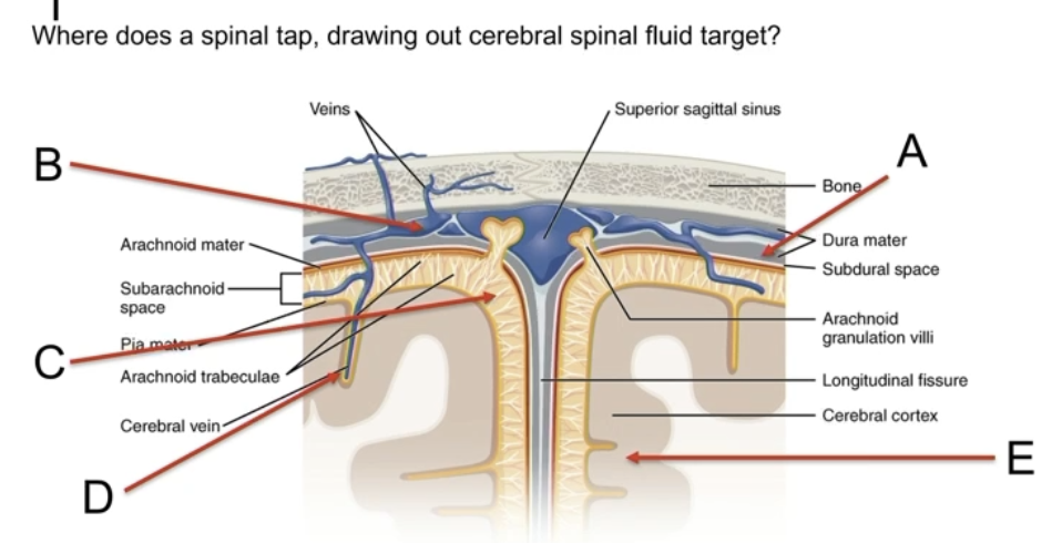
A = No fluid should be in A
B = Blood, not CSF, since veins
C = CORRECT
Includes granulosa
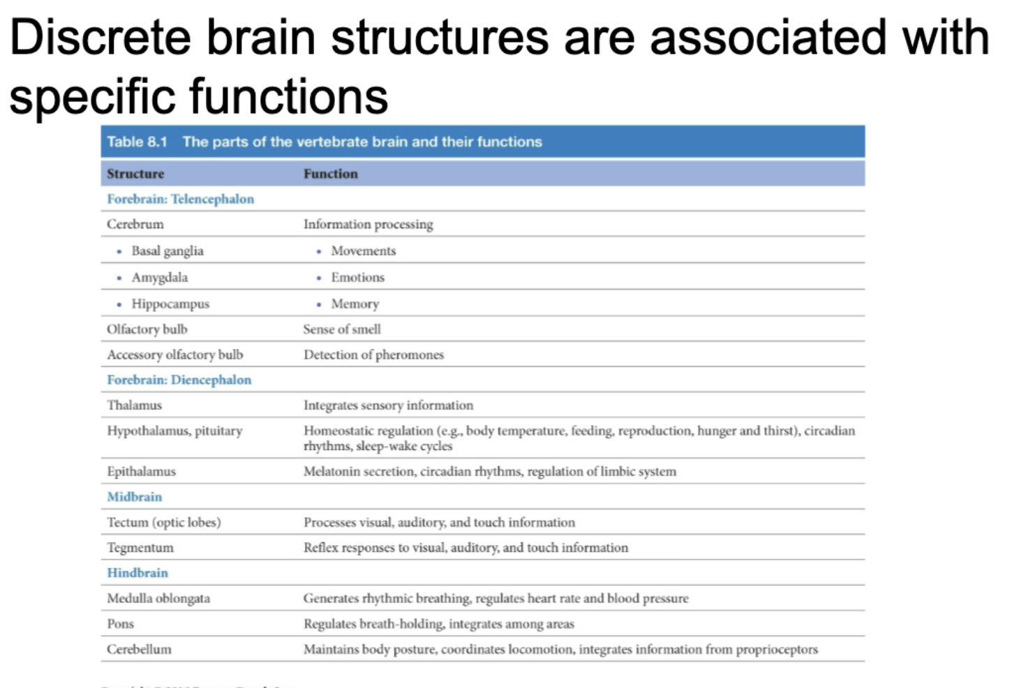
3 Fundamnetal dividison of the vertebrate brain
Hindbrain
Reflexes, non conscious movements (heartbeat)
Midbrain
Coordination
Forebrain
Cerebrum
Decision making, senses
Hypothalamus, thalamus, diencephalon
Brain development starts early
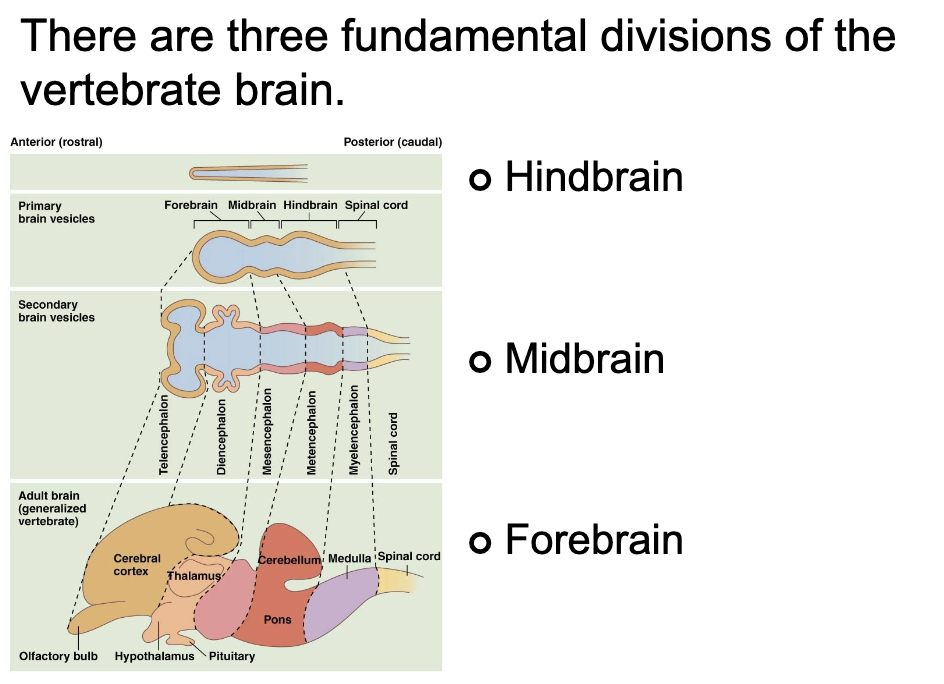
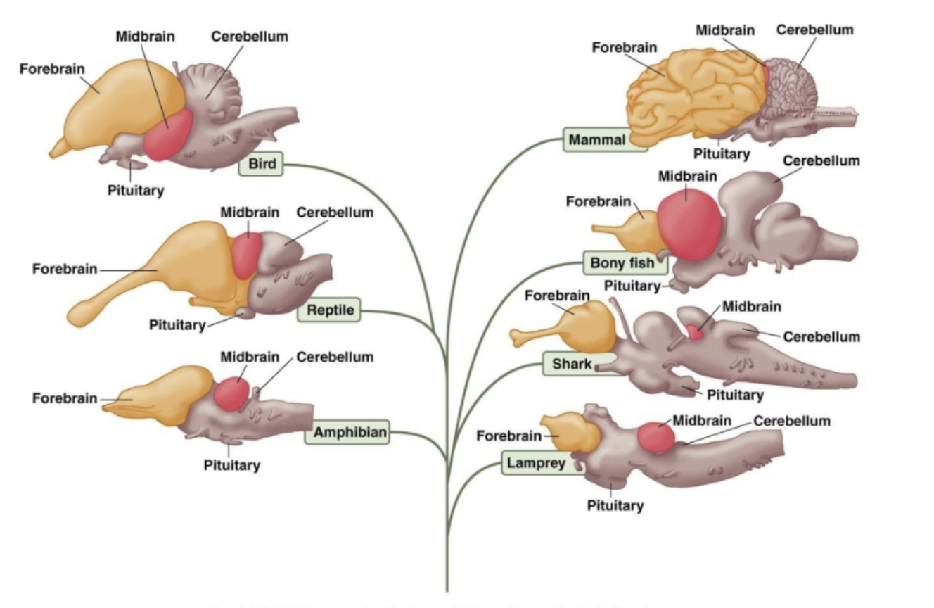
Same major brain structures found in most vertebrates
Same structures, but could have different shapes, sizes, etc
Brain chart - Parts and Functions summary slide
Different parts of the brain have specific functions
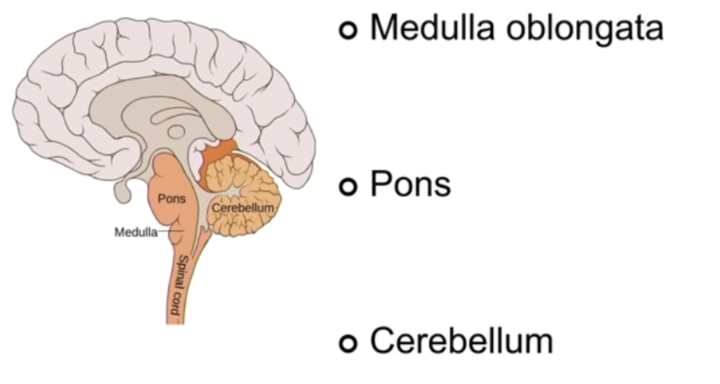
Hindbrain
Bottom of Brain + parts of Brainstem
Supports basic functions
Contains:
Medulla oblongata
Breathing, heart rate, blood pressure
Autonomic homeostasis
Pons
Communicator between medulla, cererellum, and cerebrum
Sleepy cycles
Cerebellum
Muscle movement coordination
Damage - loss of balance, coordination, language control, attention, sometimes emotion
Forebrain
Basically the Brain, which is divided into lobes
Frontal lobe
Anything in front of Central Sulcus
Voluntary control of movement
Thinking, memory, reasoning, self control
Parietal lobe
Directly behind frontal lobe / central sulcus
Knowing where you are in space (proprioception), receiving and understanding senses
Occipital lobe
At the very back
Vision
Temporal lobe
Both sides
Language, hearing, memory encoding
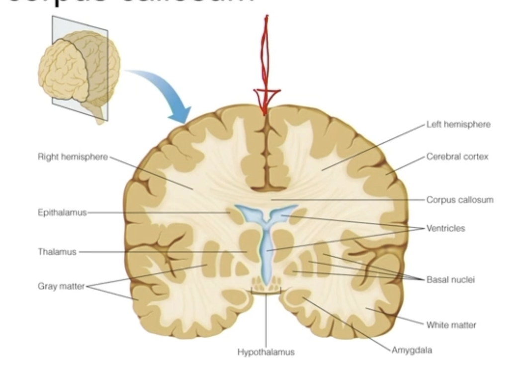
Cerebrum cross section
Split in half - 2 hemispheres, right and left
White matter in middle = Corpus Callosum
Contralateral senses - lots of crossover at corpus callosum
Functional Map of Brain
Lesioning - touch/destroy/electrical impulse/brain surgery - parts of brain
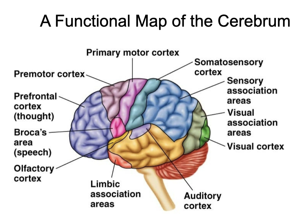
Motor areas of cerebrum - Primary motor cortex
Primary motor cortex
Controls voluntary movements of skeletal muscle
Stimulation of different regions of PMC leads to movement
Damage to PMC leads to paralysis or loss of voluntary movements but reflex remain
Damage to PMC usually results in permanent loss of these movements
In Frontal Lobe
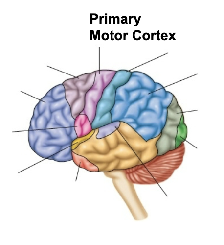
Motor areas of cerebrum - Premotor Cortex
Coordinates movements of groups muscles
PMC causes movements, PC controls them
Damage
Loss of skill
Can be relearned
In Frontal Lobe
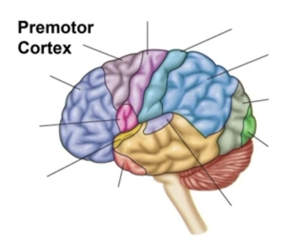
Sensory areas of cerebrum - Somatosensory cortex
Behind central sulcus
Primary input from sensory receptors in skin and muscle
Touch information from skin and muscles
Pain, temperature, vibration, proprioception
Not eyes, ears, taste
Damage leads to loss of sensation
In Parietal Lobe
Sensory areas of cerebrum - Somatosensory association area
Adjacent to primary somatosensory cortex
Interpretation of sensations
Integrating sensations with memory
Damage:
Loss of ID of sensations
In Parietal Lobe
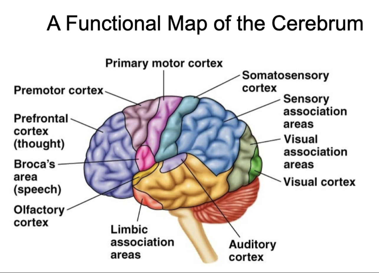
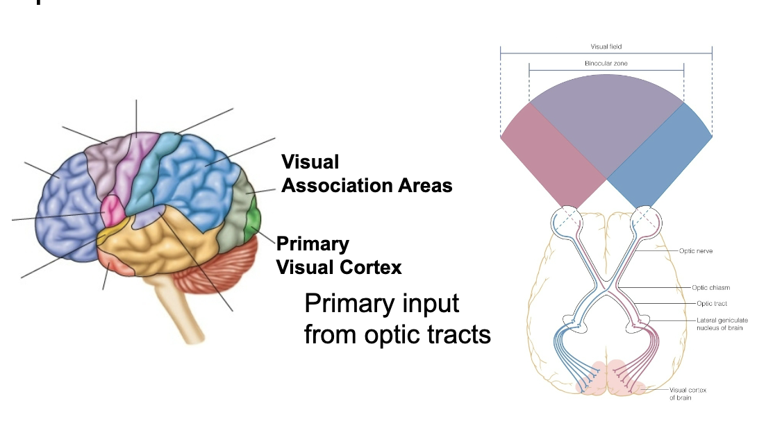
Visual areas of the cerebrum - Occipital lobe
Primary visual cortex
Receives input
Primary input from optic tracts
Visual association area
Makes sense of what is seen
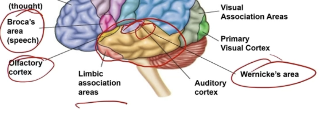
Temporal lobe parts
Wernicke’s area - speech comprehension
Broca’s area - speech production
Olfactory cortex - sense of smell
Auditory cortex
Limbic association areas - emotions, memory, motivation
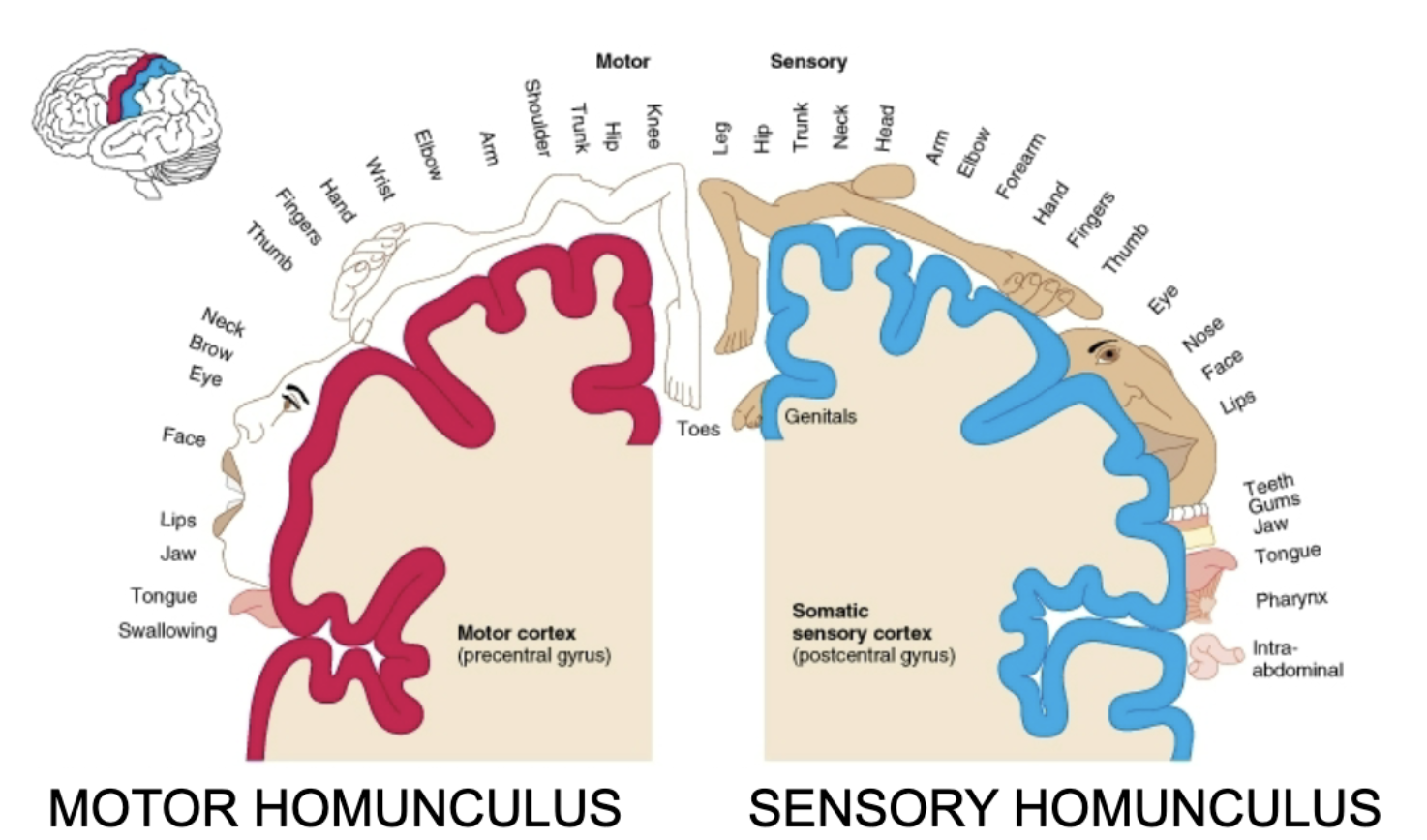
Motor and Sensory Homunculus - on both sides of brain
Distribution of amount of brain dedicated to certain movements/senses
Vary based on importance - more area on brain = more important
Ex: lots dedicated to face and fingers, little to arms and hips
Motor homunculus
Sensory homunculus
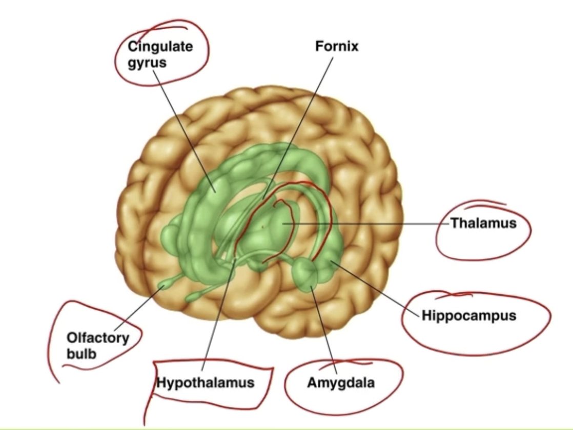
Limbic system (lizard brain)
Inside the brain, temporal lobe
Cingulate gyrus
Regulating emotions and pain
Fornix
Connector between portions of limbic system
Might have relation to episodic/short-term memory
Thalamus
Relay station - area that connects sensory organs to somatosensory organs
Receives senses, filters it, sends it out to area in brain for interpretation
All motor and sensory signals (except smell) pass through this structure
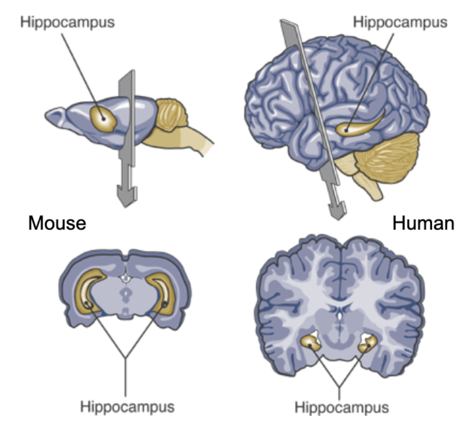
Hippocampus
Memory
Amygdala
Fear, aggression, emotions
Hypothalamus
Hormones
Olfactory bulb
Sense of smells
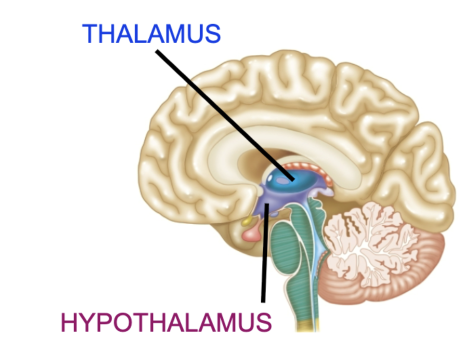
Diecephalon structures - Hypo/thalamus
Thalamus
Filters/sorts inputs, routes information
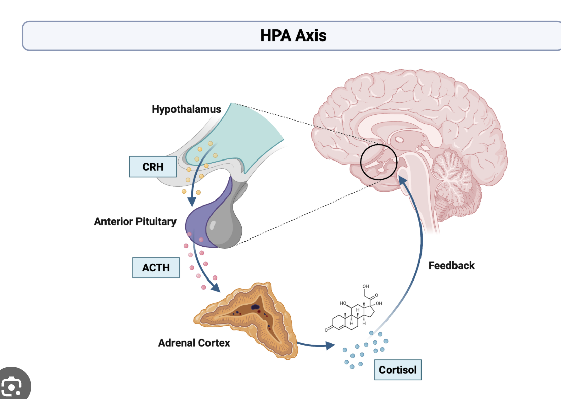
Hypothalamus
Homeostatic functions (negative feedback)
Regulates Pituitary gland
Hypothalamus → Pituitary → Other glands → Body
Hypothalamus receives input - body states (temperature, blood glucose, …) or brain states (amygdala - fear, stress ; other emotional state…)
Hypothalamus sends output to pituitary gland
Based on particular input, pituitary gland releases hormones
Hippocampus - memory
Not very big in humans
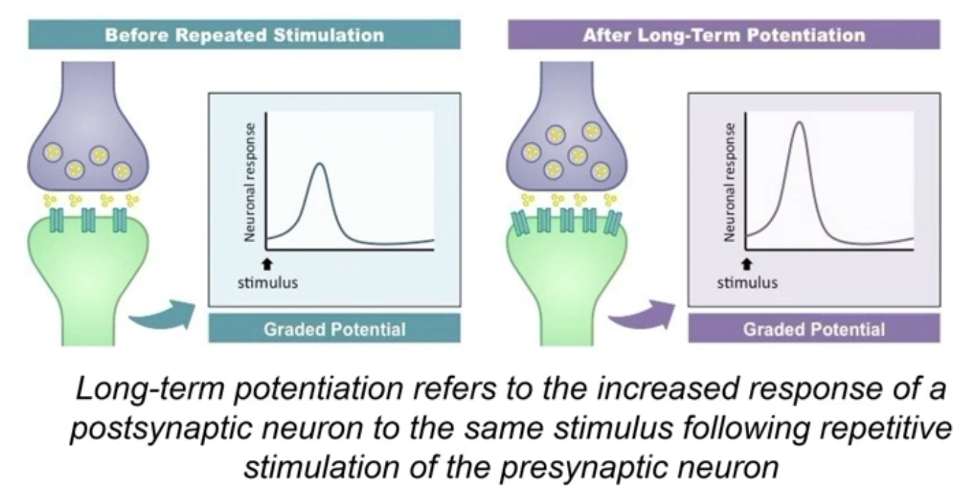
Long term potentiation - long term changes in memory = Synaptic plasticity
Causes long-lasting increases in signal transmission between neurons
Occurs at dendritic spine synapses of hippocampal neurons
Mechanism for tracking repetitive activity (salient stimuli)
Long-term potentiation
Hippocampal neuron
LTP = Repeated stimulus over and over
GRADED potentials, not action potentials
LTP vs Low-frequency stimulation
LTP = bigger graded potentials = more likely to have an action potential
More receptors
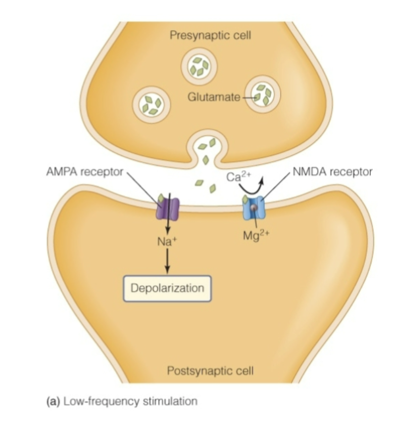
LOW FREQUENCY - Activity-dependent changes in dendritic spine synapses
Pre-synaptic cells NT = usually glutamate
NT for AMPA receptor and NMDA receptor
AMPA
Ligand-gated sodium channel (but not Na+ only)
Receives glutamate, opens, allows Na+ in, Depolarization
AP more likely to occur
NMDA
Nonselective Ligand-gated, Receives Glutamate
But Mg2+ clogs pores of receptor, doesn’t allow Ca2+ to pass through in LOW FREQUENCY
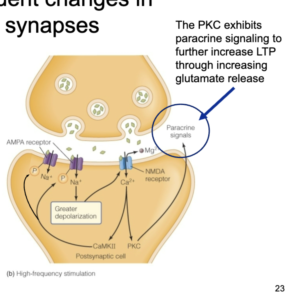
HIGH FREQUENCY - Activity-dependent changes in dendritic spine synapses
AMPA
Still receives ligand (glutamate), opens, allows Na+ in, greater Depolarization
Greater depolarization ejects Mg2+ from the NMDA receptor
NMDA
No more Mg2+ blocking
Ligand once again binds to receptor
No Mg2+ present this time, so Ca2+ can pass through
Ca2+ affects CAM-MK2 → phosphorylates AMPA receptor, opening it more (more Na+ in, more depolarization)
CAM-MK2 → leads to more active AMPA receptors and more AMPA insertion
PKC exhibits paracrine signaling, causing pre-synaptic neuron to release more glutamate = more +
More Na+ enters which causes Ca2+ enters = more positive = POSITIVE FEEDBACK
AMPA phosphorylated = More + = more AMPA receptors = More + = POSITIVE FEEDBACK
Thought to be involved in memory production
Hypothalamus
Regulates body temp
Thyroid - metabolism
Regulates food intake
Involved in the stress response
HPA (hypothalamus, pituitary, adrenal) axis
Helps maintain ion and water balance
Regulates circadian rhythms
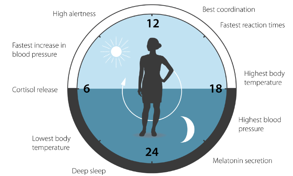
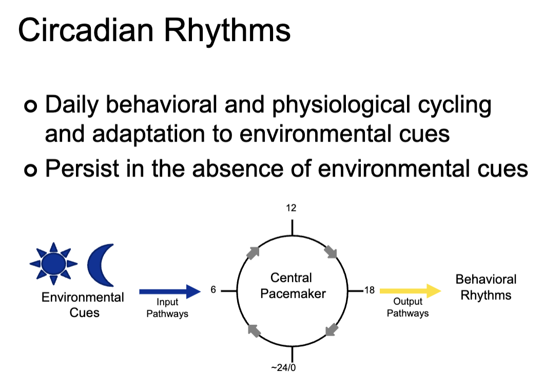
Circadian rhythms
Daily behavioral and physiological cycling and adaption to environmental cues
Environmental cue - usually Sun
Leads to input pathway
Behavioral rhythms
Persist in the absence of environmental cues
What are controlled by circadian rhythm?
Cortisol levels
Blood pressure
Body temperature
Reaction time
Blod pressure