Cell Cycle Apoptosis Mitosis and Meiosis
1/26
There's no tags or description
Looks like no tags are added yet.
Name | Mastery | Learn | Test | Matching | Spaced |
|---|
No study sessions yet.
27 Terms
List out the cell cycle and its main characteristics?

Describe the various checkpoints and characteristics for the cell cycle

What is interphase? Infra vide? What does Mitosis does not have in regards to the cell cycle?
interphase: G1, S, and G2
infra vide = mitosis’ subphases
mitosis DOES NOT HAVE CHECKS FOR DNA DAMAGE, ONLY CHECKS FOR MITOTIC SPINDLE
What is G0? What cells demonstrate this? What signals keep cells in G0? Describe these signals function
G0 = when cells are not currently undergoing cell cycle
G0 cells:
Differentiated cells
Senescent cells
Signals that can keep cells in G0:
TGF-β: Induces differentiation of cells
Contact inhibition: Cell-cell interactions through cadherin receptors
Telomere shortening: Cell senescence to avoid loss of genetic material
What happens in prophase
Chromosome condensation begins in prophase. Mitotic spindle begins to form.
What happens in prometaphase?
Nuclear membrane dissolves in prometaphase.
What happens in metaphase?
Chromosomes are most condensed in metaphase.
Mitotic spindle checkpoint.
What happens in anaphase?
Disjunction, or separation of sister chromatids at the
centromere
What happens in telophase?
Decondensing begins and nuclear membrane begin to reform in telophase.
Describe the function of cyclins? How are they regulated?
proteins that regulate the cell cycle by activating cyclin-dependent kinases (CDKs) which phosphorylate target proteins
Regulation: on the transcription level and via proteolytic degradation.
What does cyclin D do?
ctivates cyclin dependent kinases 4 and 6, and they phosphorylate Rb.
Describe the three main molecular components of the regulation of cell cycle
cyclins (inducible)
Cyclin Dependent Kinase (constitutive)
Cyclin dependent Kinase Inhibitors (inducible)
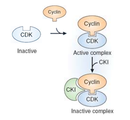
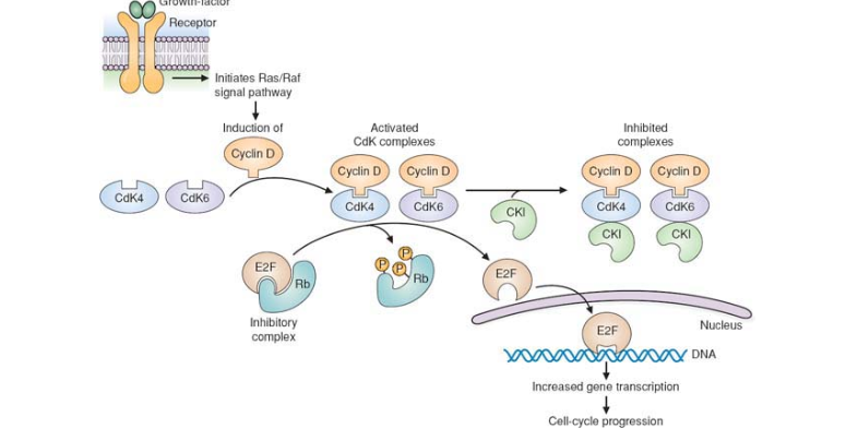
How does the number of Cyclin D increases? Explain the mechanism in which Cyclin D regulates the transiction from G1 to S phase;
Adequate cell growth (in G1) must precede DNA replication (in S)
cell growth = gradual increase in cyclin D
G1/S transition controlled by restriction point
only passed with adequte Cyclin D levels
The exact mechanism of Cyclin D:
Cyclin D activates cdk complexes (4/6) → CDK complesex phosphorylates Rb-E2F complex →dissociation of Rb (Retinoblastoma protein) from E2F (transcription factor) → E2F go inside nucleus and increases gene transcription → cell-cycld progression
What is p53? How is it activated? Explain its effect
p53 is a transcription factor (known as guardian of the genome)
Activated via DNA damage or phosporylation
How p53 protects the cell
initiates transcription of p21 which is a CKI and arrests the cell cycle
initiates the transcription of DNA repair enzymes (GADD45).
If the DNA damage is not repaired, apoptosis is initiated by IGF-BP3 (inhibits anti-apoptotic cell signals) and Bax (activates apoptosis) .
How is apoptosis utilized? What are the two major pathways? Compare and contrast the two pathways. What is the result of apoptosis?
Utlized for:
embryogenesis
maintenance of proper cell number
removal of injured cells
Can be initiated by two major pathways:
Death receptor pathway
Mitochondrial pathway
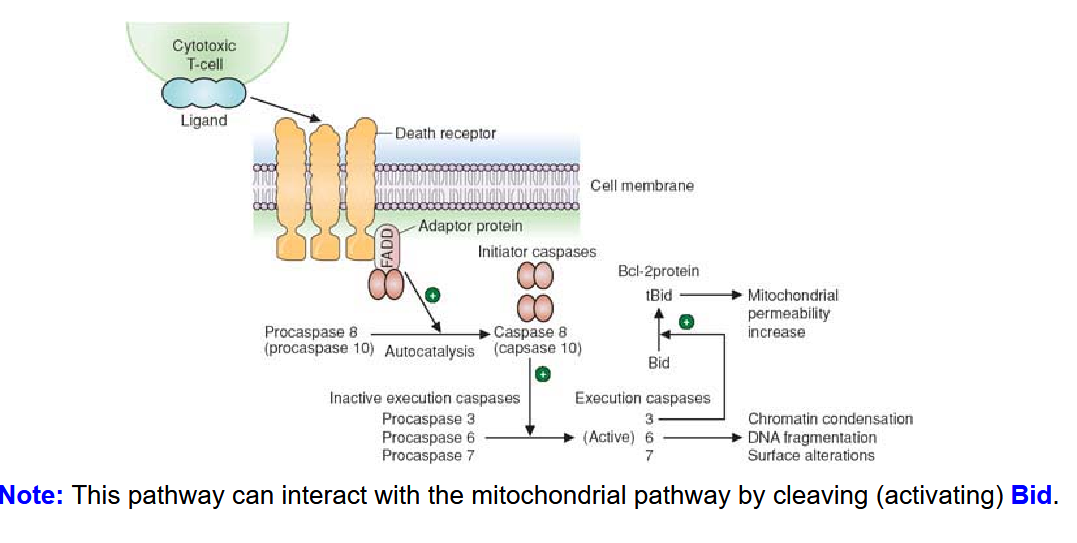
Both pathways= activate caspase cascade; Mitochondria: Death from within; Death receptor: Death from outside the cell
Effect:
chromatin condenses,
DNA fragmented,
cell shrinks and breaks up
apoptotic bodies are cleared out by macrophages.
What are Caspases? How are they produced? Describe the two types and their functions
Cysteine proteases; Produced as proenzymes; activated by proteolytic cleavage.
Initiator caspases (caspase 8, 9, 10)
Activated directly by the death cell receptor and the mitochondrial pathways.
Execution caspases (caspase 3, 6, 7)
Activated by initiator caspases.
Cleave many different proteins in the cell, including actin, proteins of the nuclear envelope, DNA repair enzymes, and the inhibitor of caspase dependent endonuclease.
Draw out the Death Receptor Pathway for Apoptosis; How can this pathway interact with the mitochondrial pathway?
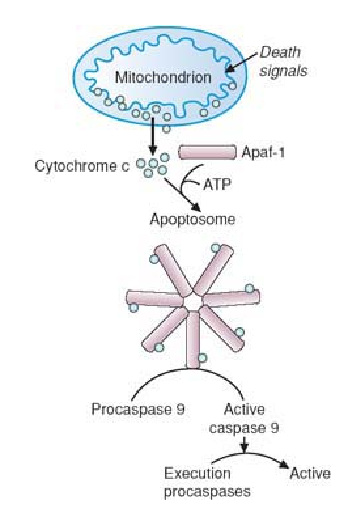
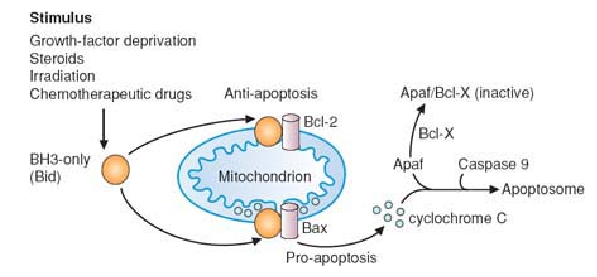
Draw out the Mitochondrial Pathway of Apoptosis. What are the mitochondrial death signals?
Mitochondrial death signals
higher than normal intracellular calcium levels
cell injury
lack of growth factors
List out the pro-apoptotic and anti-apoptotic factors
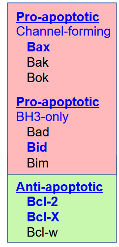
What do pro-apoptotic channel-forming factors do? What are they dependent on?
Form channels in the outer mitochondrial membrane to release cytochrome c.
Function is dependent on binding to the BH3-only
members.
What do Pro-apoptotic BH3-only factors do?
Regulate the activity of the channel forming pro-apoptotic factors
What TWO THINGS DO Anti-apoptotic factors do?
Sequesters BH3-only members away from the channel forming pro-apoptotic factors.
Can bind and inactivate Apaf-1.
Describe how apoptosis is regulated
regulated by ratio of pro/anti- apoptosis factors
Bid combines with Bax (stabilization) = cytochrome C release
Bcl-2 sequesters Bid away from Bax
What are doubled in S-phase? What happens to nucleosome as replication fork advance?
S phase = double DNA and Histones
Nucleosome disappears as replication fork advance
List out the enzymes activated in DNA replication
DNA polymerase:
polymerase delta and epsilon = major replicative polymerase
also function in DNA repair
some can digest DNA; some can replicate while bypasing DNA damage
Primase synthesizes RNA primers and hydrolases remove them.
Helicases unwind parental DNA strands.
Single strand-binding proteins prevent single strands of DNA from reassociating .
Topoisomerases relieve torsional strain caused by unwinding.
DNA ligase joins two adjacent DNA strands.
PCNA holds polymerases together to enhance the process.
Compare and contrast Spermatogenesis and Oogenesis
Sperm:
begins and proceed at puberty; 64 days to complete
4 sperms per meiotic division
Oogenesis:
Begins at birth. Millions of secondary
oocytes are present, but only ~400 will
eventually mature.Halted at meiosis I until puberty; at Metaphase II until fertilization (polar bodies can’t be fertilized)
Meiosis produces 1 ovum
At fertilization, what forms? What happens to sperm’s mitochondria?
At fertilization, the sperm and egg form pronuclei.
Sperm mitochondria and its mtDNA is lost
ONLY MATERNAL MTDNA IS INHERITED