Lower Appendicular Skeleton (BYU EXSC 440 Lab)
1/118
There's no tags or description
Looks like no tags are added yet.
Name | Mastery | Learn | Test | Matching | Spaced | Call with Kai |
|---|
No analytics yet
Send a link to your students to track their progress
119 Terms
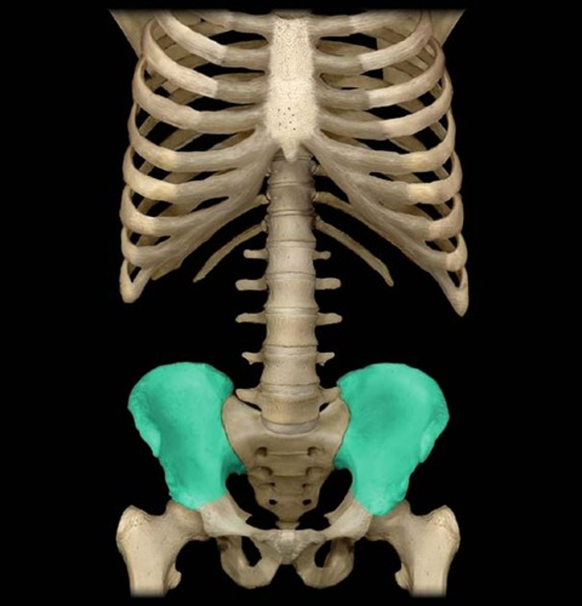
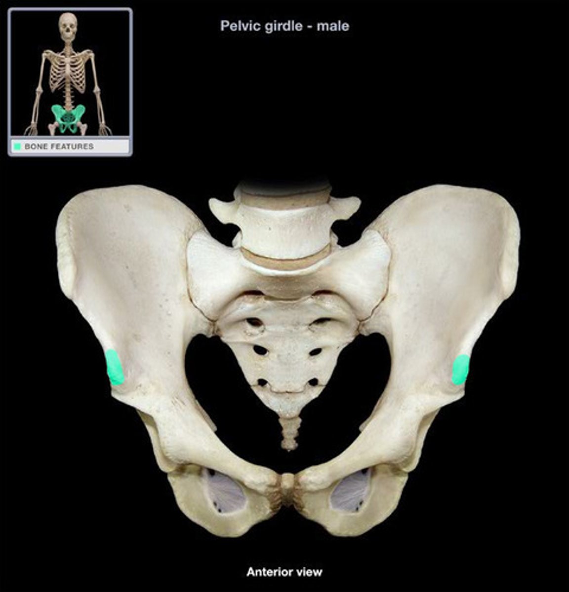
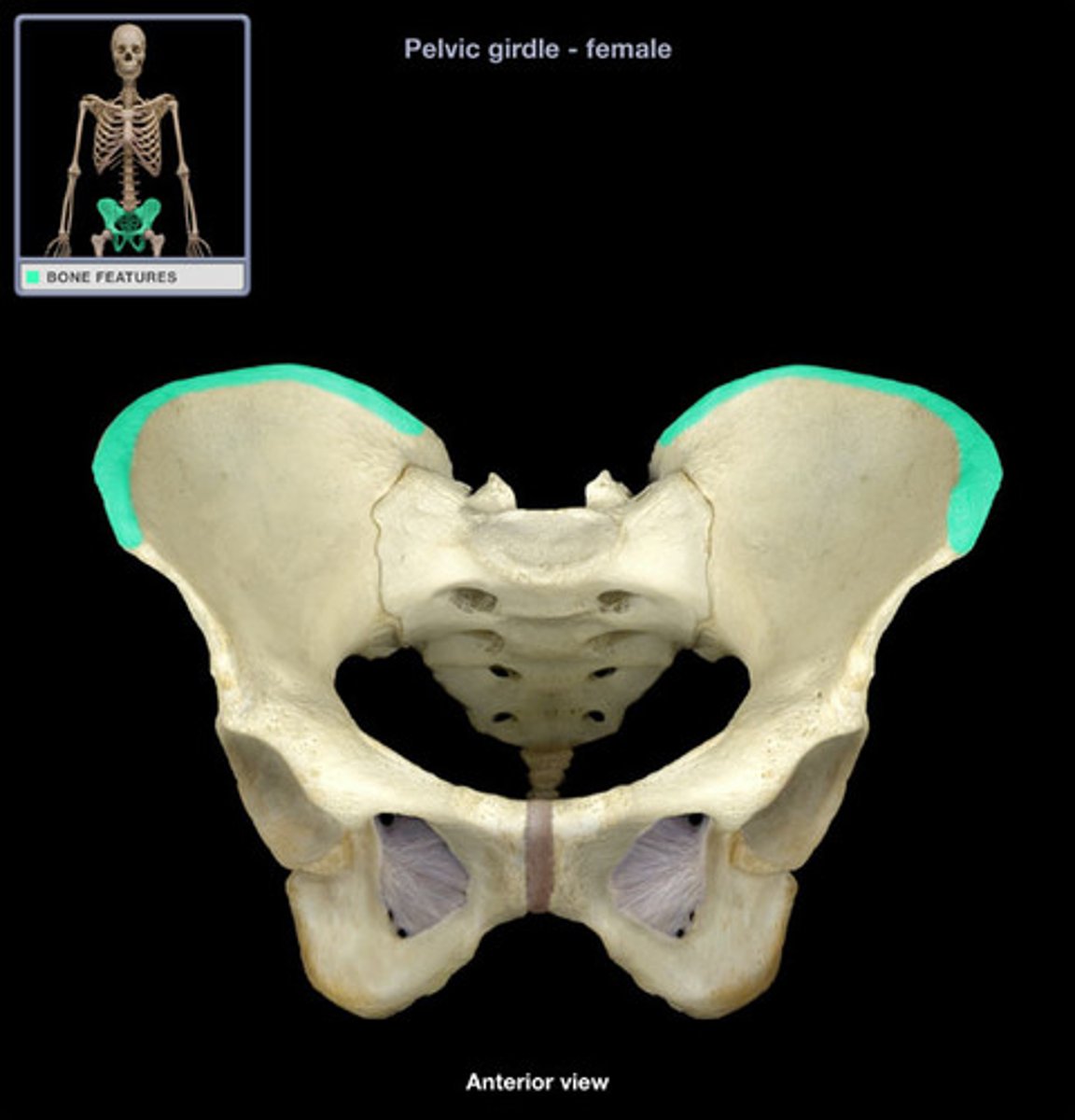
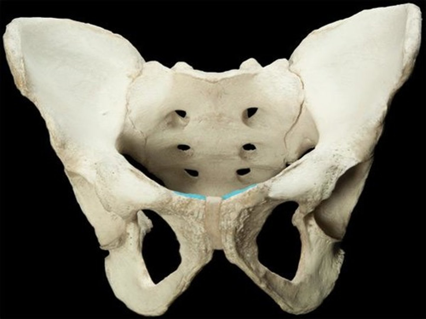
Os Coxa
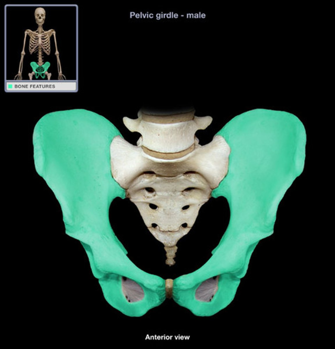
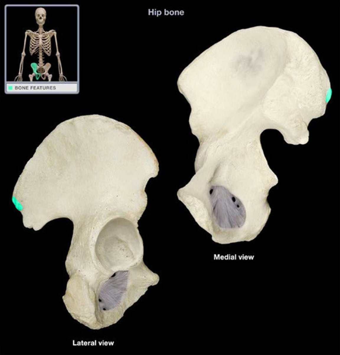
Ilium
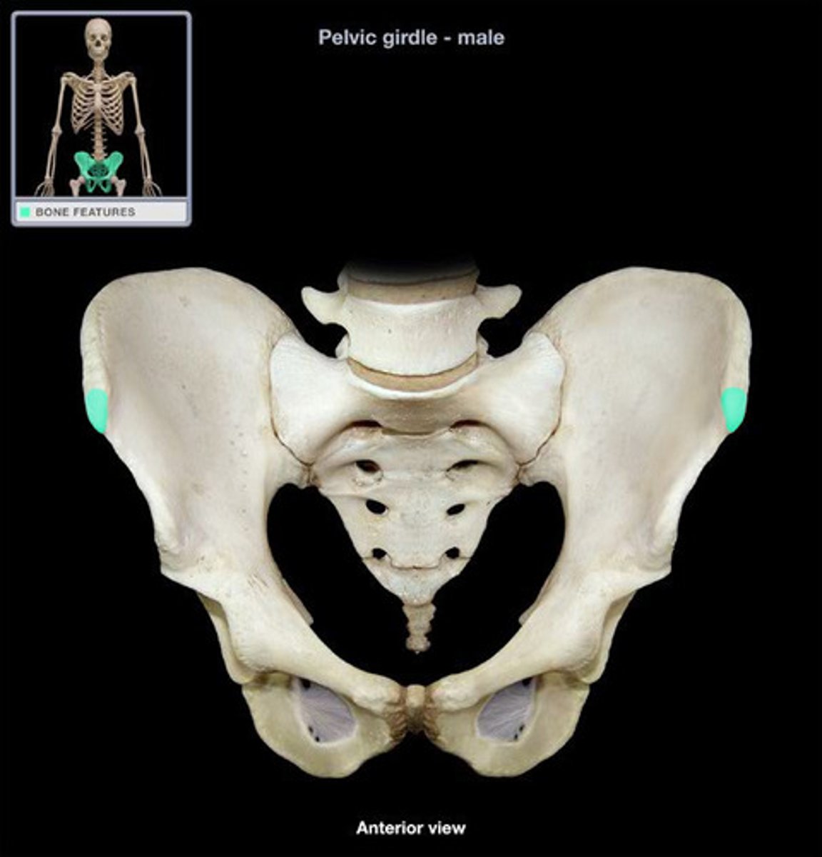
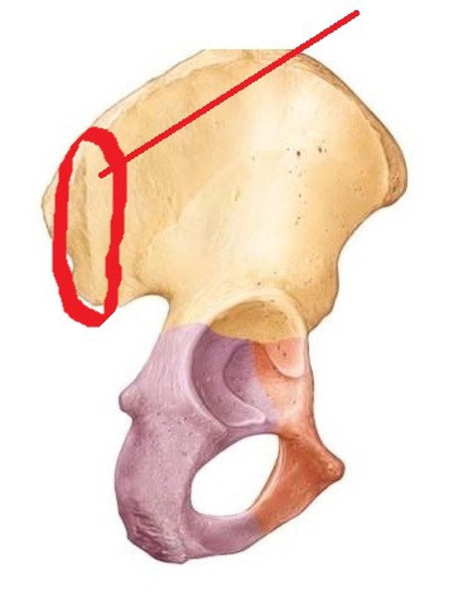
Anterior superior iliac spine (ASIS)
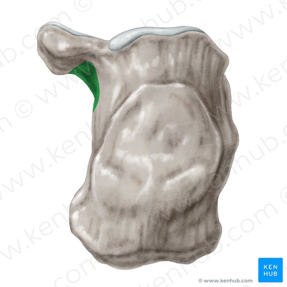
Anterior inferior iliac spine (AIIS)
Iliac crest
Posterior Superior Iliac Spine (PSIS)
Posterior inferior iliac spine (PIIS)

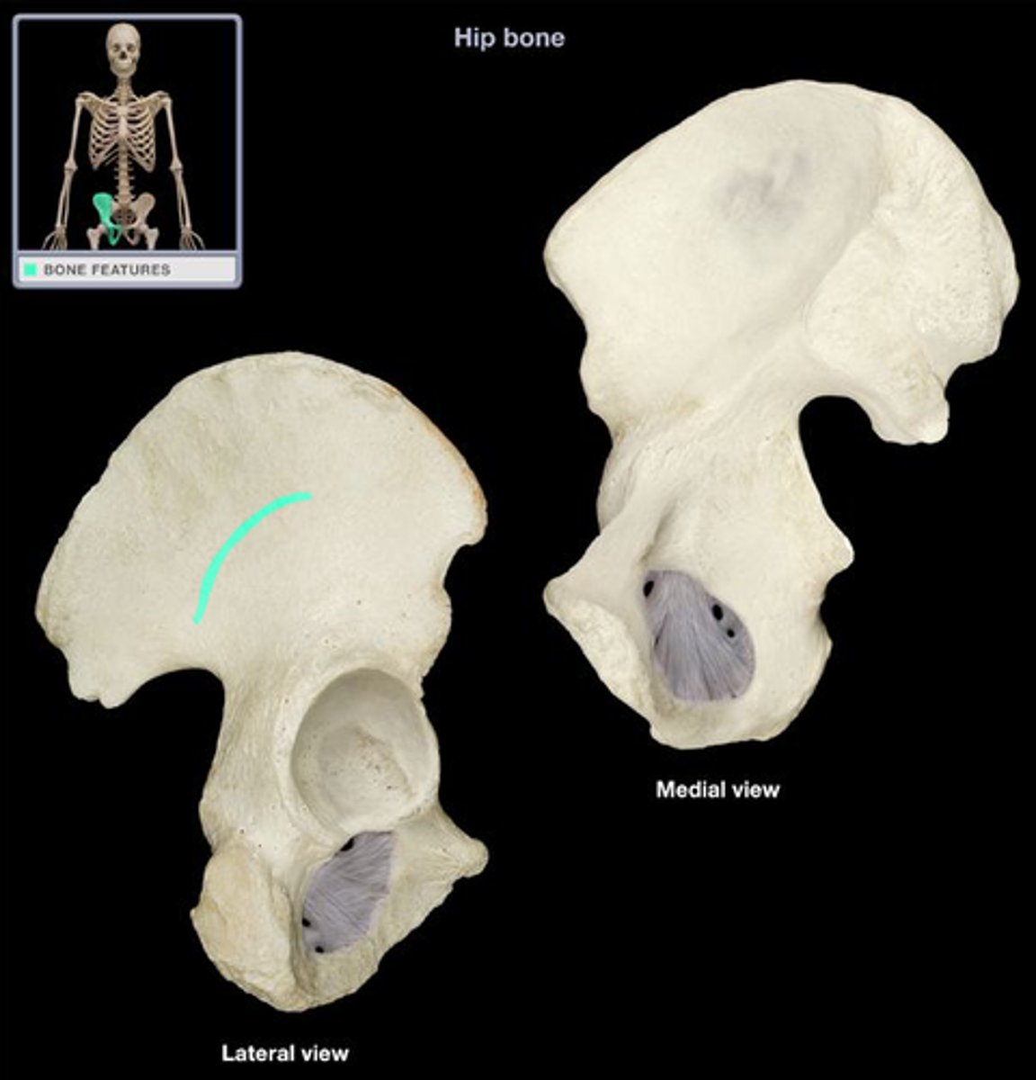
Anterior gluteal line
Posterior gluteal line
Inferior gluteal line
Auricular surface of ilium
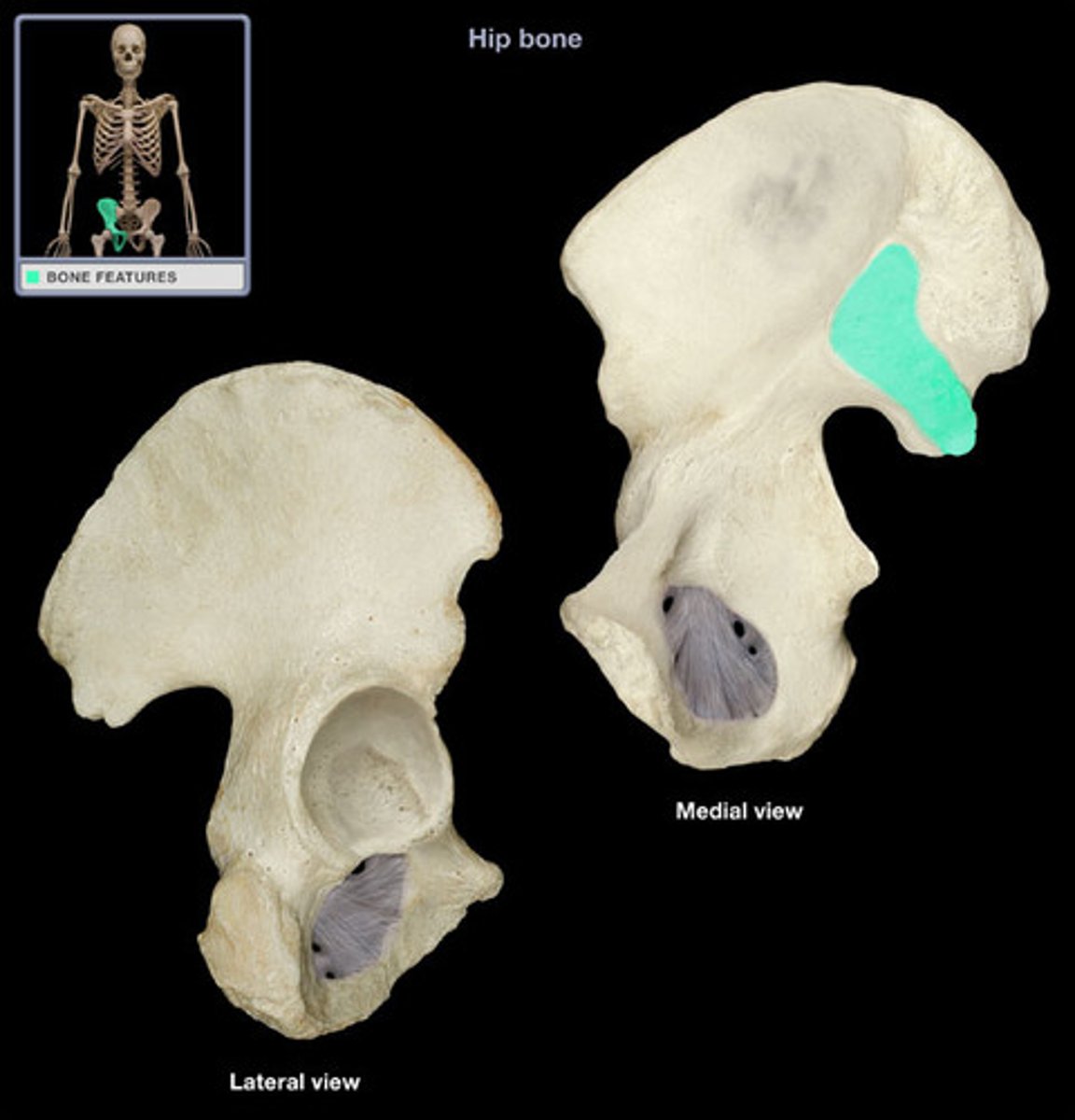
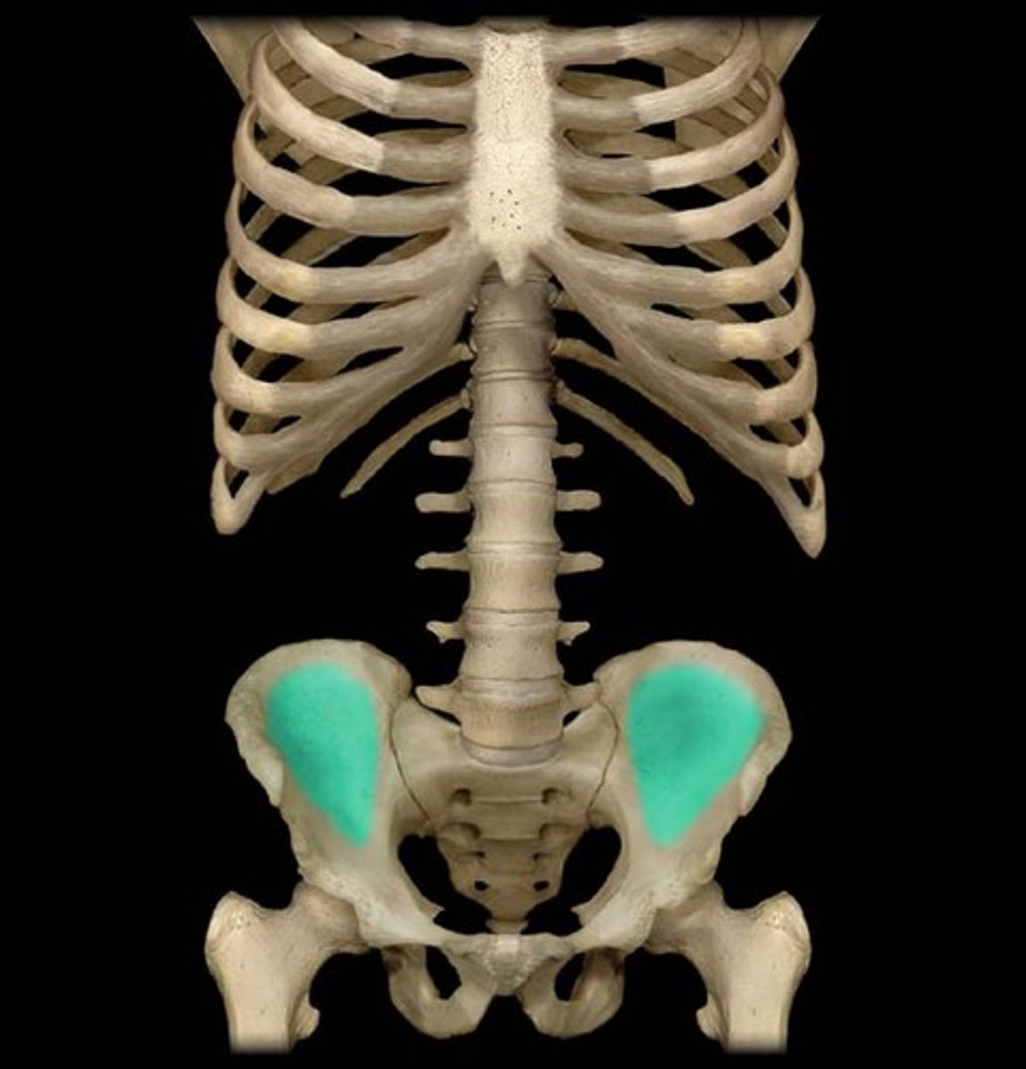
Iliac fossa
The broad, slightly concave inner surface of the ilium.
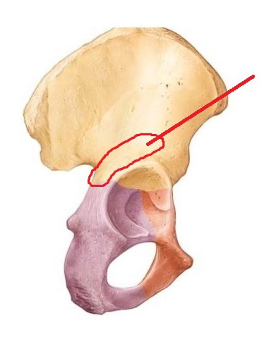
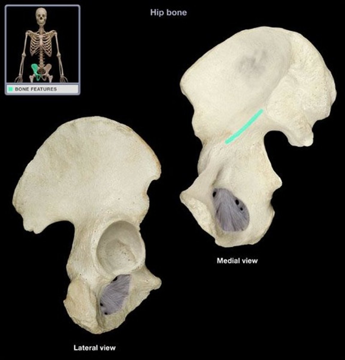
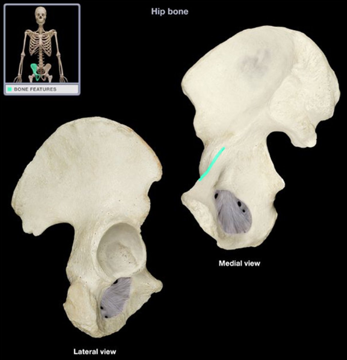
Arcuate line
a ridge of bone that runs inferiorly and anteriorly from the auricular surface
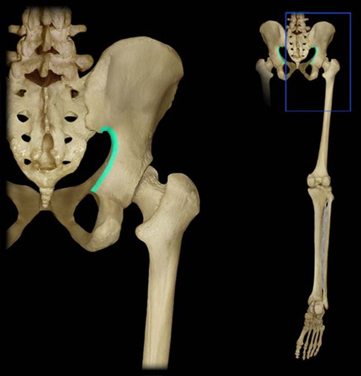
Greater sciatic notch
allows blood vessels and the large sciatic nerve to pass from the pelvis posteriorly into the thigh
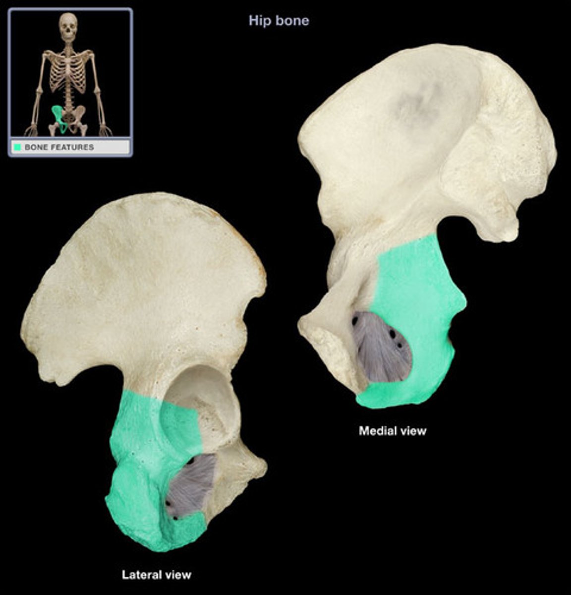
Ischium
Lesser sciatic notch
inferior to ischial spine

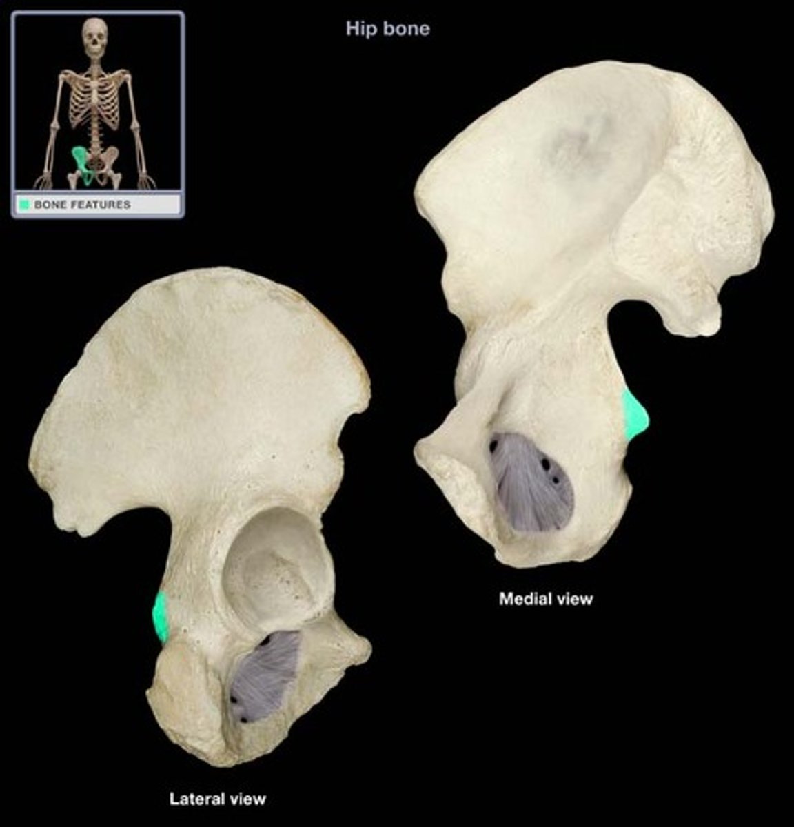
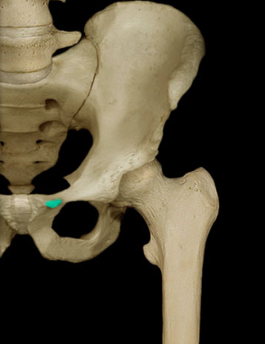
Ischial spine
located superior to the ischial tuberosity and projects medially into the pelvic cavity
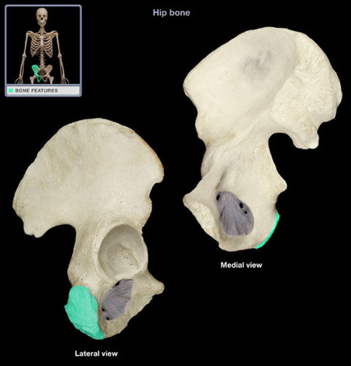
Ischial tuberosity
receives the weight of the body when sitting
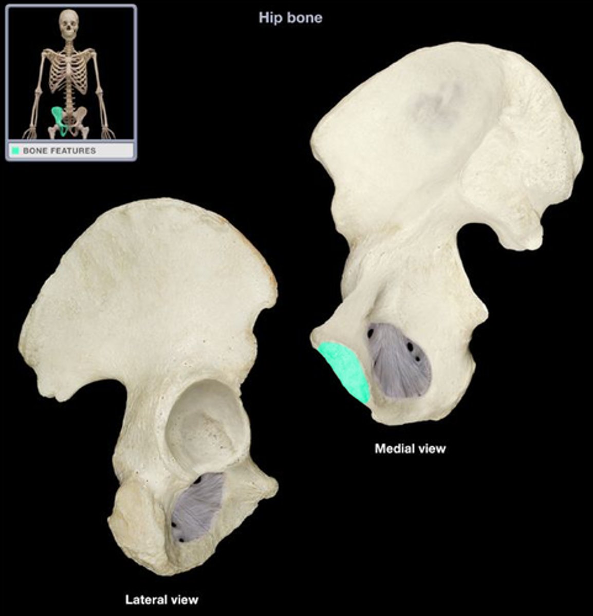
Ischial ramus
bony extension projecting anteriorly and superiorly from the ischial tuberosity; joins with the inferior pubic ramus to form the ischiopubic ramus

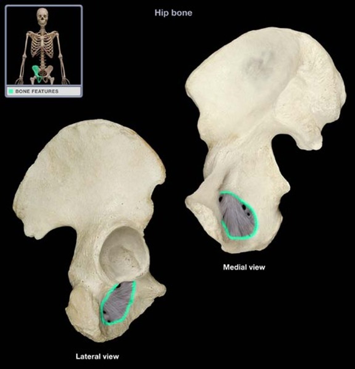
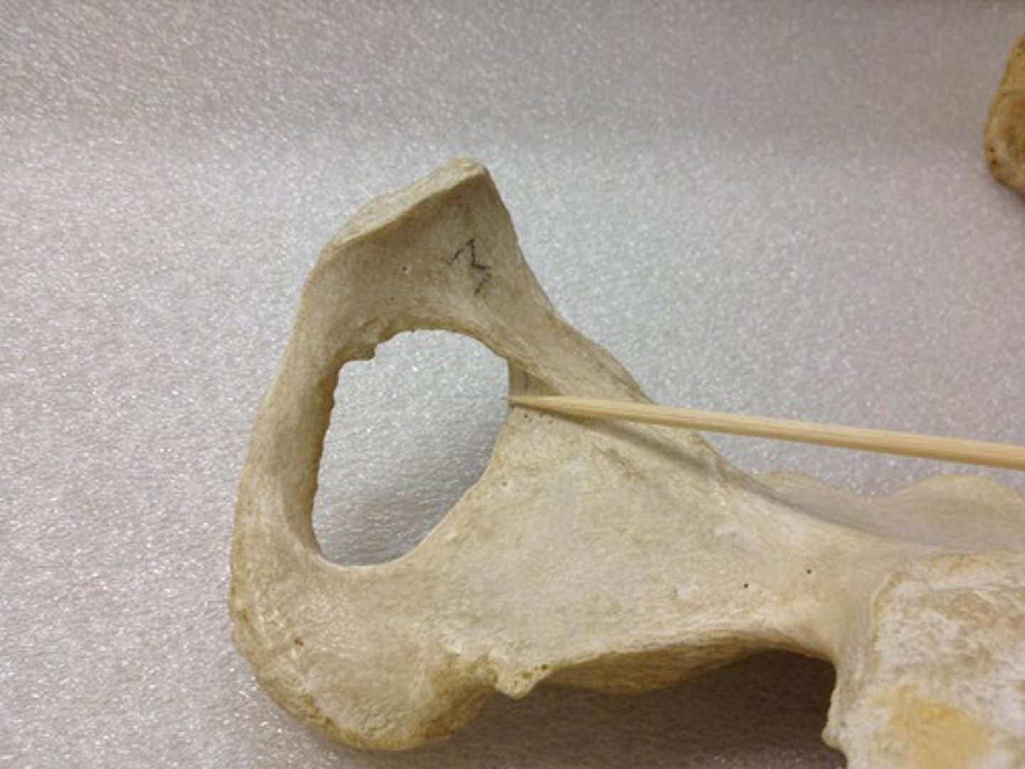
Obturator foramen
Pubis
The medial anterior portion of the pelvis
Pubic tubercle
Pubic crest
thick anterior border
Superior pubic ramus
superior extension of the body of the pubis
Pectineal line of pubis
Obturator groove
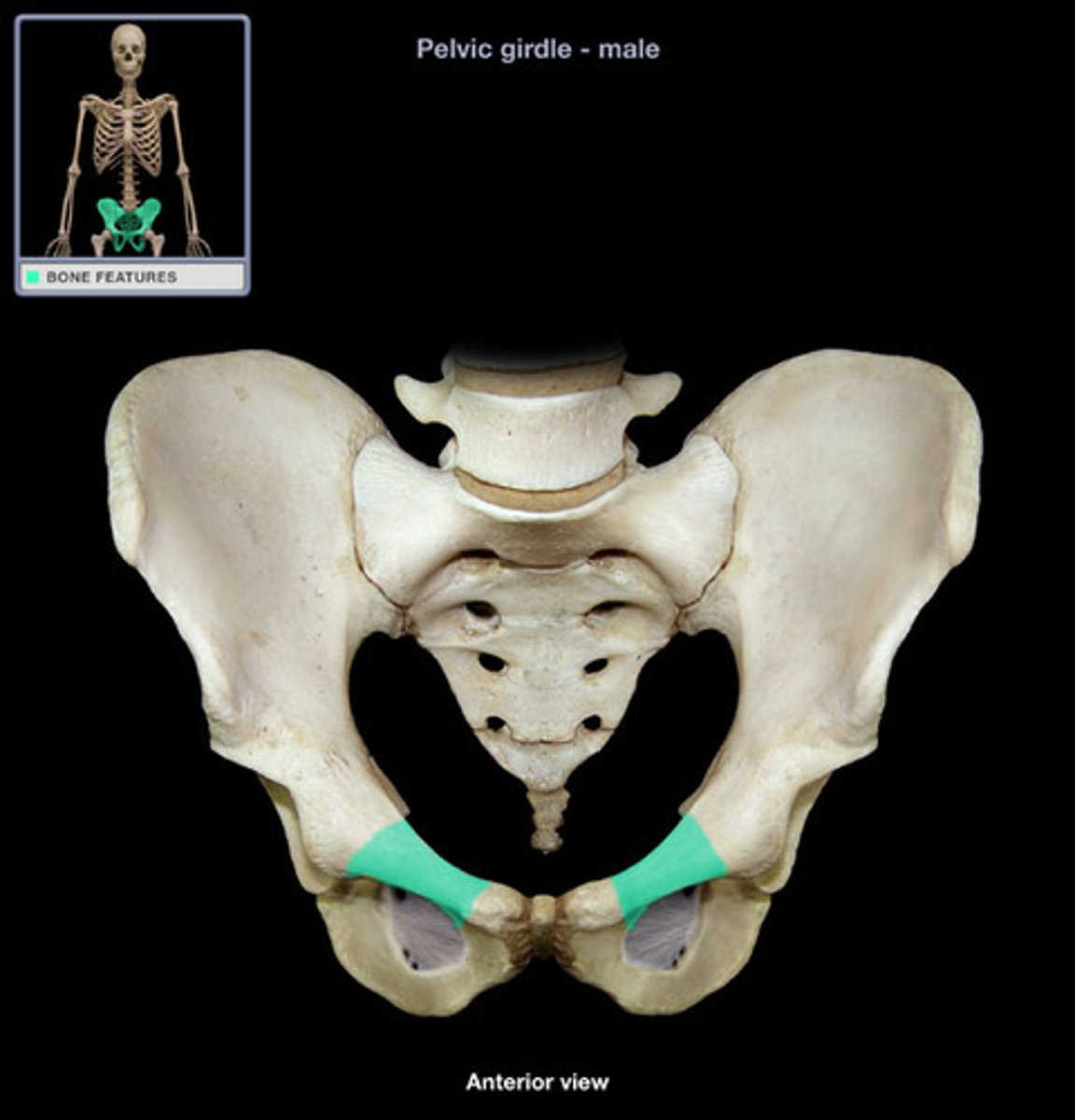
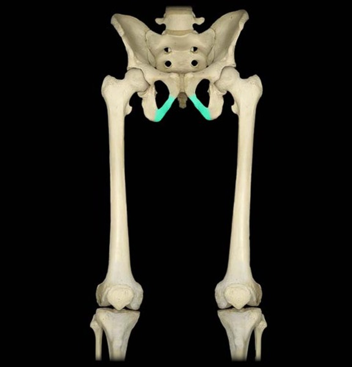
Inferior pubic ramus
inferior extension of the body of the pubis; articulates with the ischium
Symphyseal surface
medial articulating surface between 2 pubis bones
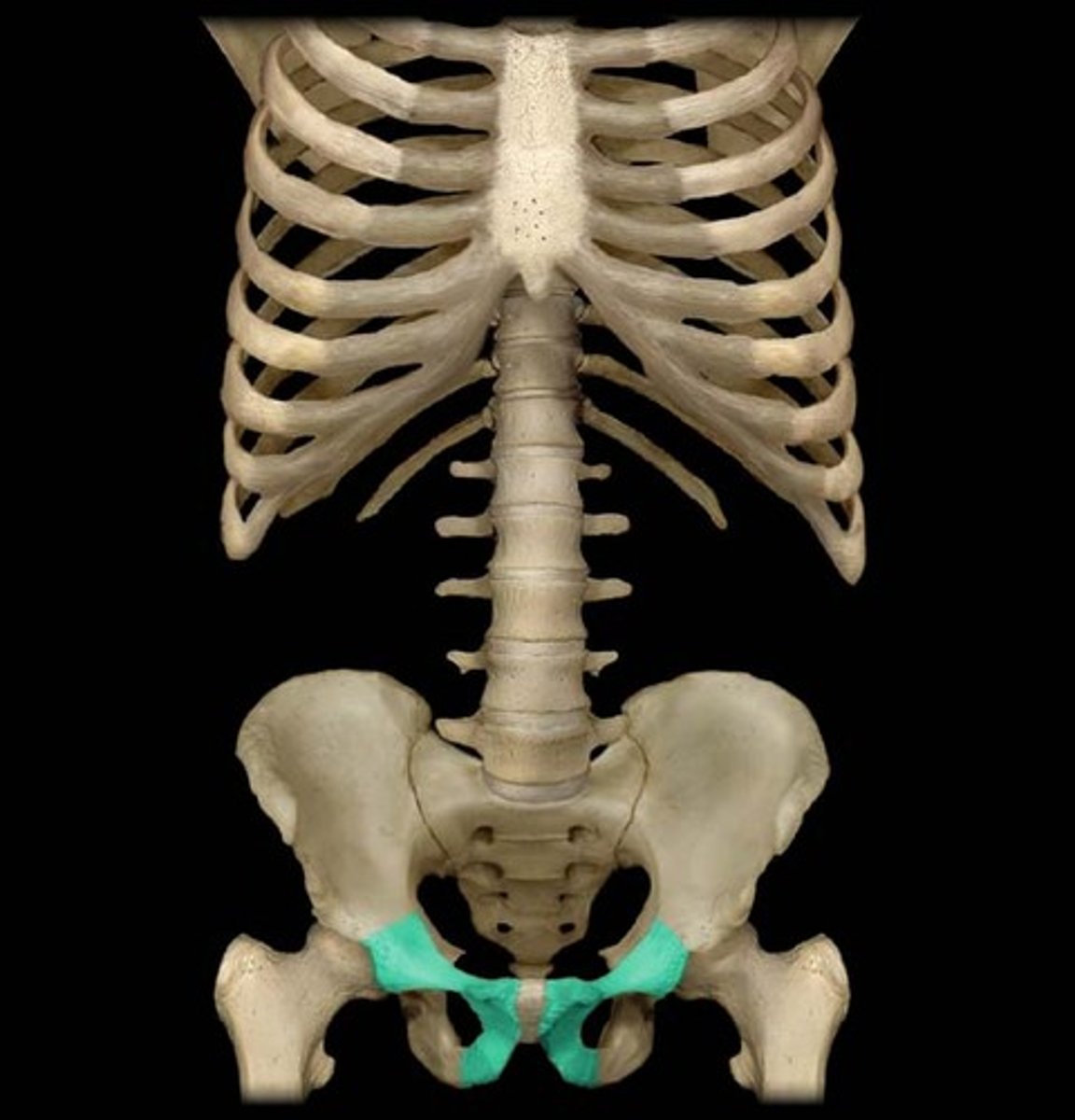
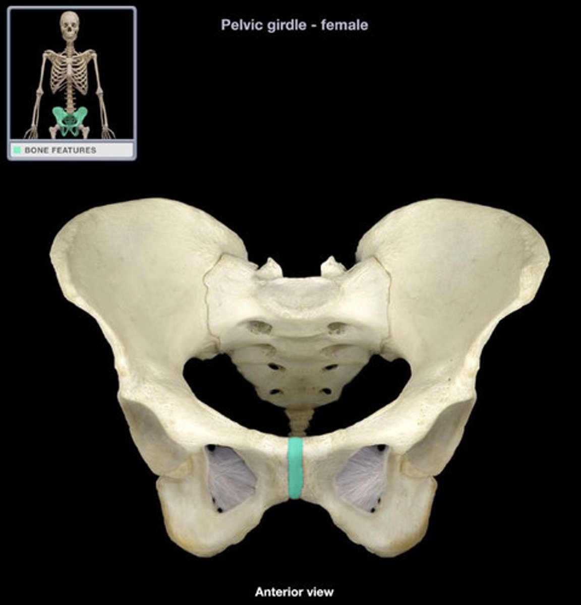
Pubic symphysis
cartilaginous joint at which two pubic bones fuse together
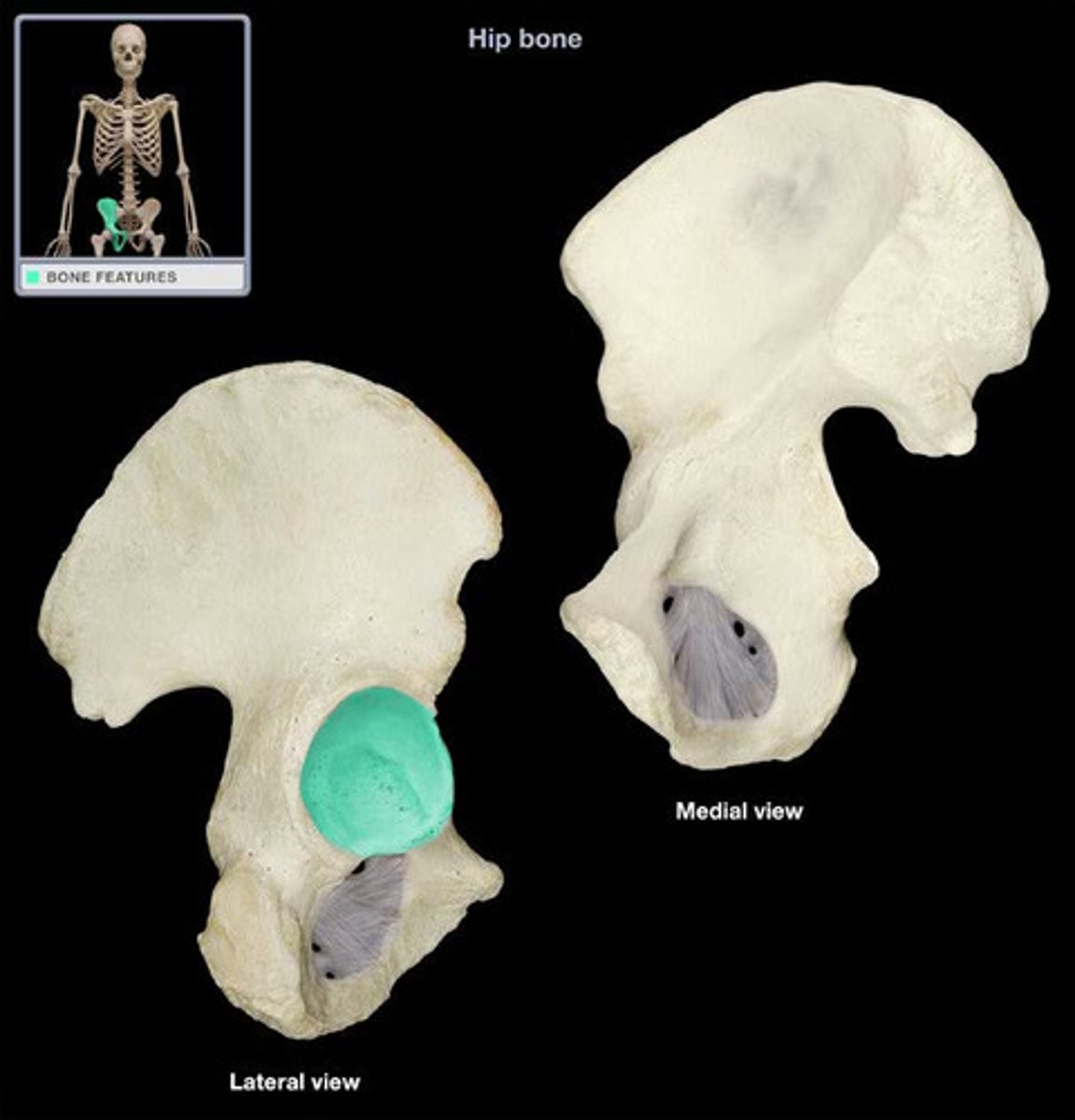
Acetabulum
large socket in the pelvic bone for the head of the femur
Acetabular rim
outside edge of acetabulum
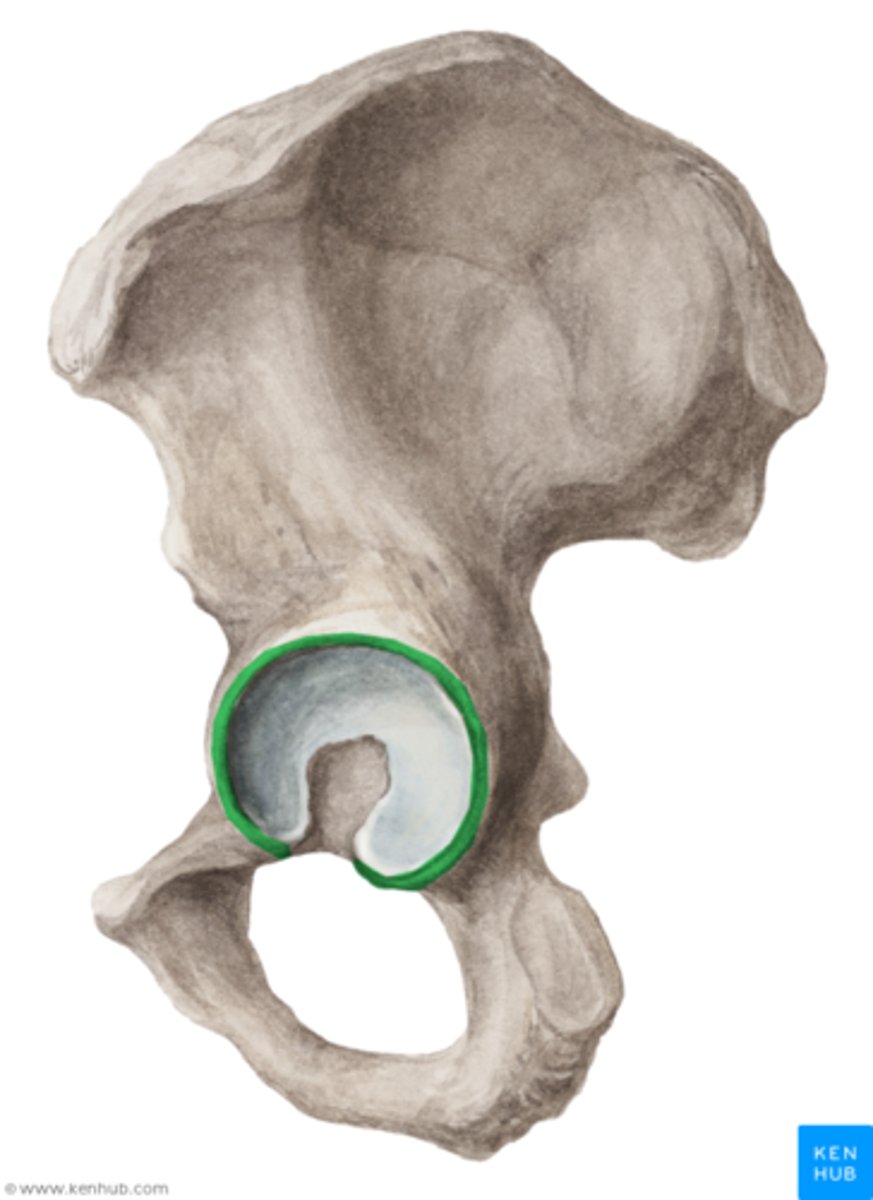
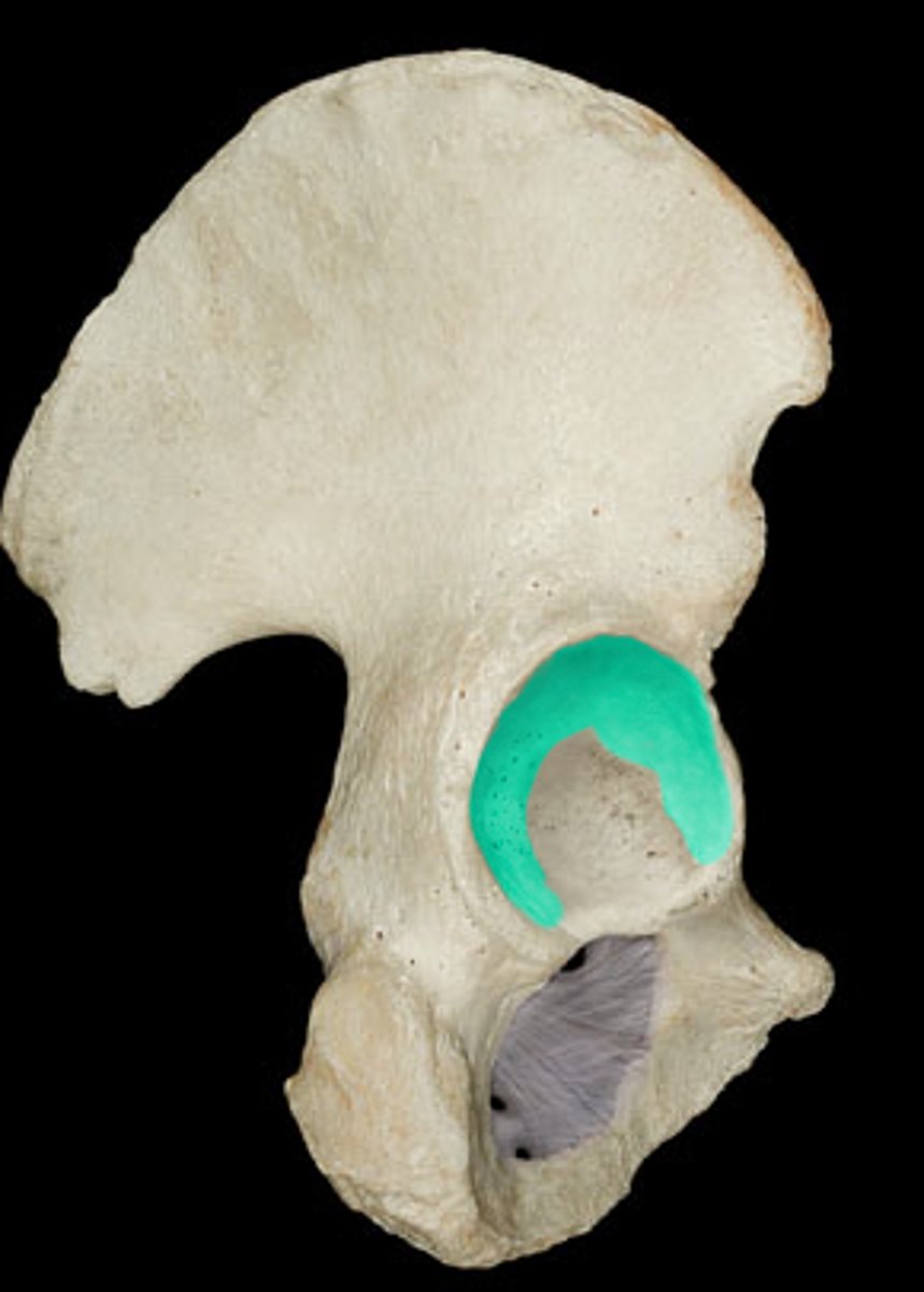
Lunate surface
Smooth articulating surface on the periphery of the acetabulum
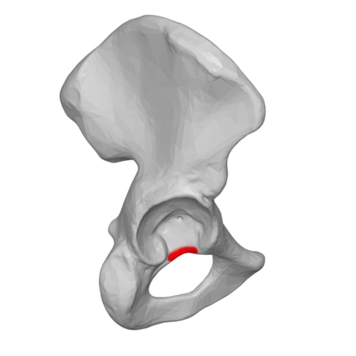
Acetabular notch
deep notch in the inferior part of the brim
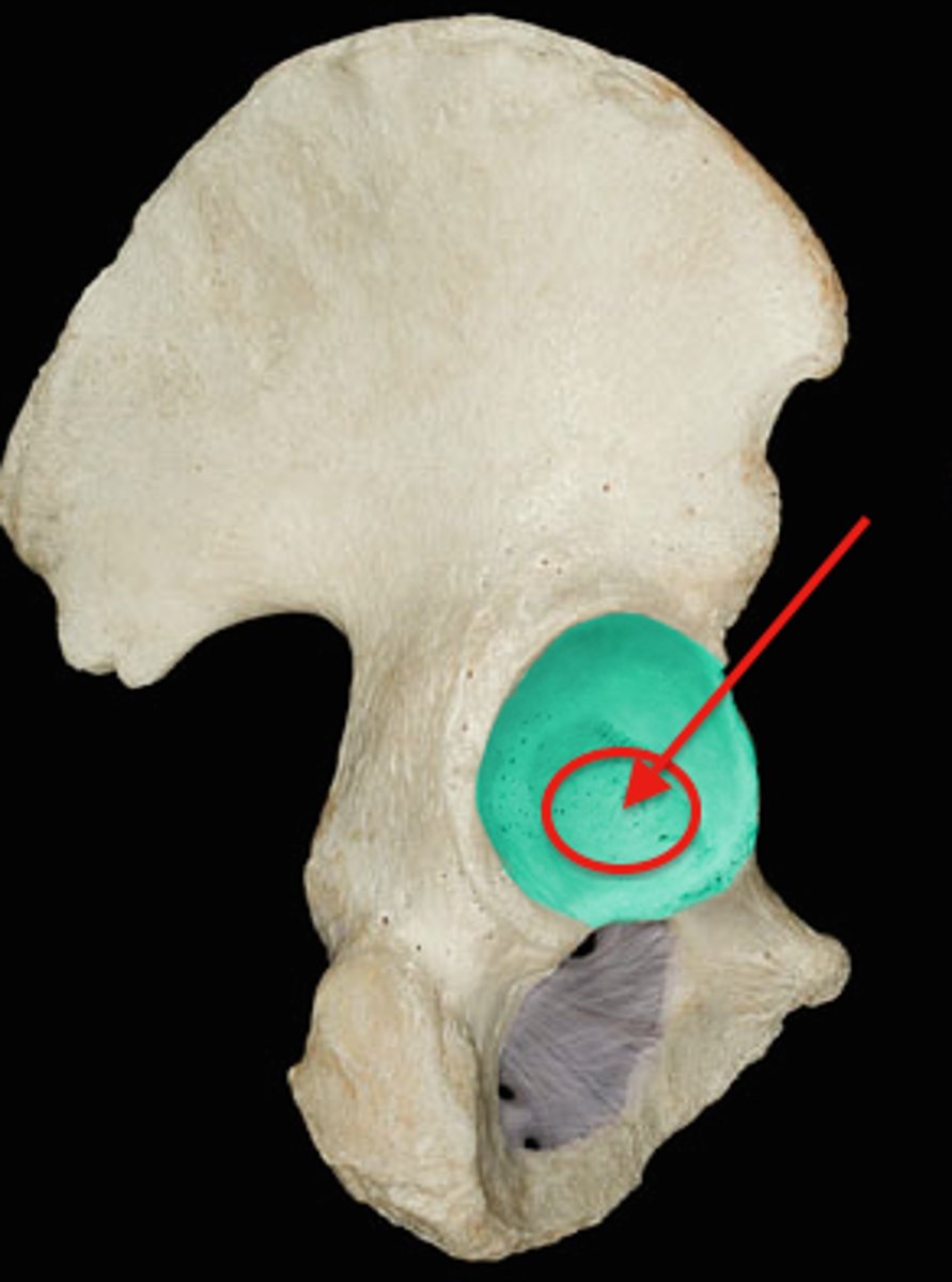
Acetabular fossa
circular depression located deep in the acetabulum
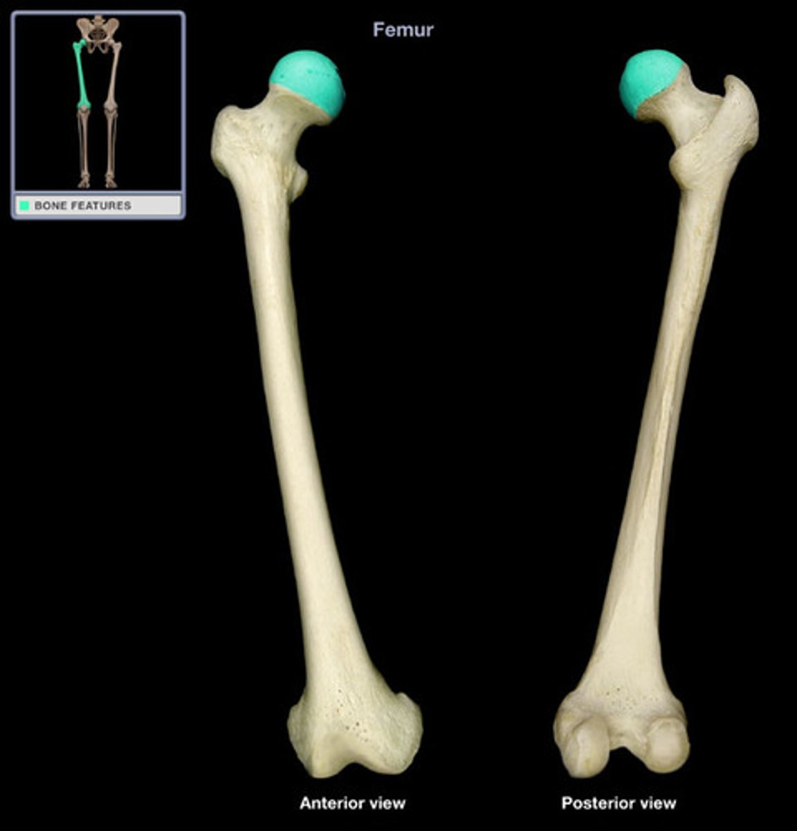
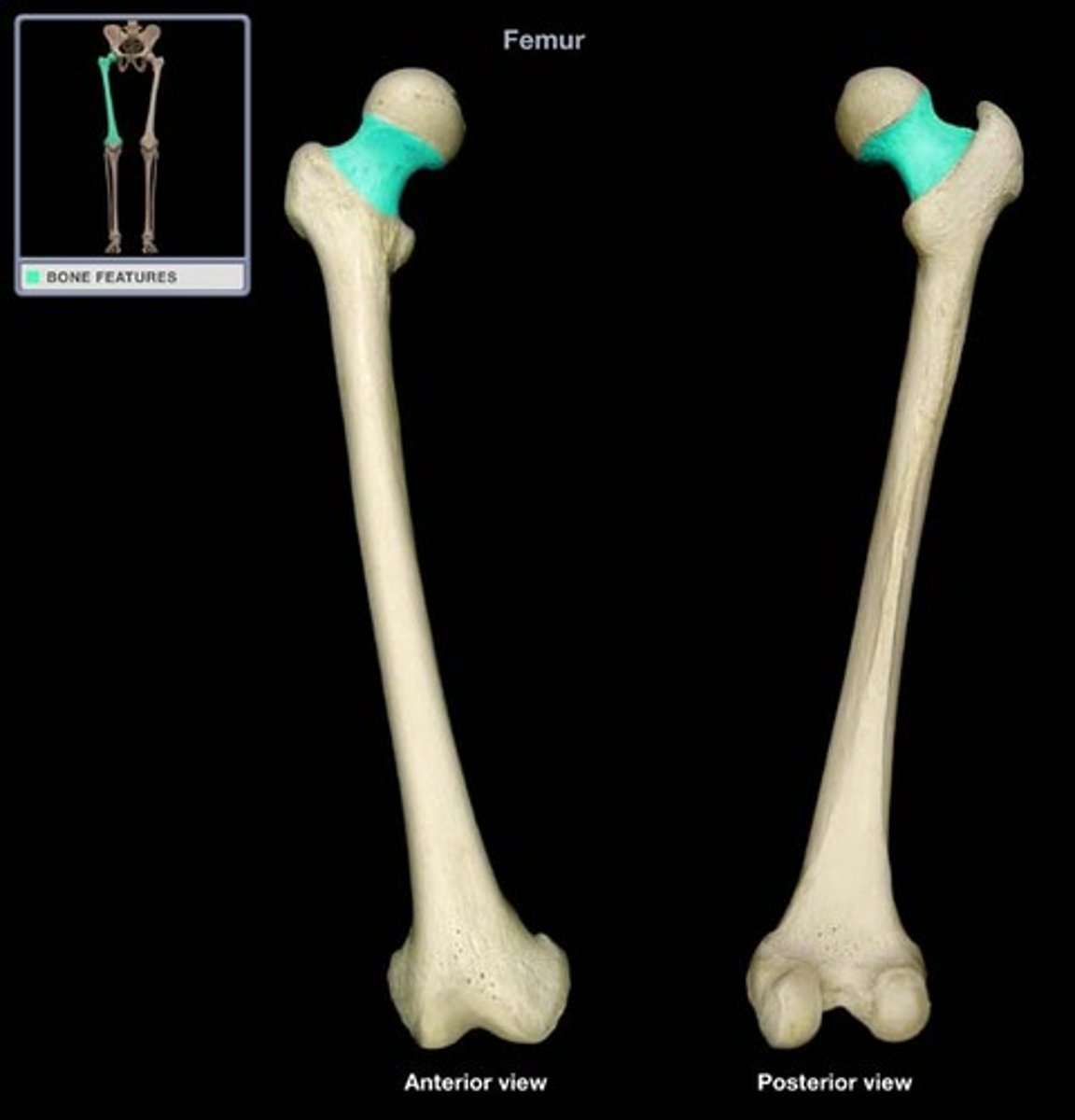
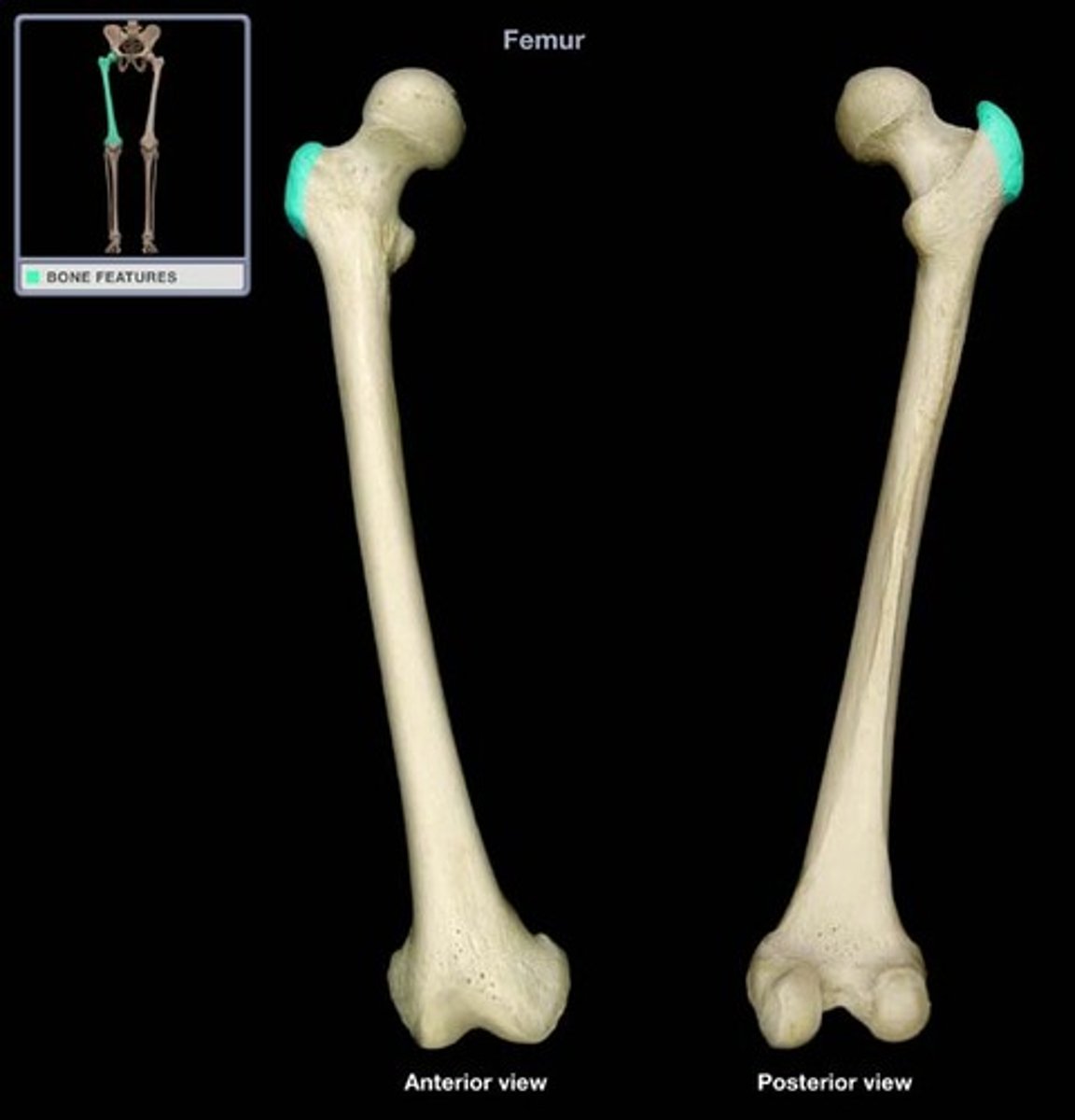
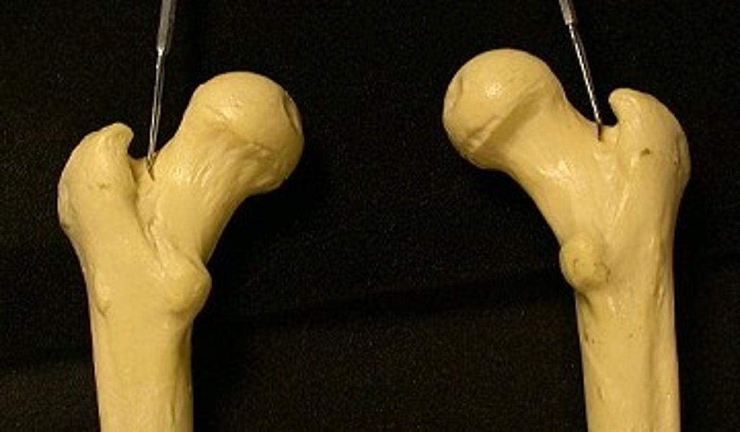
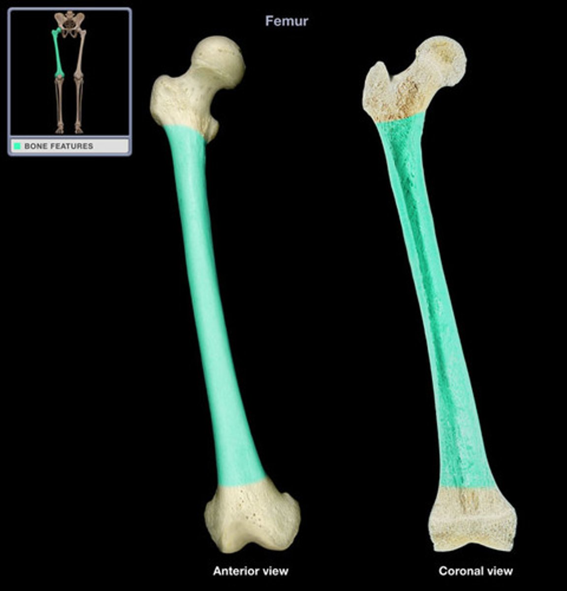
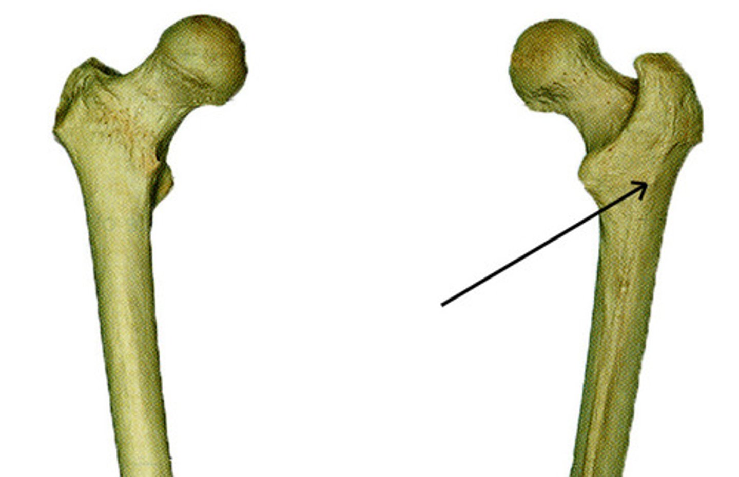
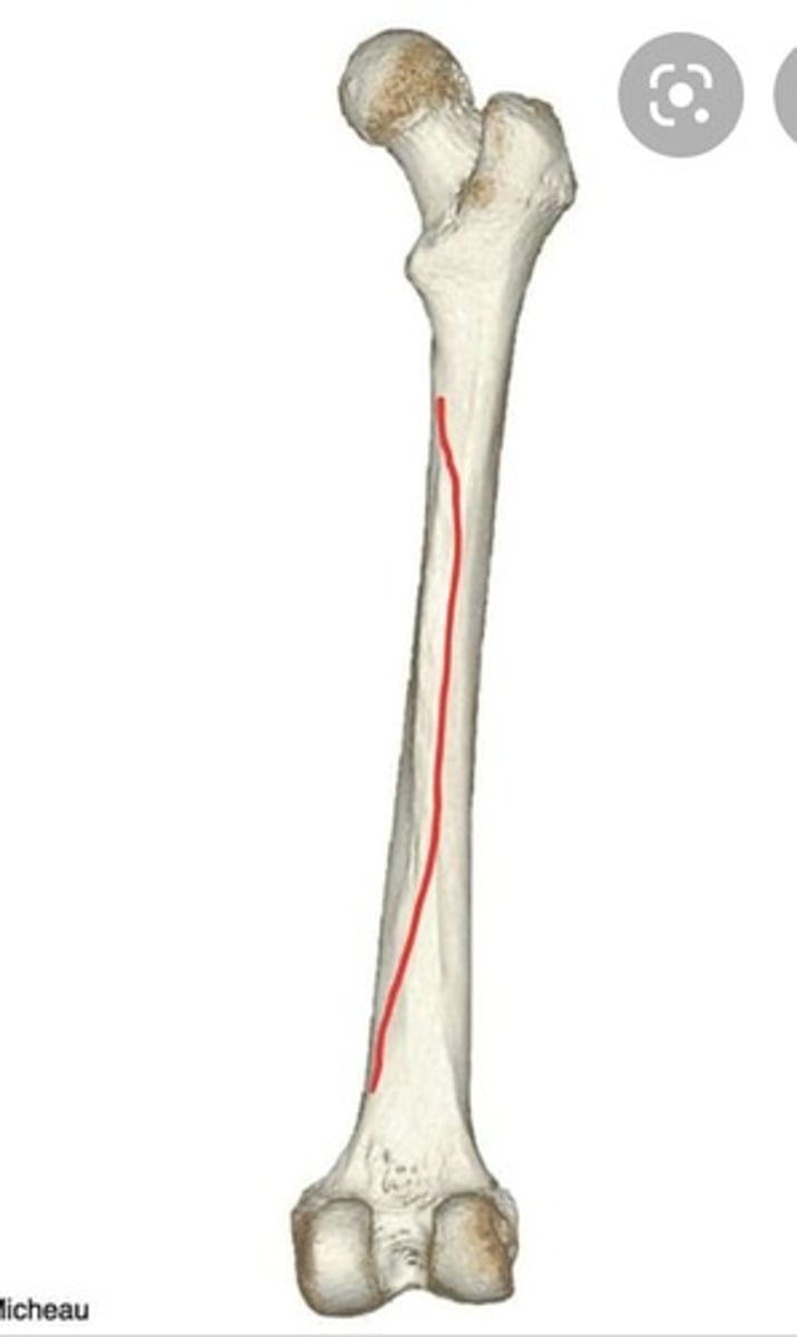
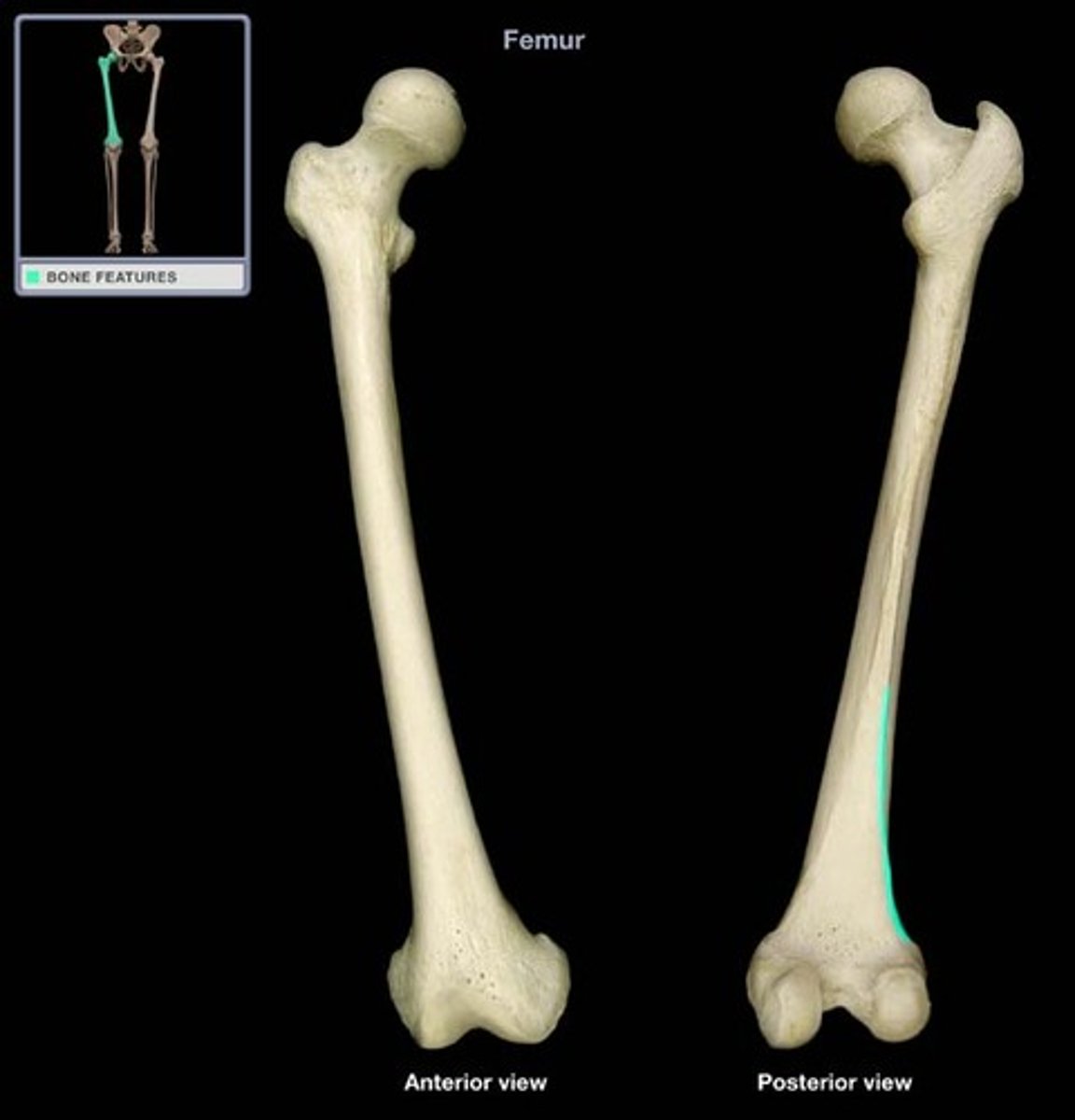
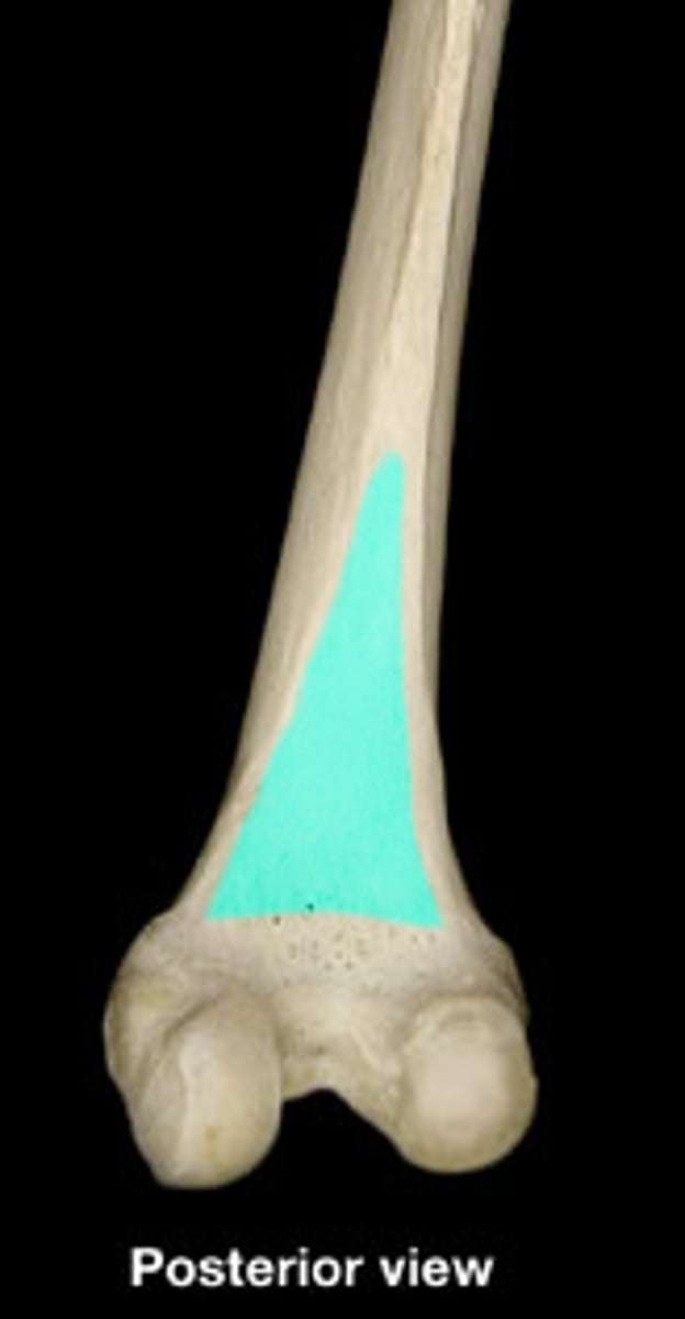
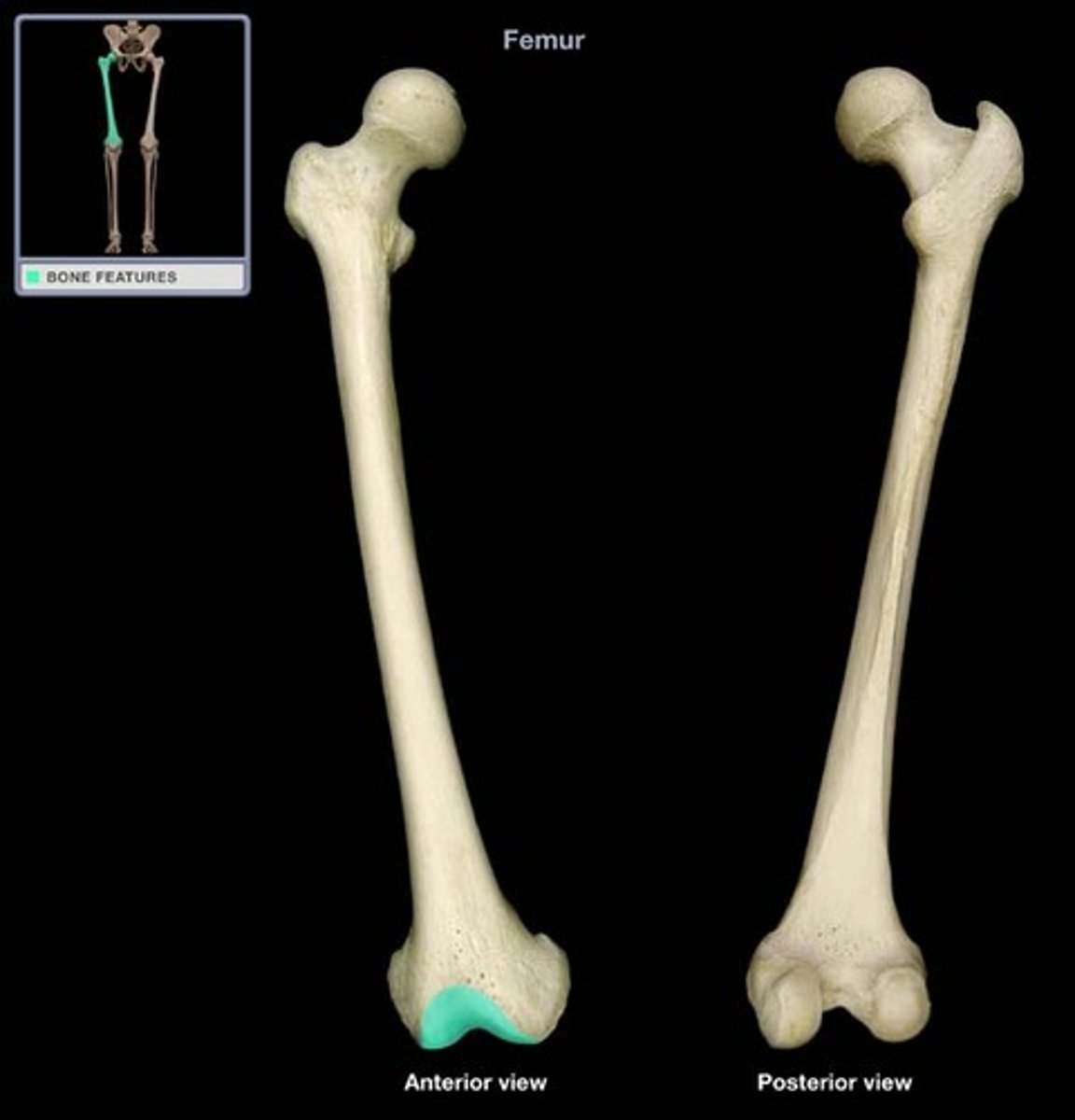
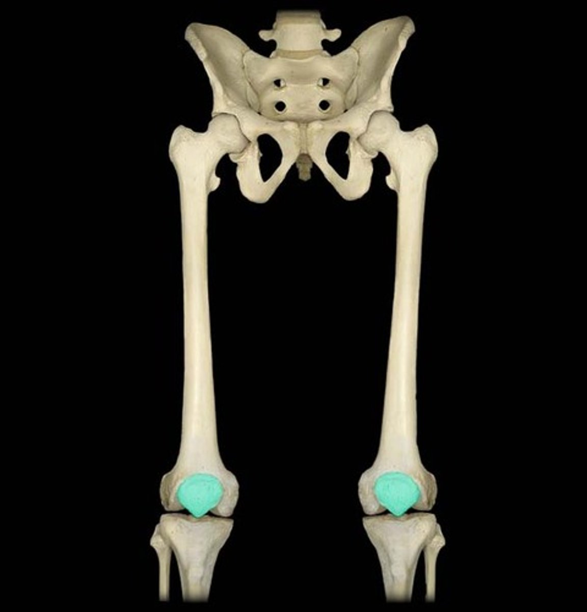
Femur
thigh bone
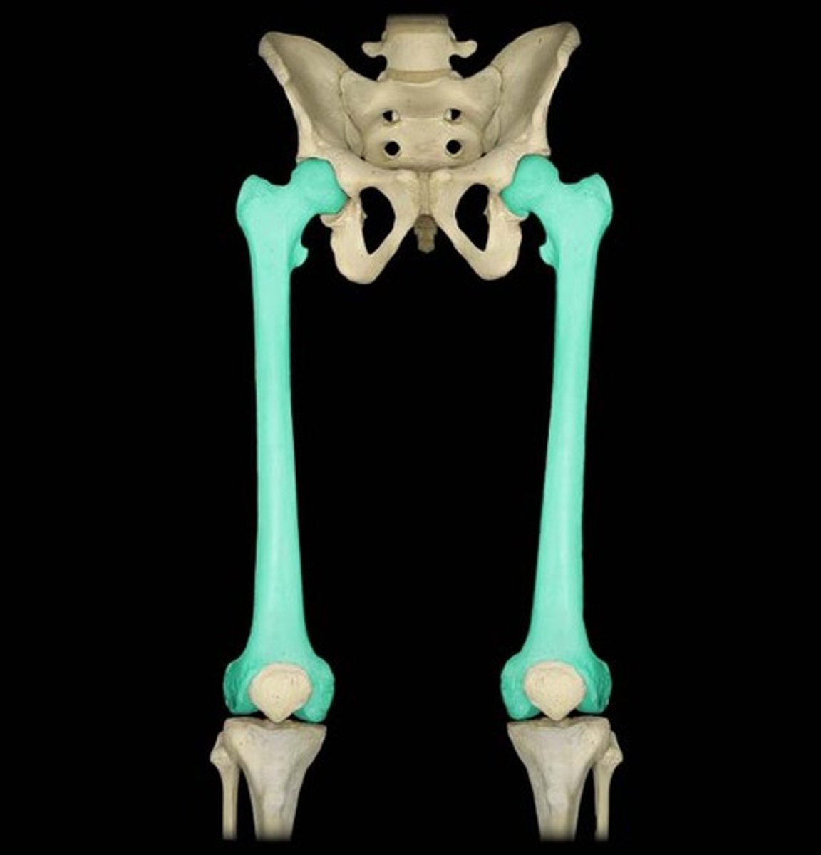
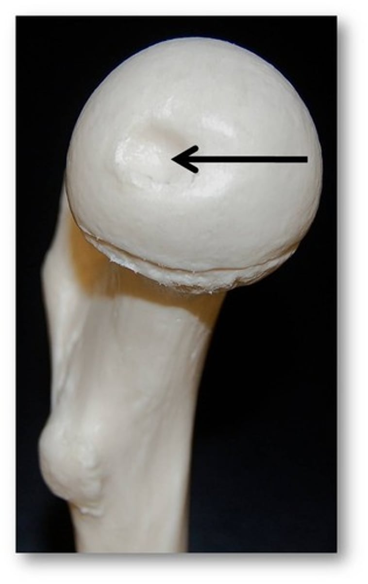
Head of femur
Fovea capitis
pit in the head of a femur
Neck of femur
Greater trochanter
A bony prominence on the proximal lateral side of the thigh, just below the hip joint.
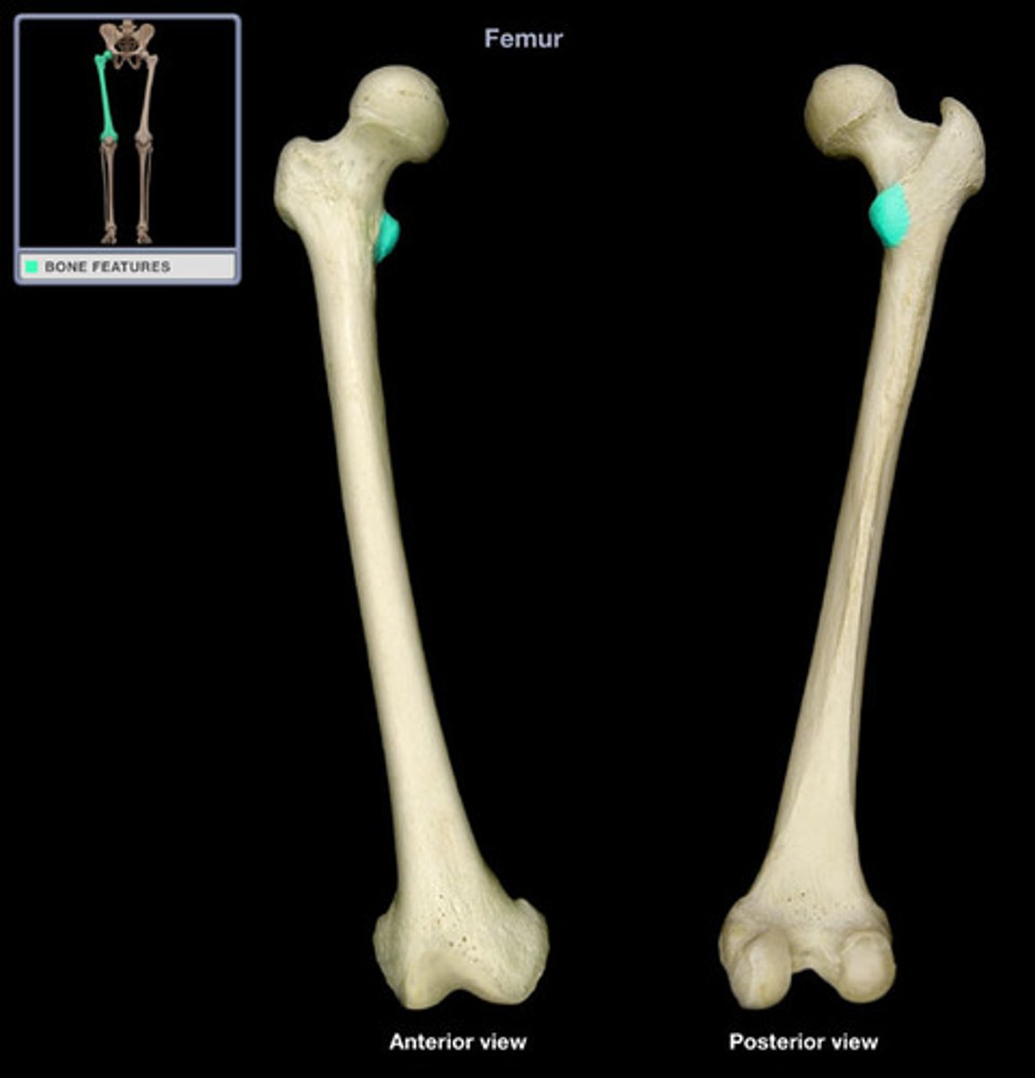
Lesser trochanter
The projection on the medial/superior portion of the femur.
Pectineal line of femur

Intertrochanteric line
region formed anteriorly between the greater and lesser trochanters

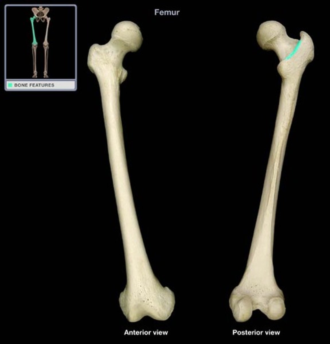
Intertrochanteric crest
region formed posteriorly between the greater and lesser trochanters
Quadrate tubercle
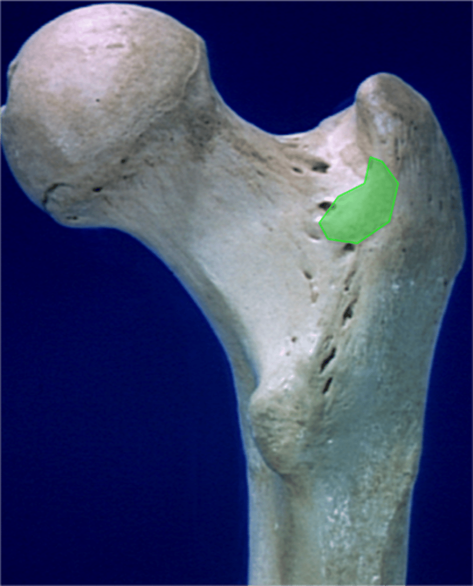
Trochanteric fossa
depression on medial surface of greater trochanter
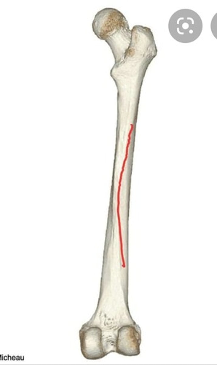
Shaft of femur
Gluteal tuberosity
back of femur; bump above the linea aspera that is on the upper portion of femur
Medial lip of linea aspera
Lateral lip of linea aspera
Medial supracondylar line
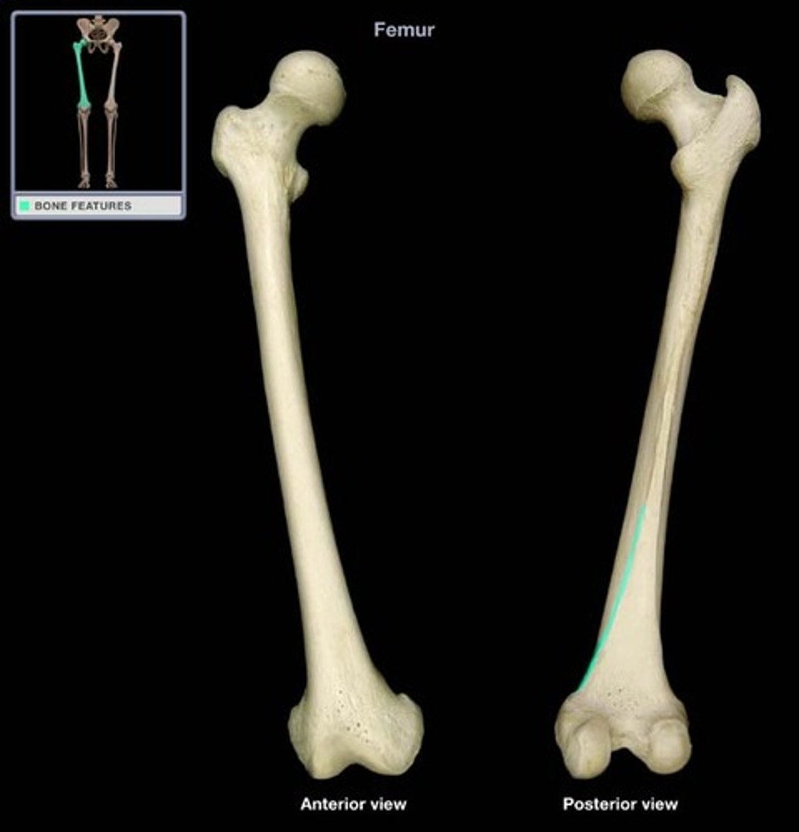
Lateral supracondylar line
Adductor tubercle
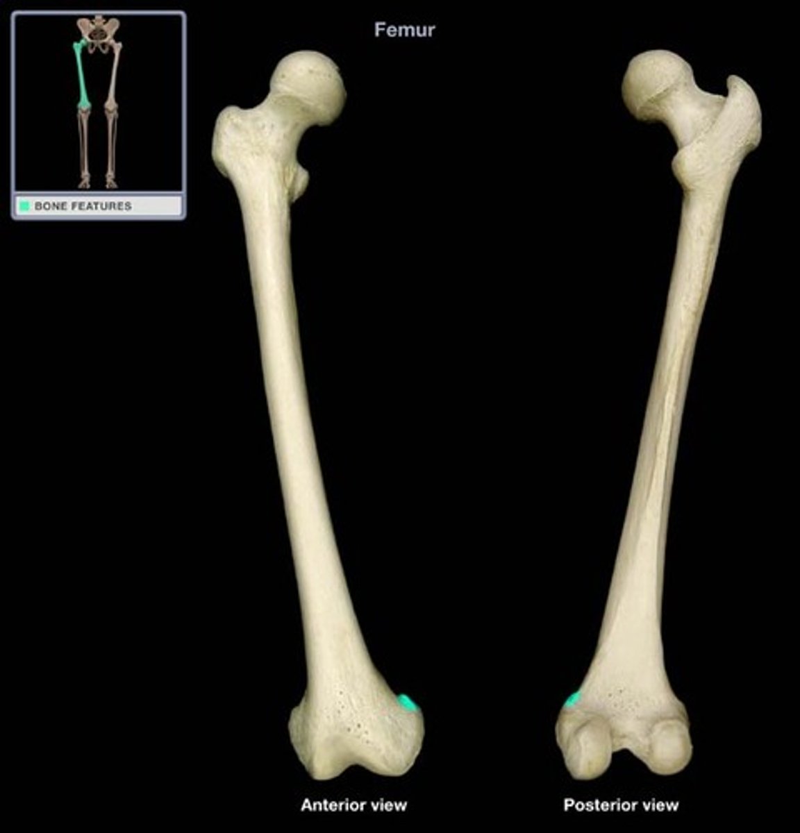
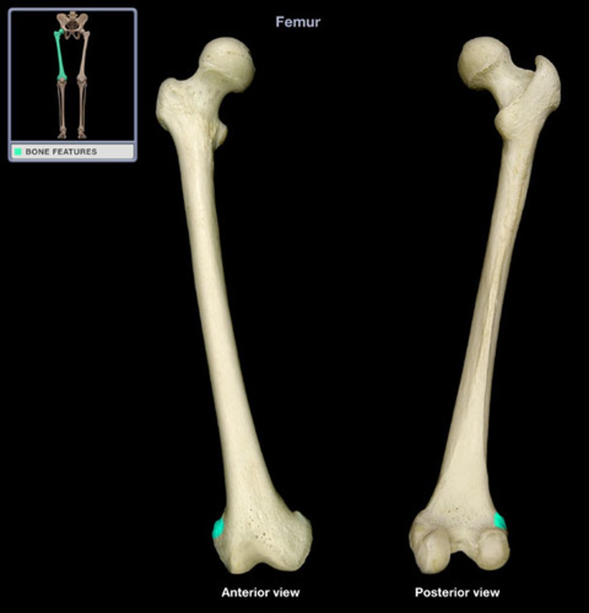
Medial epicondyle of femur
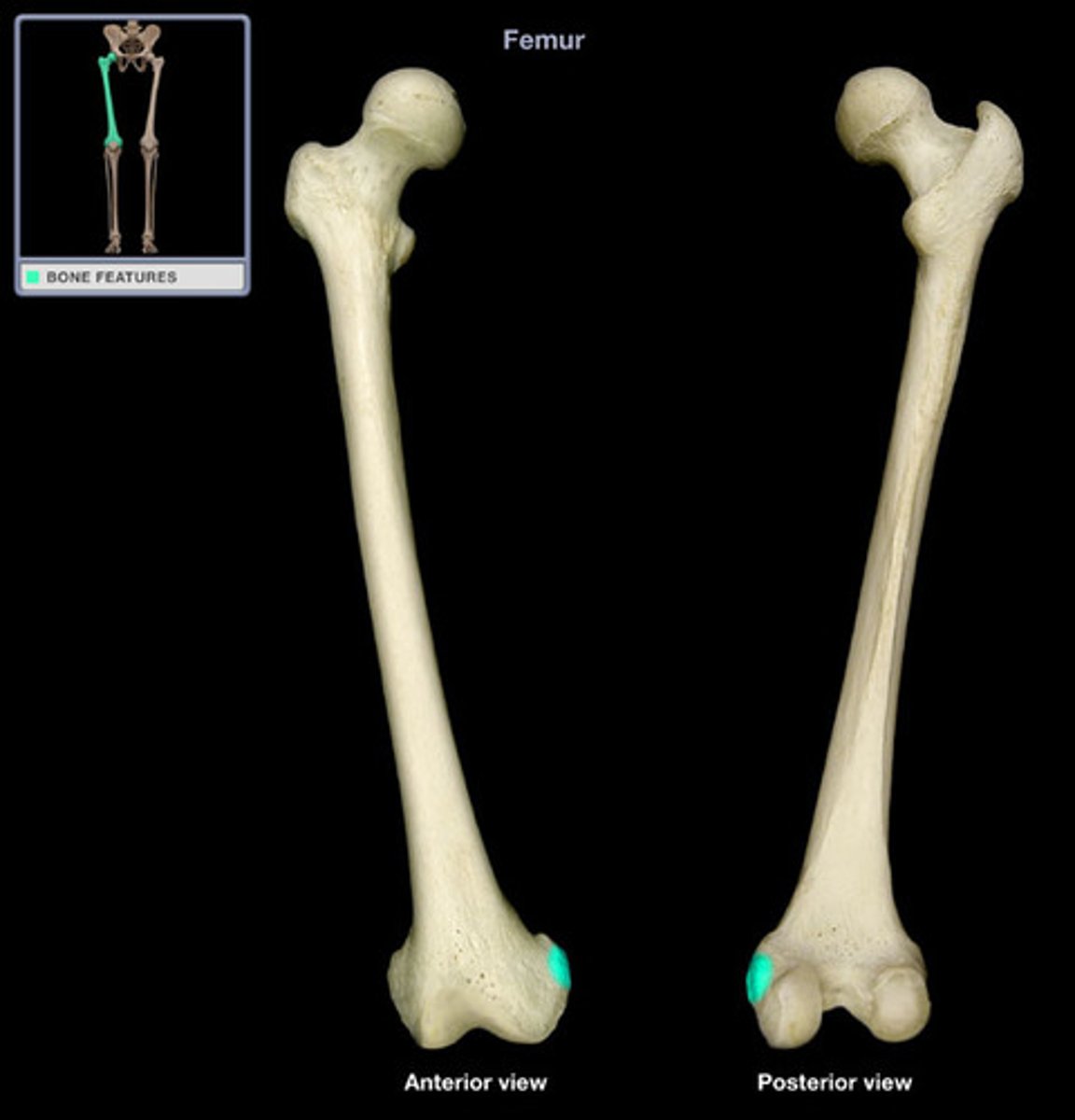
Lateral epicondyle of femur
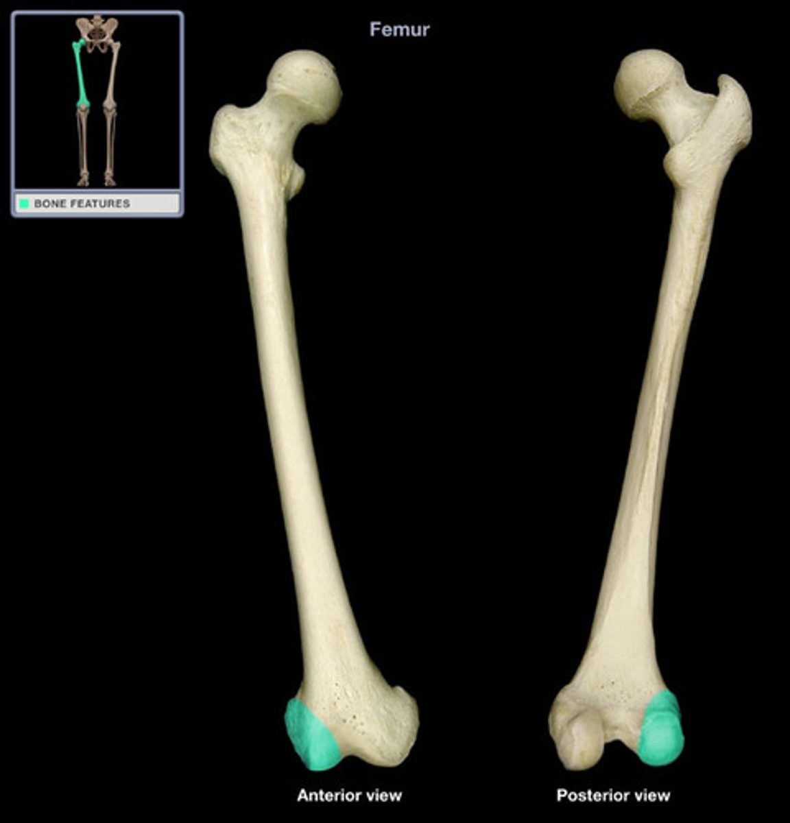
Medial condyle of femur
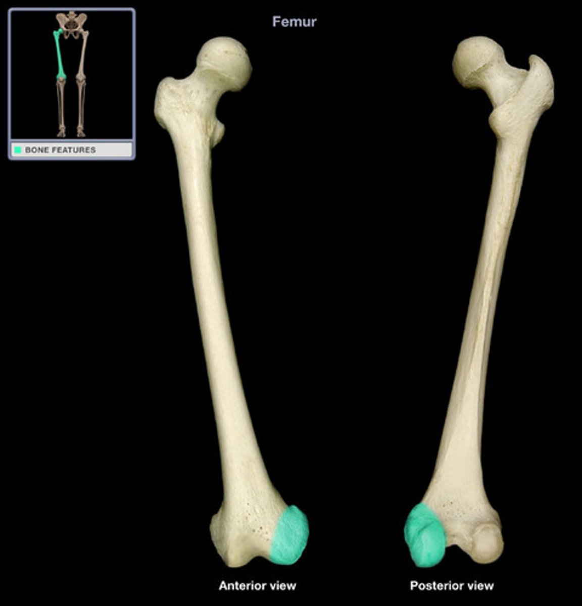
Lateral condyle of femur
Intercondylar notch
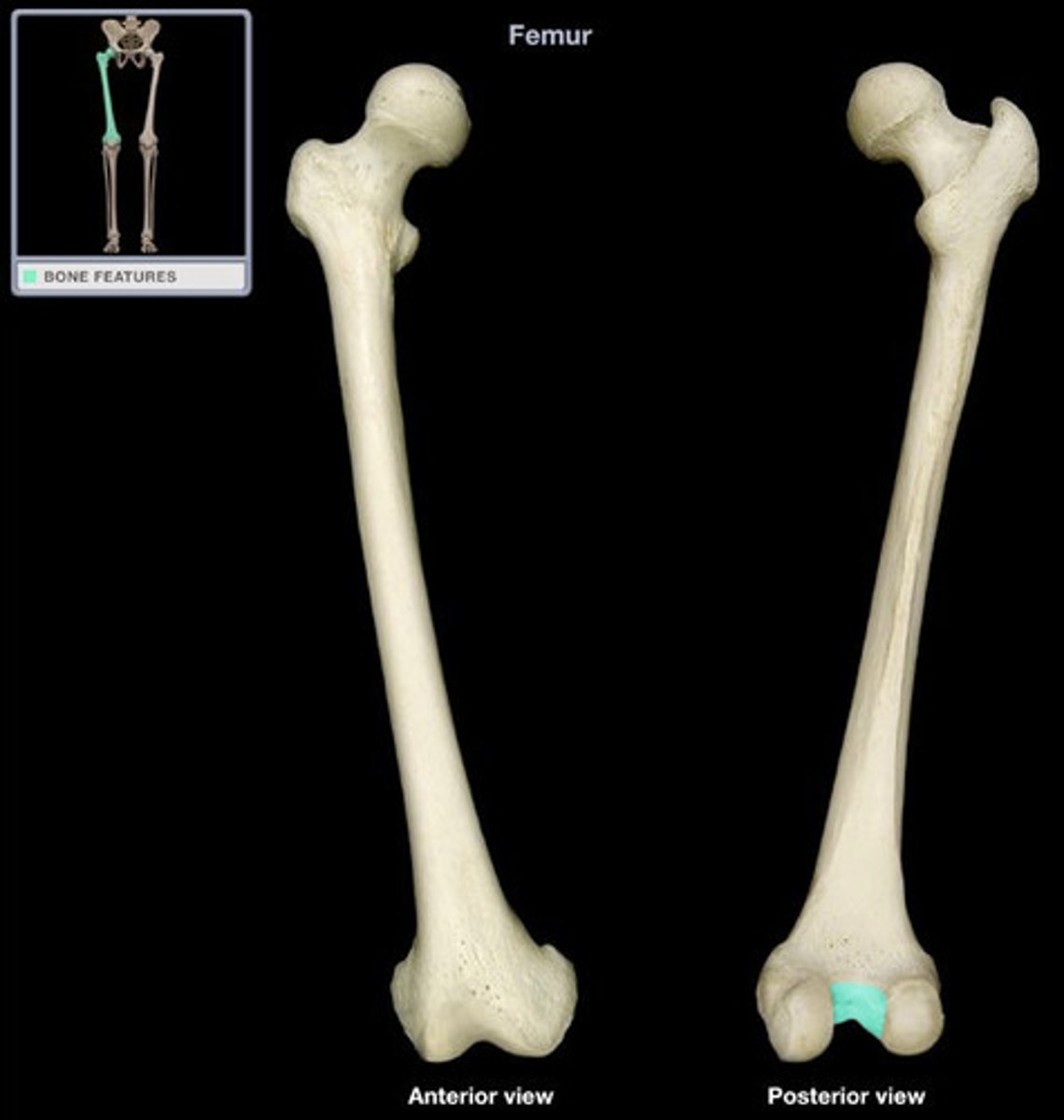
Popliteal surface
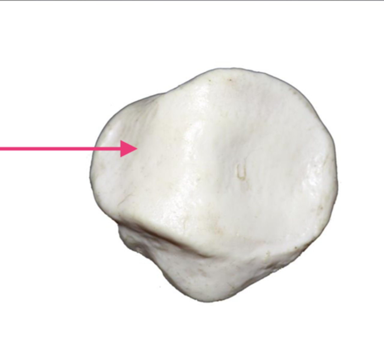
Patellar surface
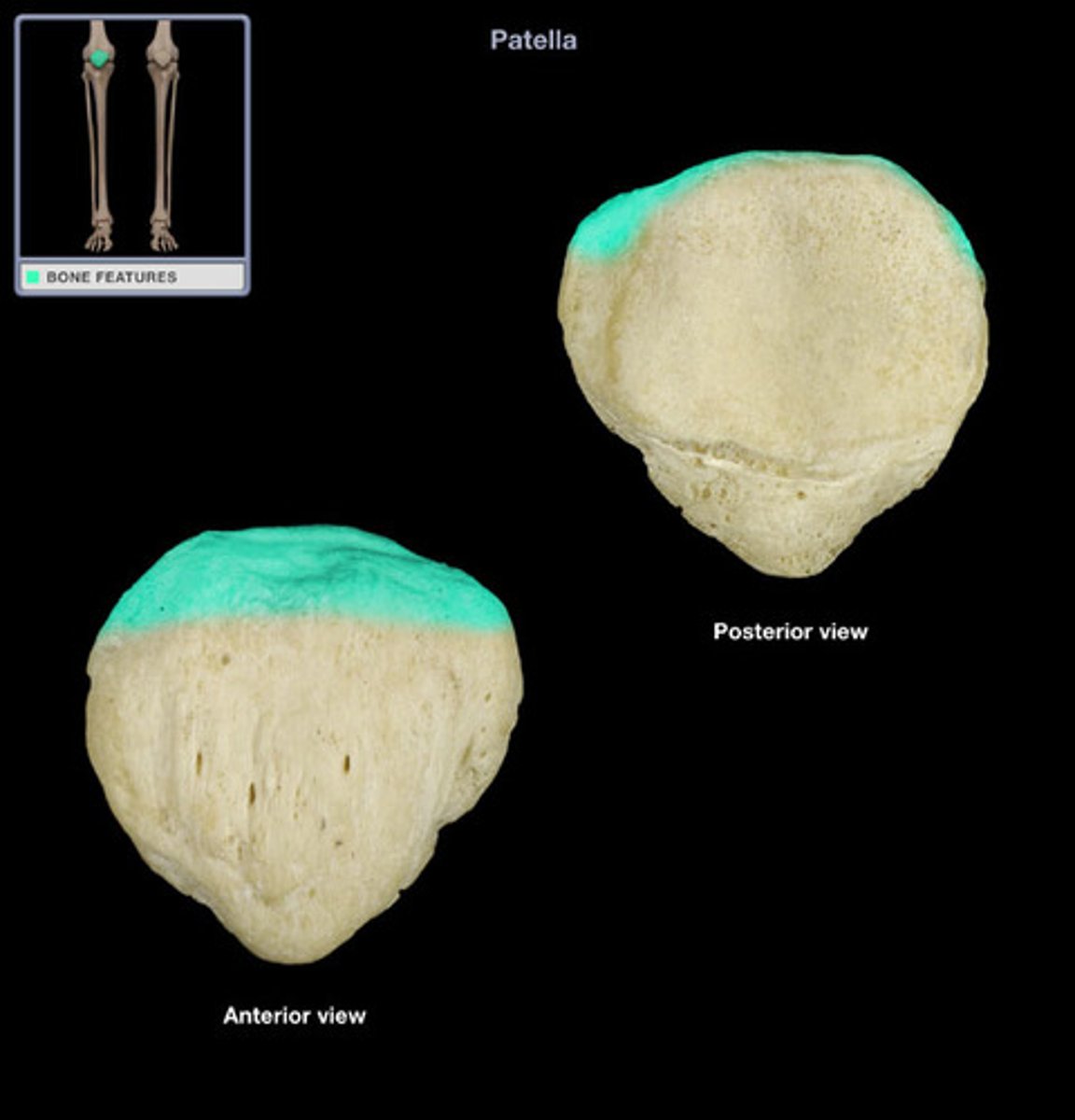
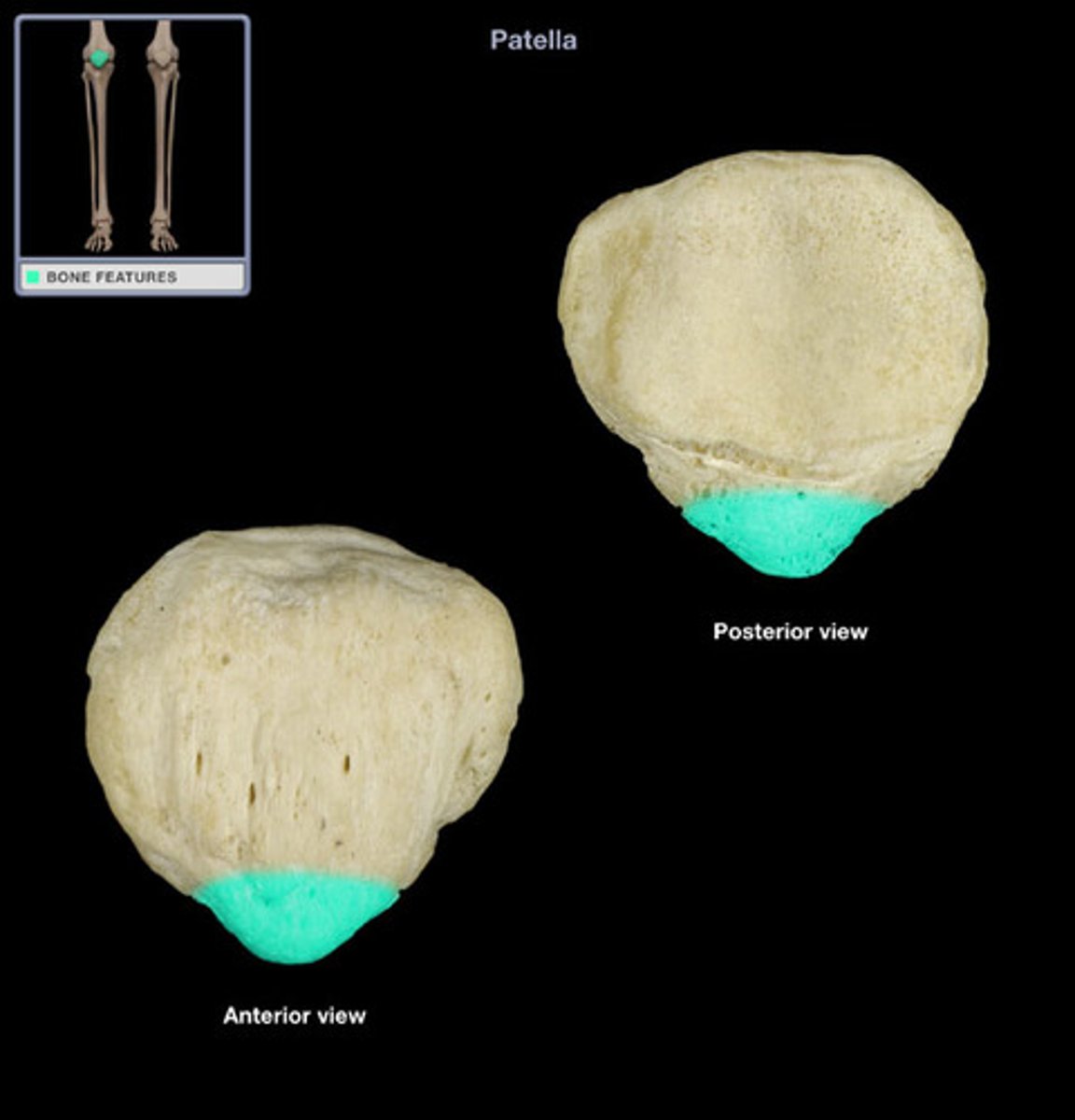
Patella
Base of patella
broad, flat surface at the proximal end
Apex of patella
point at the distal end
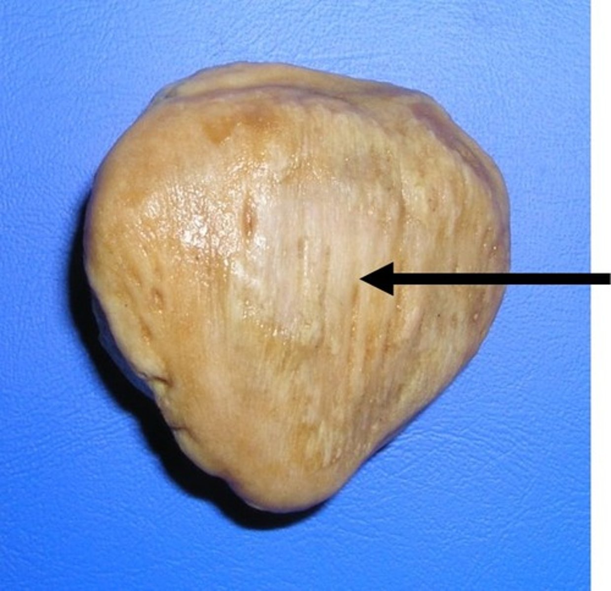
Anterior surface of patella
convex (bump upward) and rough
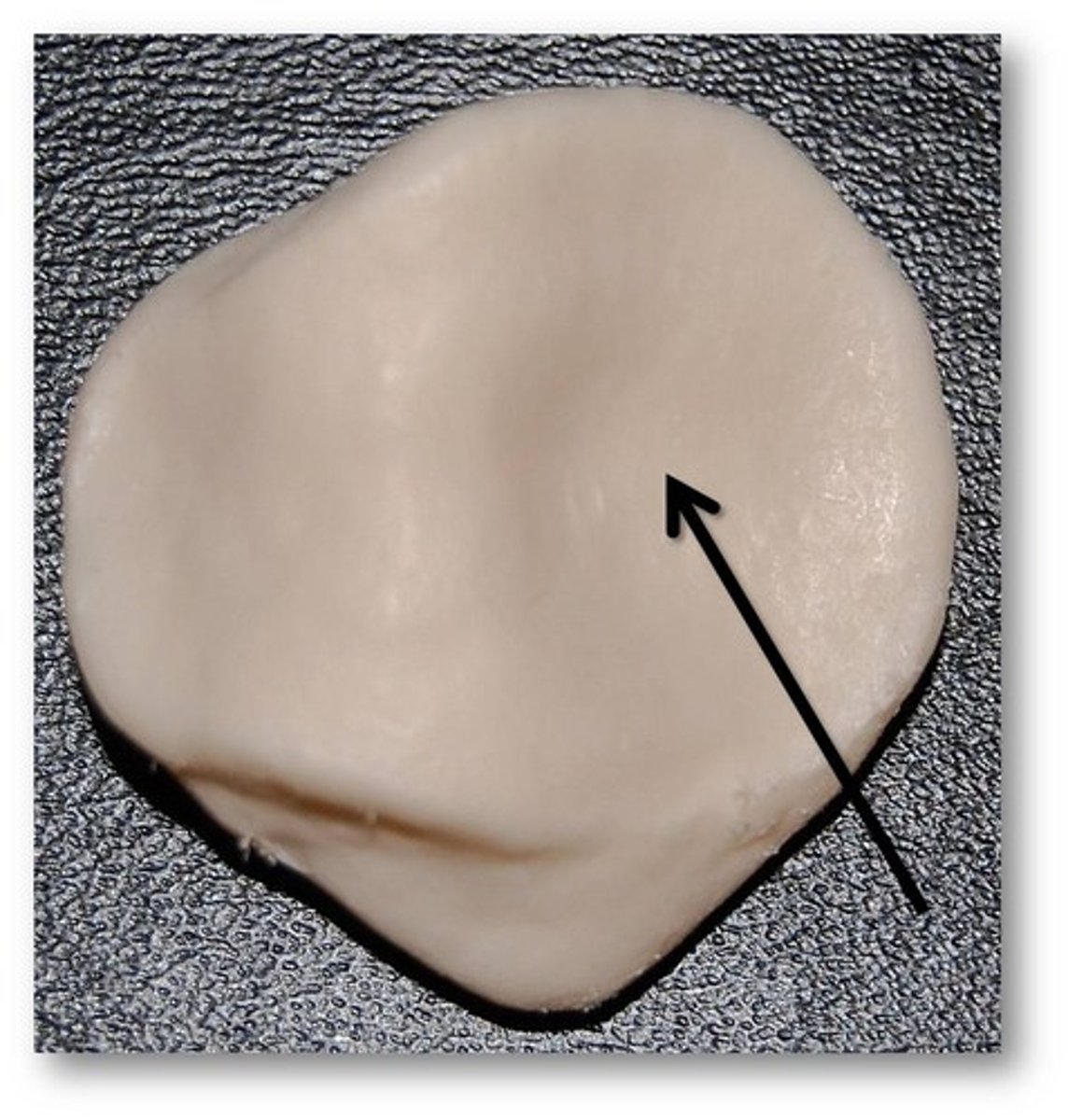
Articular surface of patella
articulates with the femur's patellar surface
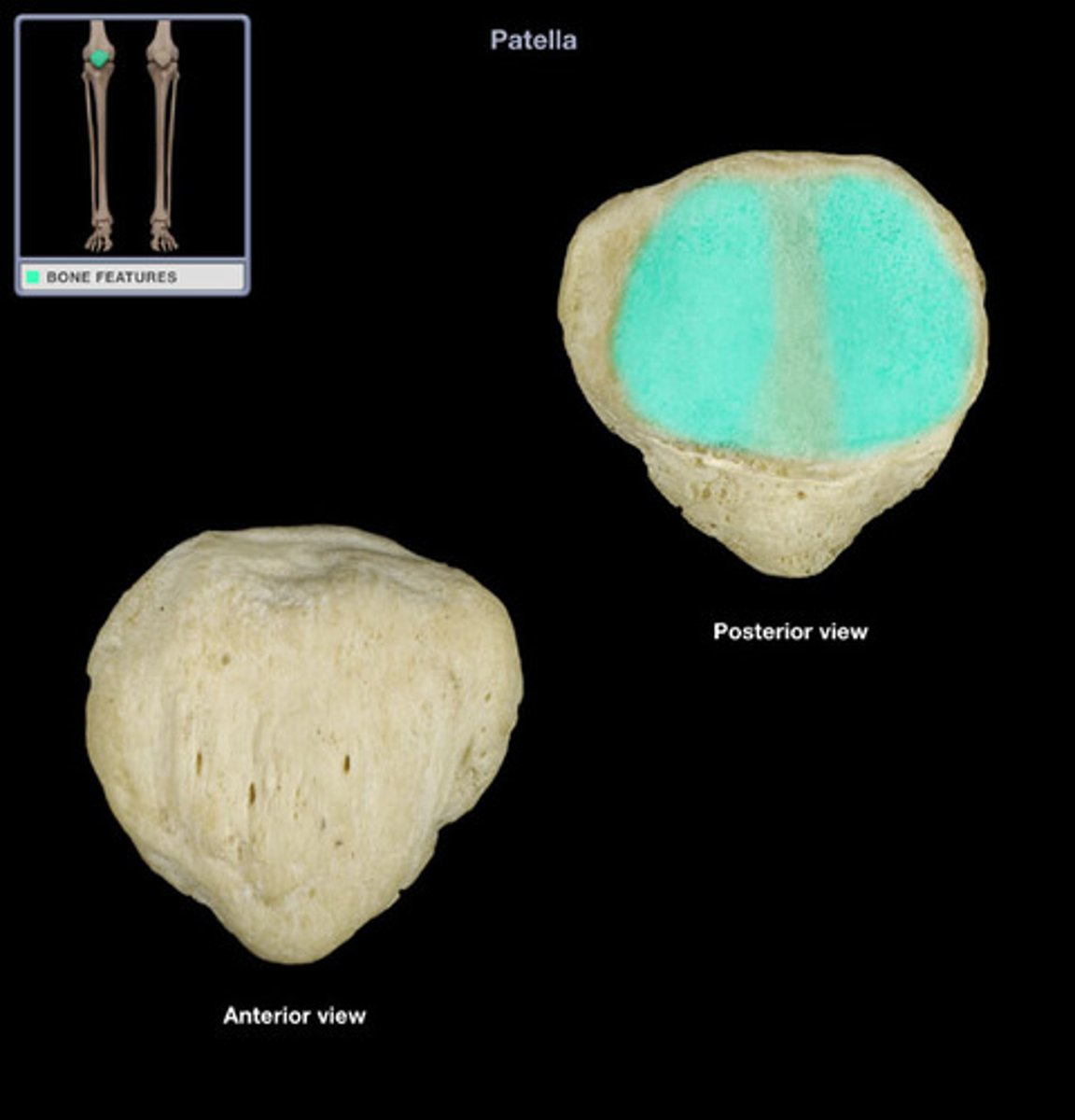
Medial facet of articular surface of patella
Lateral facet of articular surface of patella
Always rests on the larger lateral side
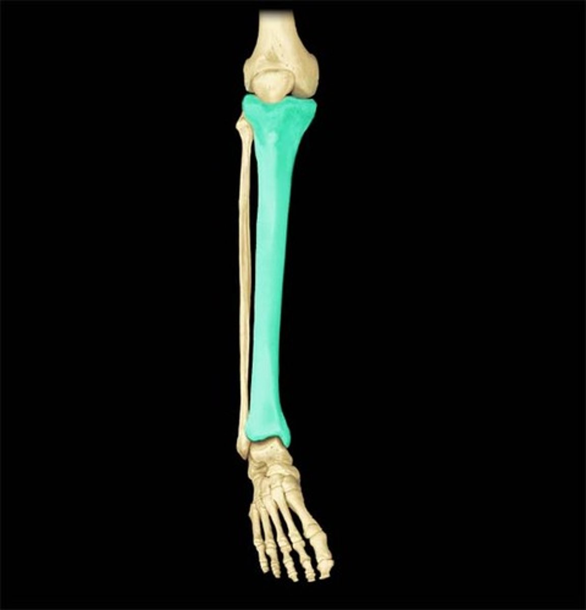
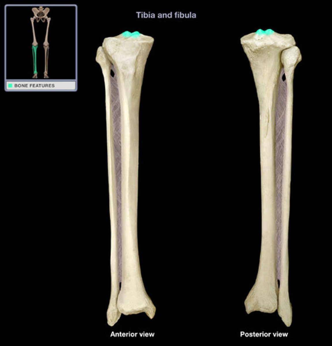
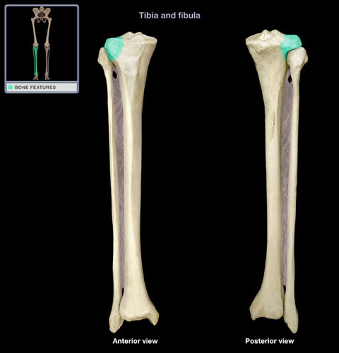
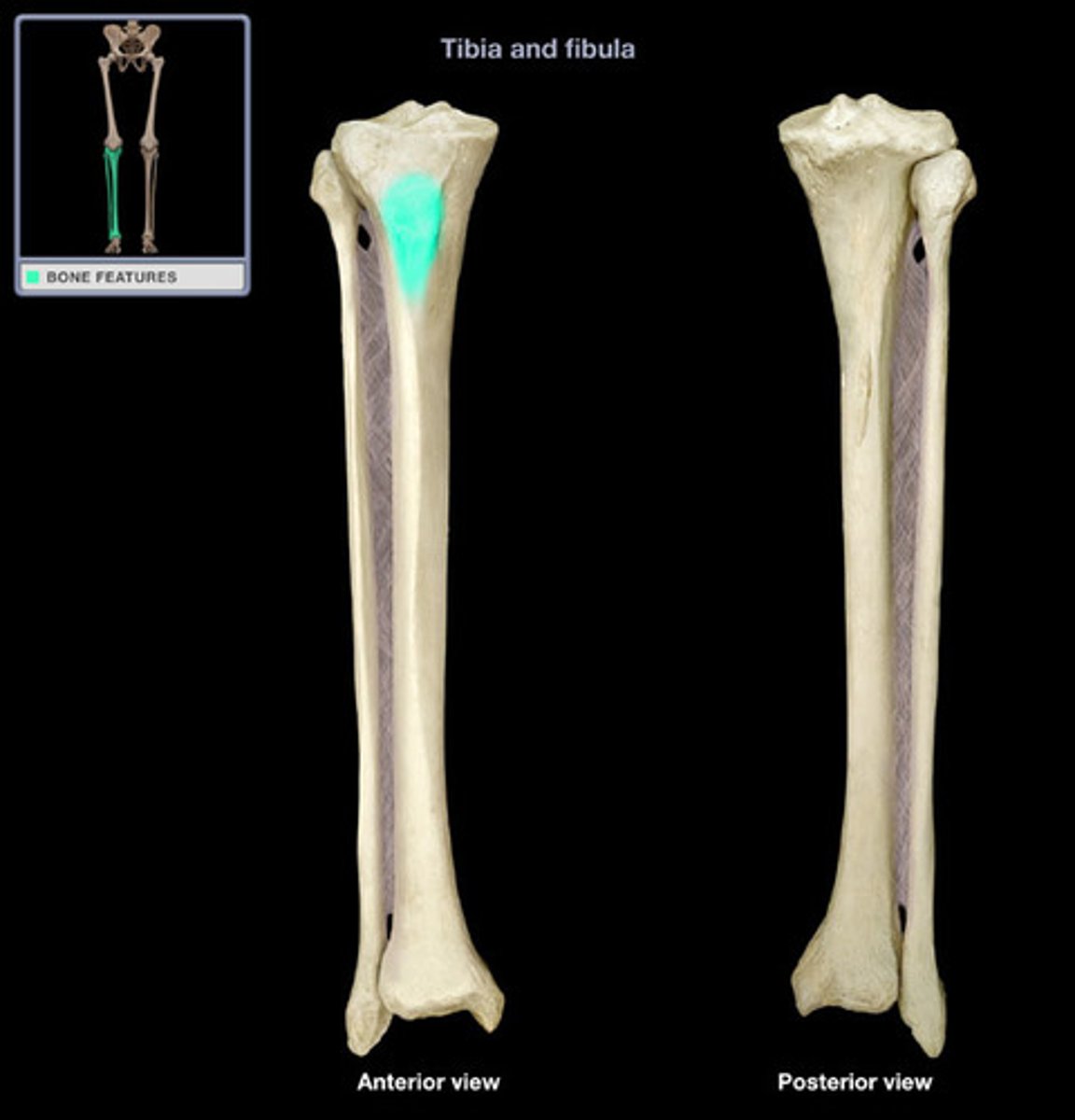
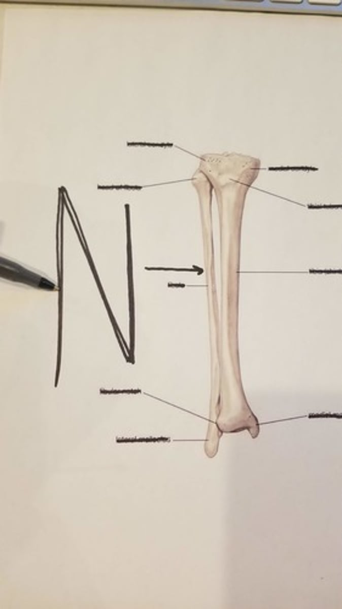
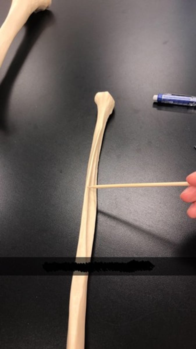
Tibia
shin bone
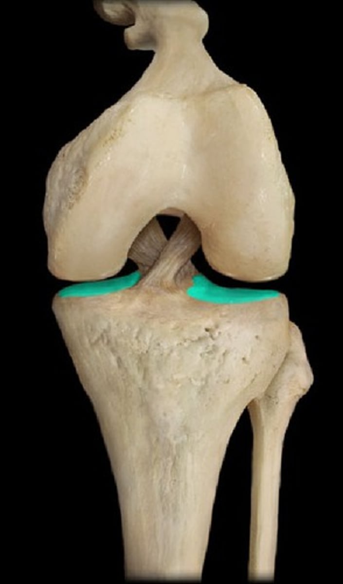
Tibial plateau
the top, flat portion of the tibia
Intercondylar eminence
Medial condyle of tibia
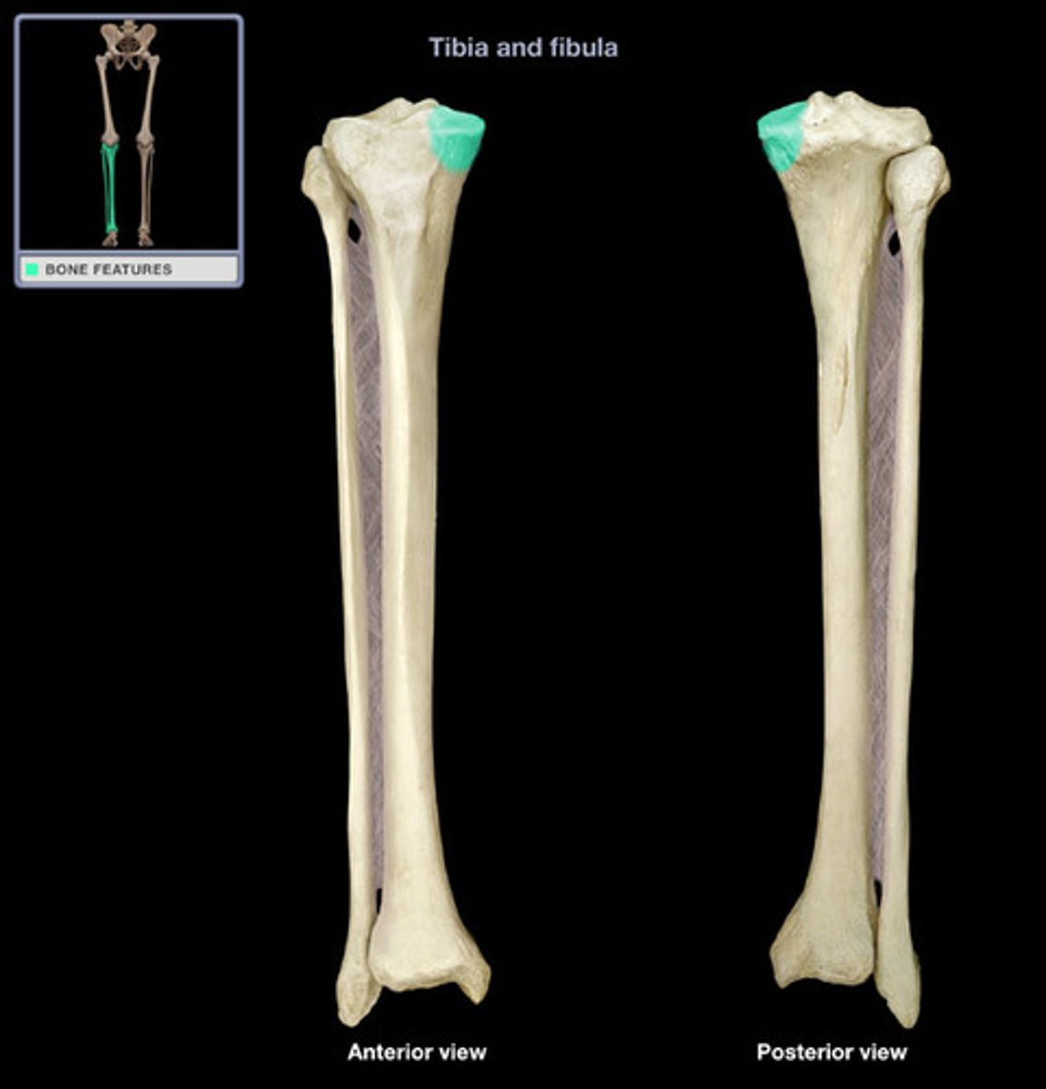
Lateral condyle of tibia
articulates with lateral condyle of femur
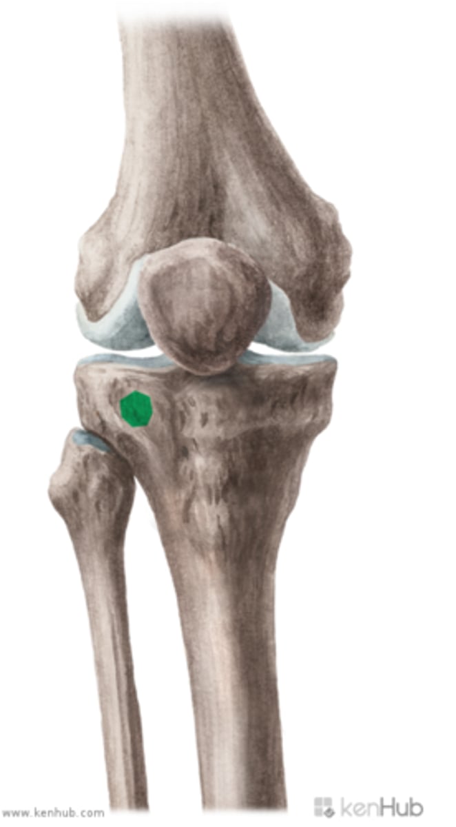
Tibial tuberosity
point where the patellar ligament attaches
Gerry's tubercle
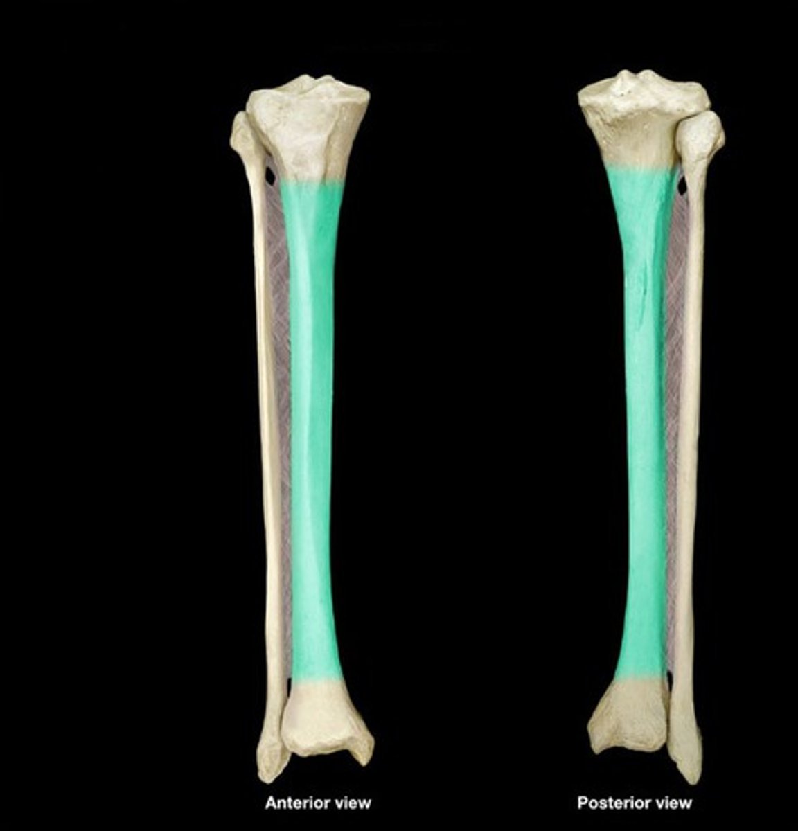
Shaft of tibia
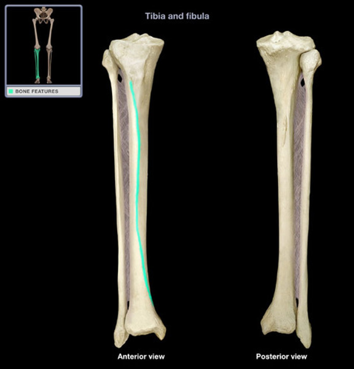
Anterior border of tibia
sharp ridge of bone easily palpated because it is close to the surface
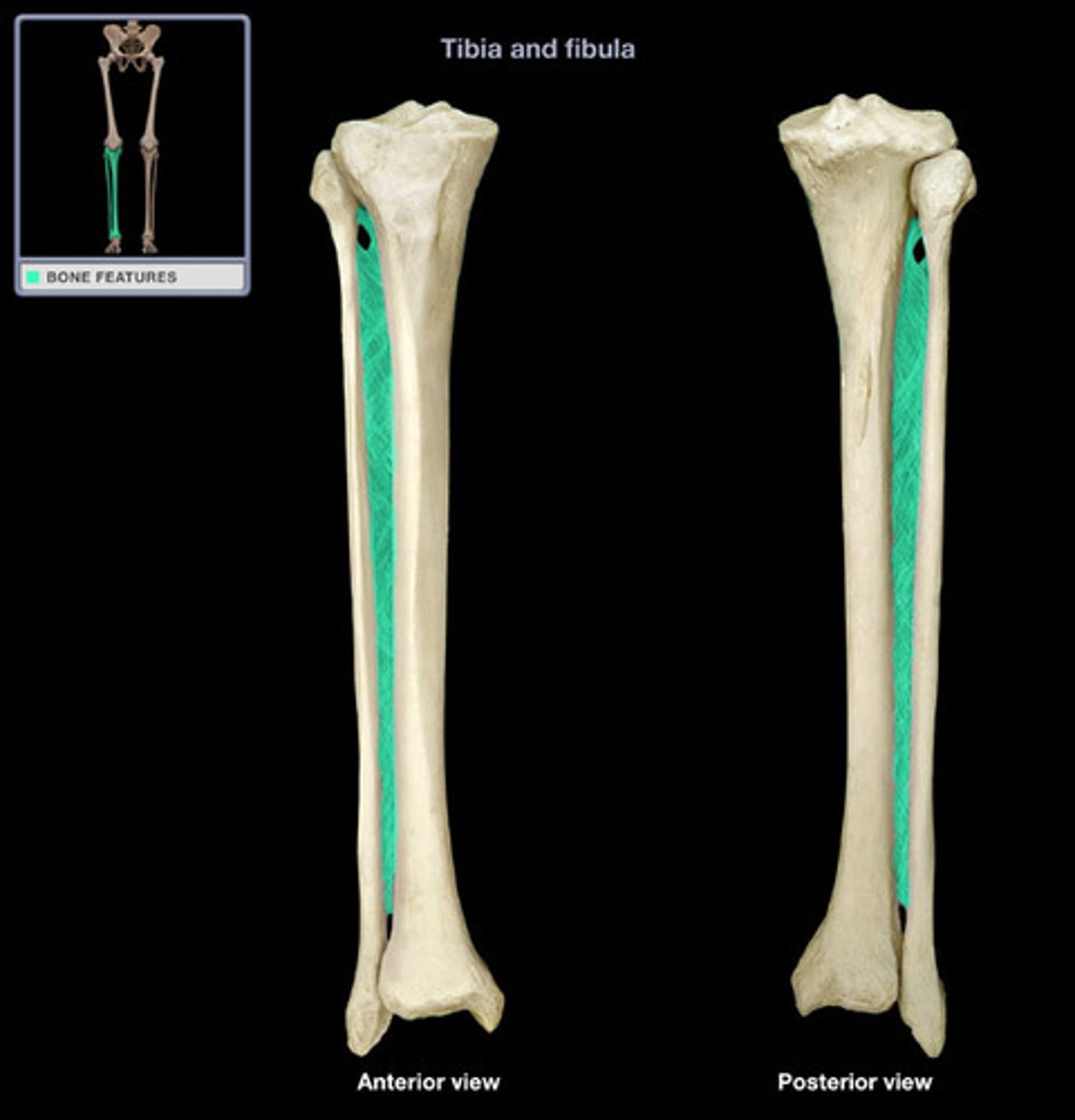
Interosseous border of tibia
small ridge running down the lateral side of the tibial shaft; for attachment of the interosseous membrane between the tibia and fibula
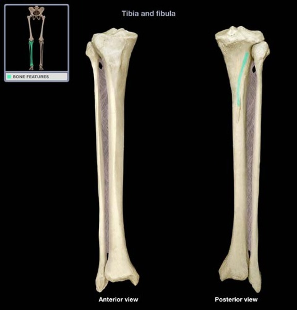
Soleal (popliteal) line
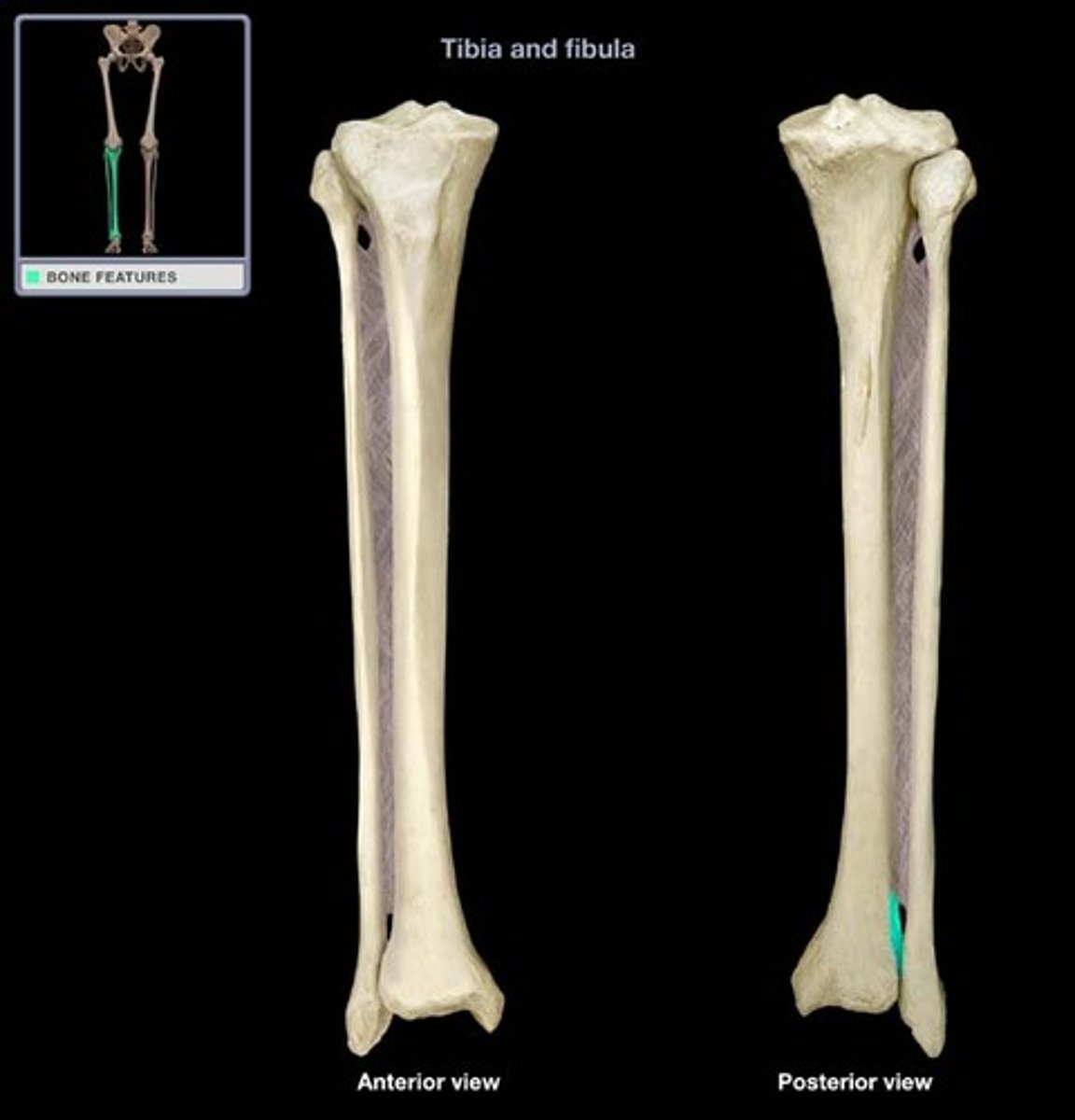
Fibular notch
lateral notch on the distal end tibia where tibia and fibula articulate
Medial malleolus

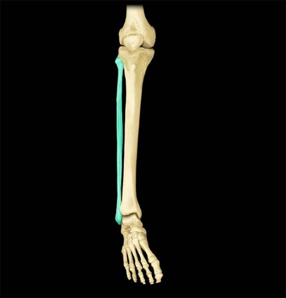
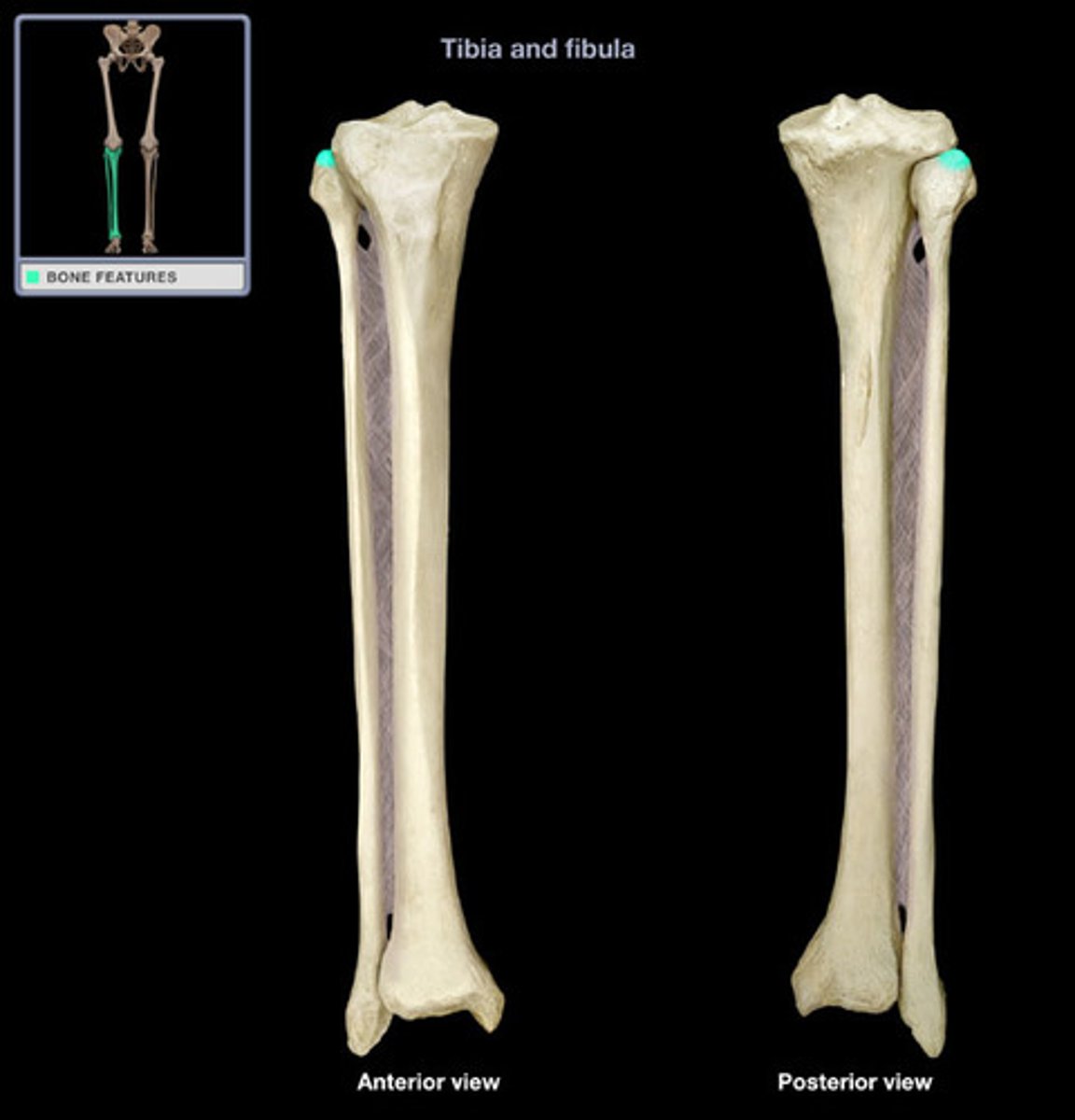
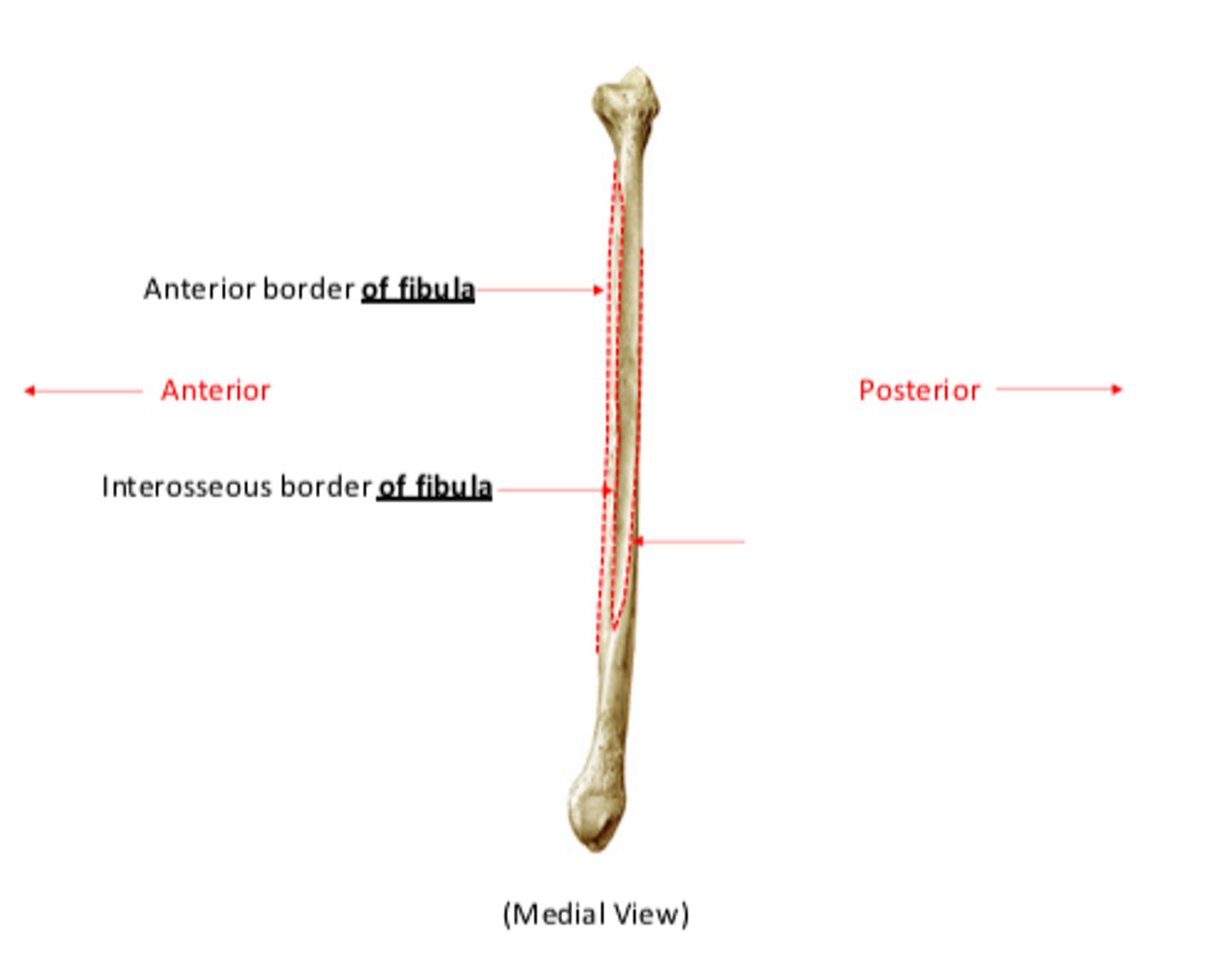
Fibula
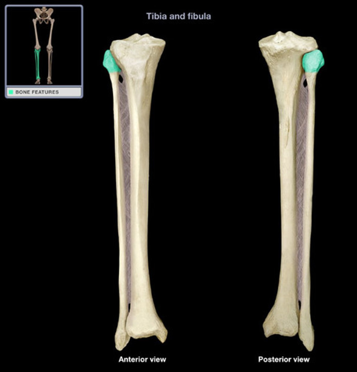
Head of fibula
proximal end of fibula
Styloid process of fibula (aka the apex of the fibula)
sharp pointy end (lateral malleolus side)
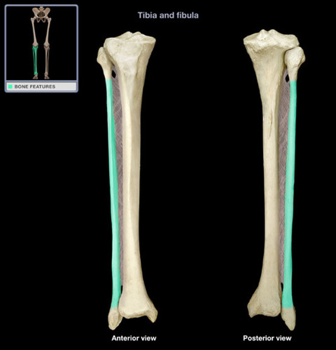
Shaft of fibula
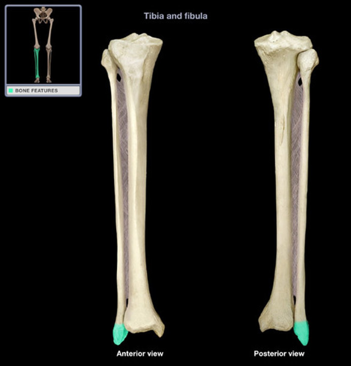
Lateral malleolus
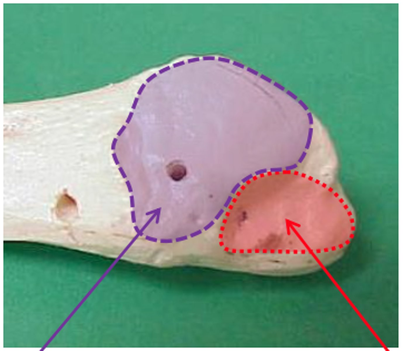
Articular facet of lateral malleolus
Purple

Lateral malleolar fossa
red
Anterior border of fibula
Interosseous border of fibula
Medial border of fibula
blank red arrow
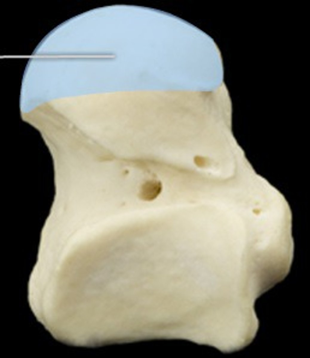
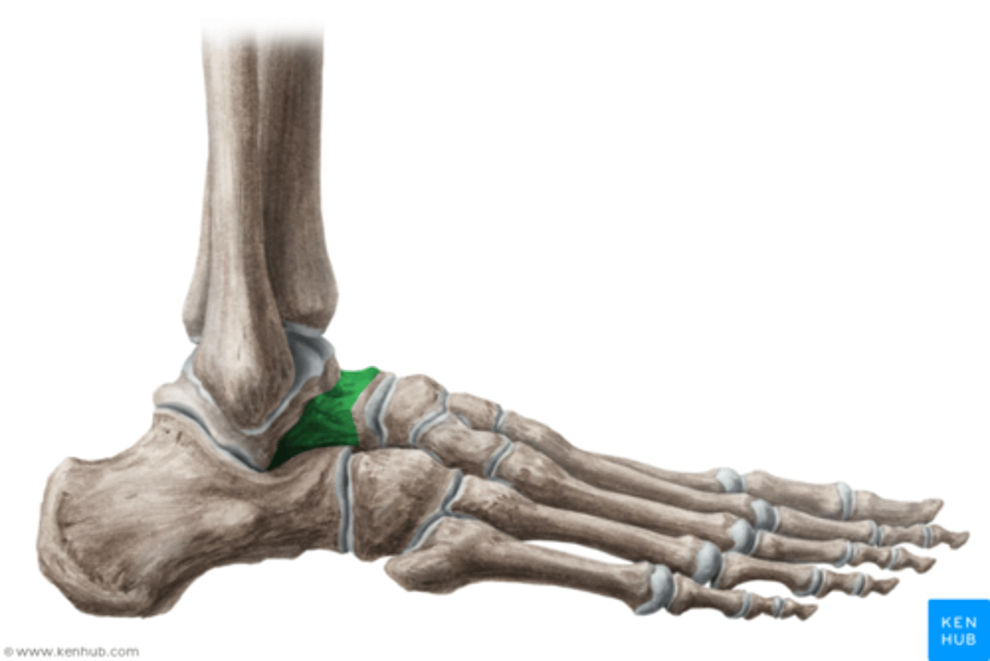
Talus
Talus (disarticulated)
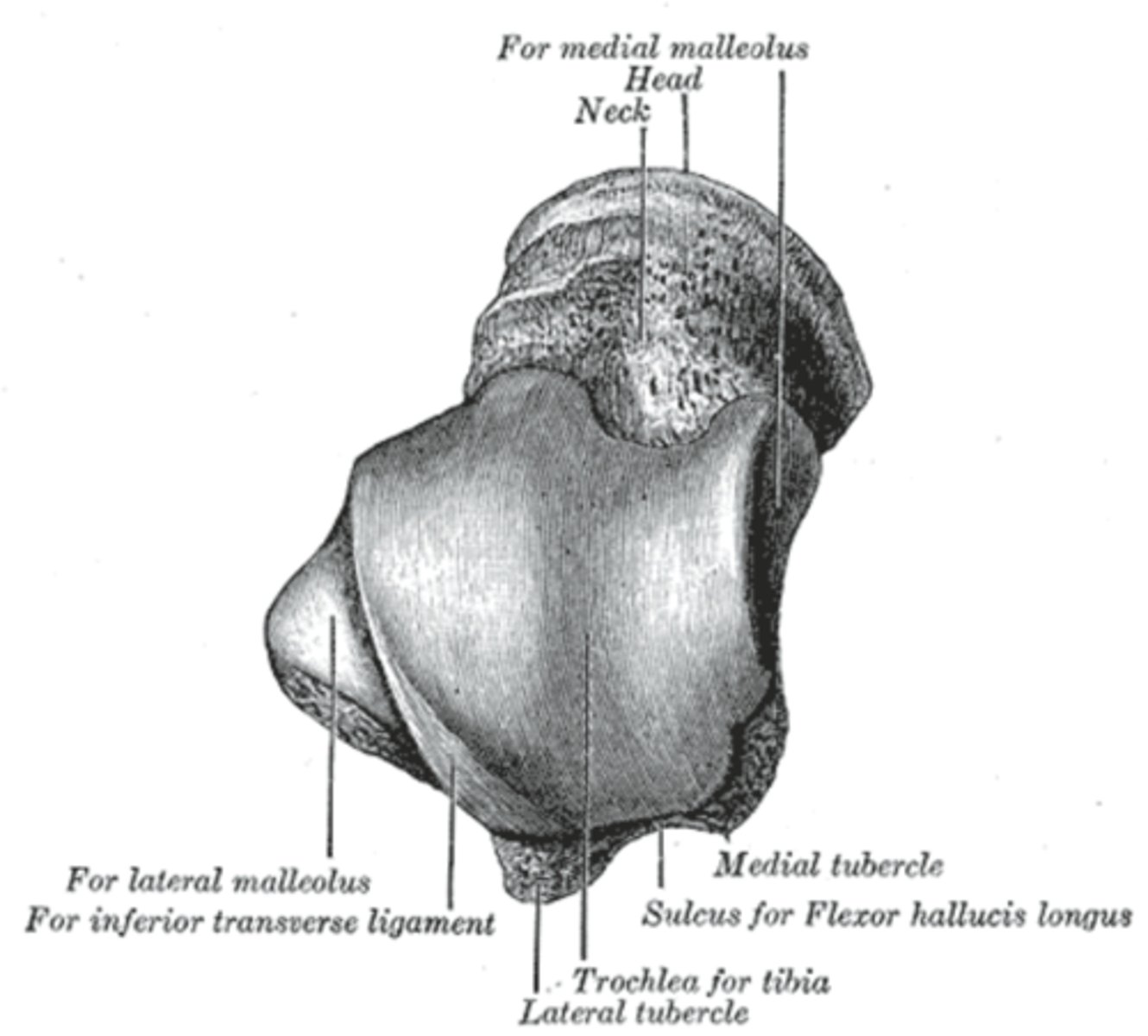
Head of talus
Neck of talus
Trochlea of talus
articulates with tibia
Medial tubercle
1
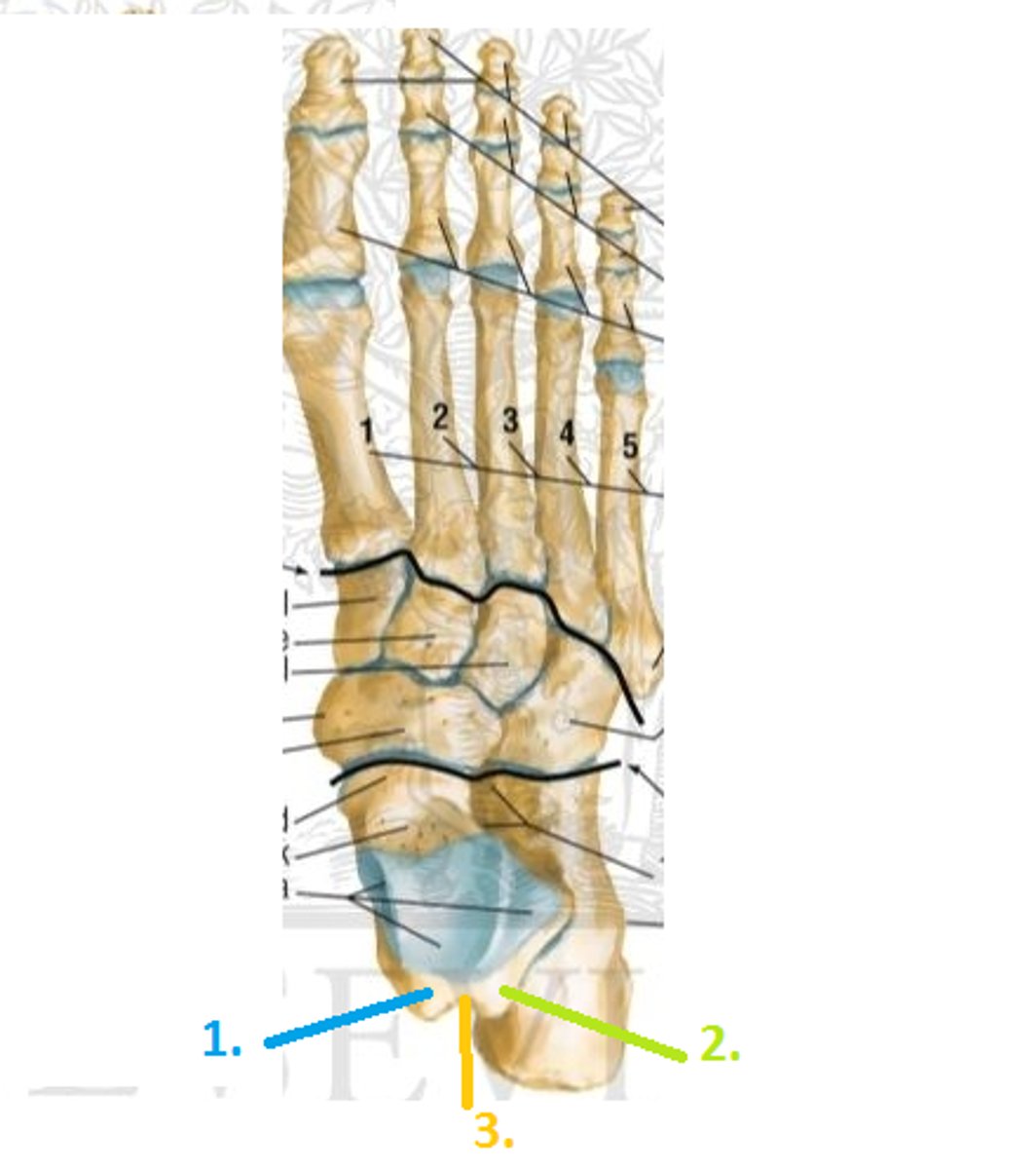
Lateral tubercle
2

Groove for flexor hallucis longus tendon on talus
3

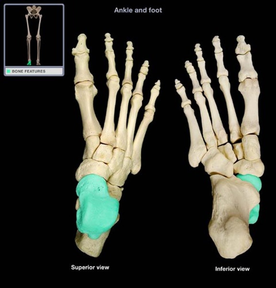
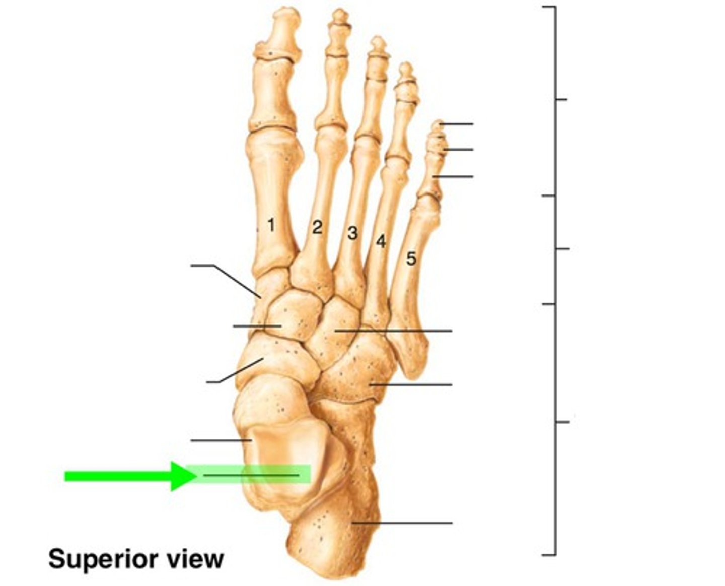
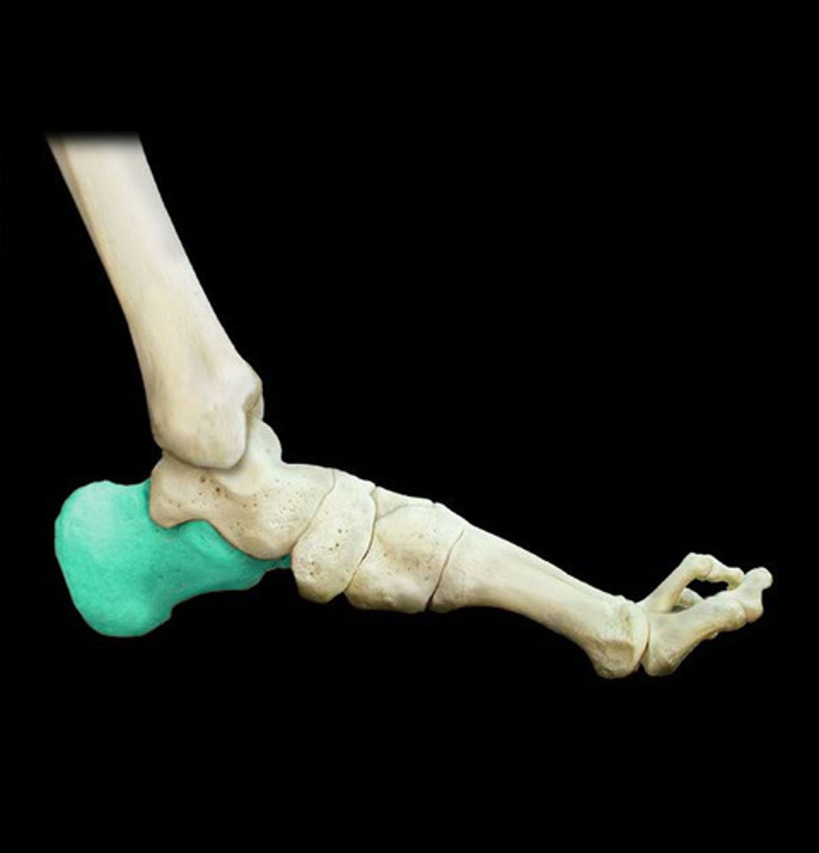
Calcaneus
heel bone
Sustentaculum tali
The anteromedial surface of the calcaneus that largely supports the talus.
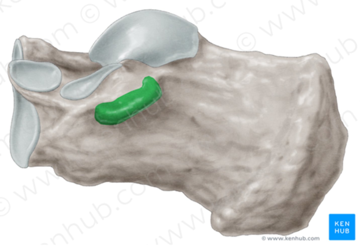
Groove for flexor hallucis longus tendon on calcaneus