Chapter 11: Serology Techniques: Past, Current, and Future
11.1: Introduction to Forensic Serology
- Forensic serology is the component of forensic biology that deals with the examination and identification of biological evidence.
- It focuses on determining the presence and identification of various bodily fluids such as blood, semen, and saliva in a questioned sample.
Class Characteristics and Individual Characteristics of Biological Evidence
- The identification of an unknown fluid sample is based on a comparison of the class characteristics of a sample with known standards of its class.
- Forensic identification typically involves bodily fluids, such as blood, semen, and saliva.
- If the presence of the bodily fluid is confirmed, the individual characteristics of the biological evidence are then determined to find out whether or not a bodily fluid sample has come from a particular individual.
- Short Tandem Repeats (STR): The most commonly utilized forensic DNA analysis used for human identification.
- Y-chromosomal STR analysis is often utilized for the investigation of sexual assault crimes.
- Mitochondrial DNA analysis is used for the identification of human remains.
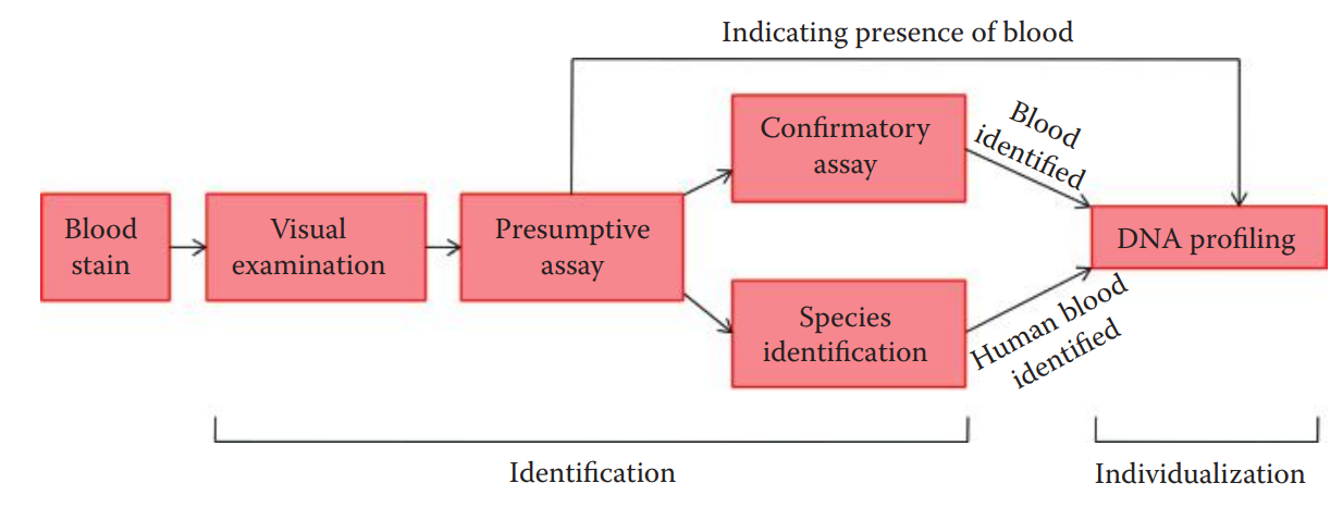
- Establishing the probative value of a sample requires both the identification of its class characteristics and the individualization of its contributor.
Presumptive and Confirmatory Assays
- The identification of bodily fluids can be carried out using presumptive and confirmatory assays to identify the type of bodily fluid in question.
- Presumptive Assays
- Positive assay indicates the possibility of the presence of the bodily fluid in question.
- Negative assay indicates that the questioned bodily fluid is absent.
- Confirmatory Assays
- These assays are utilized to identify bodily fluids with higher certainty than presumptive assays.
- These are performed when a sample has to be identified as blood.
Primary and Secondary Binding Assays
- Primary Binding Assays
- It involves the initial binding between a single epitope of an antigen and a single binding site of an antibody.
- They are very sensitive assays.
- Secondary Binding Assays
- They are less sensitive and easier to perform than primary binding assays.
- Precipitation-based assays have been used for species identification.
- Agglutination-based assays are more sensitive. It detects antigens located on the surface of cells or carriers, which are normally applied to blood group typing.
11.2: Primary Binding Assays
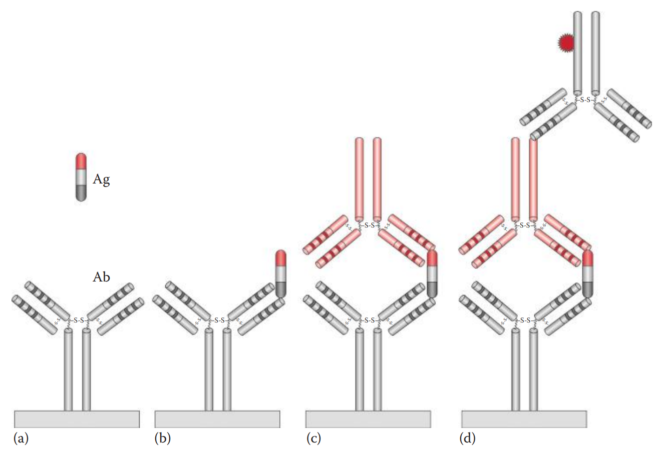
Enzyme-Linked Immunosorbent Assay (ELISA)
- It is an immuno-enzyme assay that can be used to detect and measure the antibody or antigen in question.
- Antibody-sandwich ELISA: The most common ELISA that is used in forensic serology.
- It is utilized to detect the prostate-specific antigen (PSA) to identify seminal stains and amylase for the identification of saliva.
- An antibody coating is formed by nonspecific adsorption onto a solid phase such as the wells of a polystyrene plate.
- Several enzymes such as alkaline phosphatase and horseradish peroxidase have been used as reporting enzymes to label the antiglobulin for ELISA.
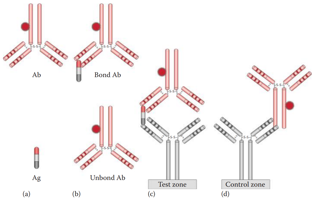
Immunochromatographic Assays
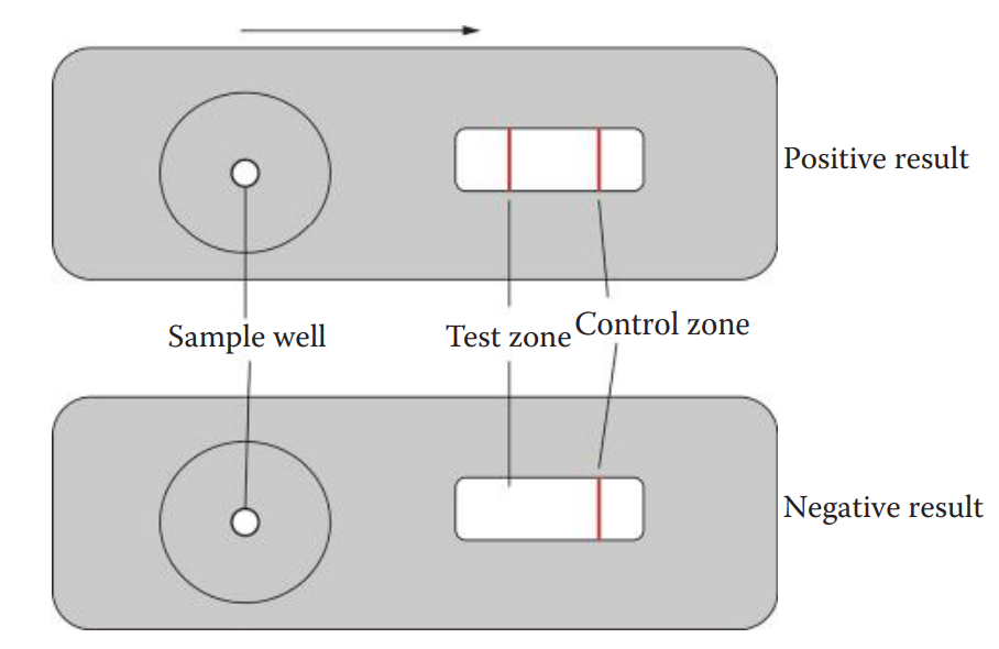
- The assay is carried out by loading a sample into the sample well.
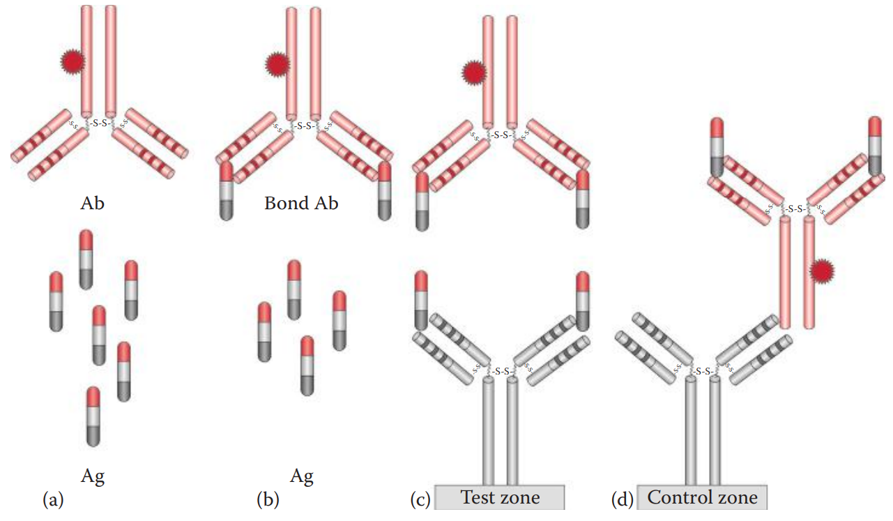
- The antigen in the sample binds to the dye-labeled antibody already in the sample well to form an antigen–antibody complex.
- The complex then diffuses across the nitrocellulose membrane until it reaches the test zone.
- The antibody immobilized at the test zone traps the antigen–antibody complex to form an antibody–antigen–antibody sandwich.
- The presence of the antigen in the sample results in a colored vertical line at the test zone.
- The immunochromatographic device also utilizes a control zone to ensure that the device works properly and that the sample has diffused completely along the test strip.
Common Immunochromatographic Assays for Forensic Applications
| Assay | Antigen | Labeled Antibody | Immobilized Antibody | Forensic Application |
|---|---|---|---|---|
| ABAcard®; HemaTrace® | Hemoglobin (Hb) | Monoclonal anti-human Hb antibody | Polyclonal anti-human Hb antibody | Blood and species identification |
| RSID-Blood | Glycophorin A (GPA) | Monoclonal antihuman GPA antibody | Monoclonal antihuman GPA antibody | Blood and species identification |
| RSID-Saliva | Human salivary α-amylase (HAS) | Monoclonal antihuman HAS antibody | Monoclonal antihuman HAS antibody | Saliva identification |
| One-Step ABAcard PSA® | Prostate-specific antigen (PSA) | Monoclonal antihuman PSA antibody | Polyclonal antihuman PSA antibody | Semen identification |
| RSID-Semen | Semenogelin (Sg) | Monoclonal antihuman Sg antibody | Monoclonal antihuman Sg antibody | Semen identification |
11.3: Secondary Binding Assays
Precipitation-Based Assays
- Immunodiffusion
- A passive method in which an antigen or an antibody or both are allowed to diffuse and therefore a gradient, from low to high concentration, is established for an antigen or an antibody or both.
- Single Immunodiffusion: A concentration gradient is established for either an antigen or an antibody.
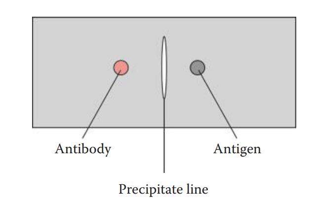
- Double Immunodiffusion: A concentration gradient is established for both an antigen and an antibody.
- The Ouchterlony assay is named after the Swedish immunologist, Örjan Ouchterlony, who developed it. The assay can be performed in an agarose gel supported by a glass slide or polyester film
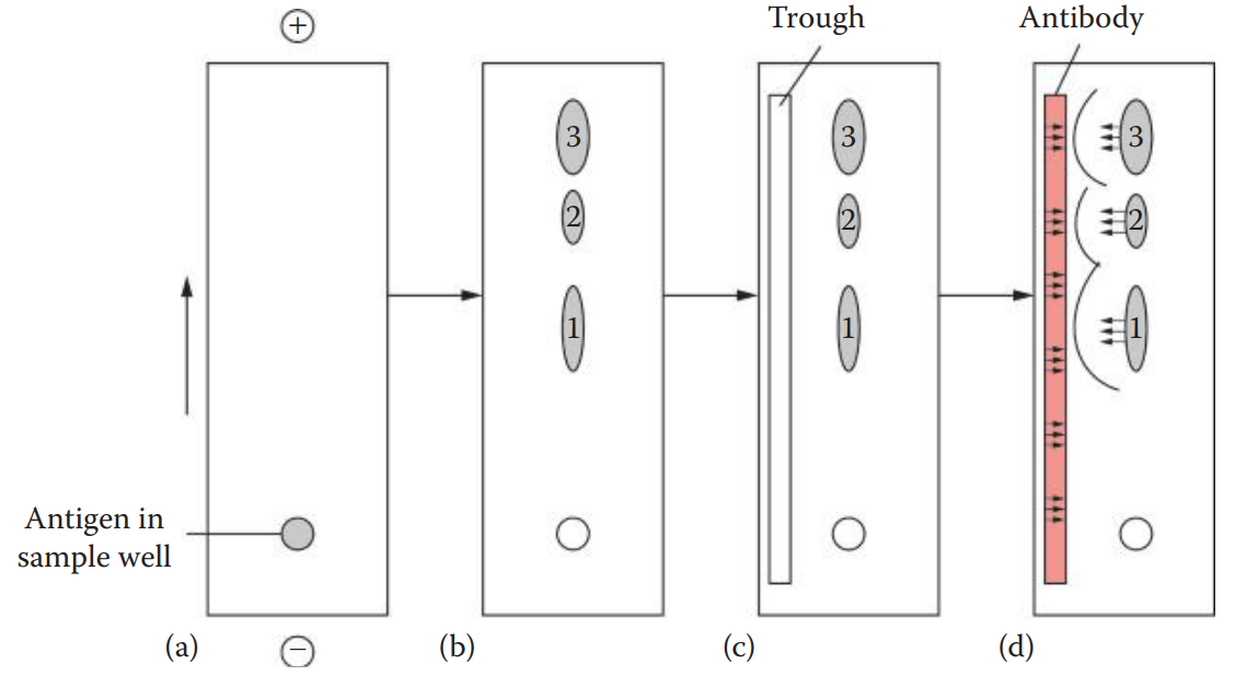
- Immunoelectrophoretic Methods
- Immunoelectrophoresis (IEP): This technique uses electrophoresis to separate the antigen mixture before immunodiffusion.
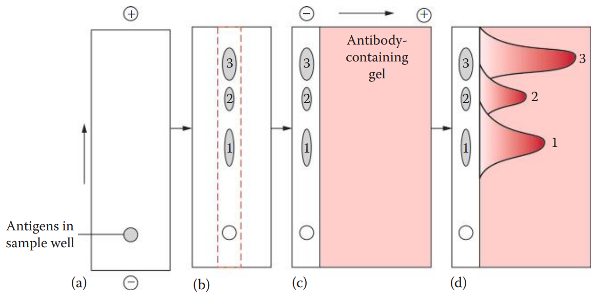
- Crossed Immunoelectrophoresis (CRIE): Also known as two-dimensional IEP, is a modification of IEP.
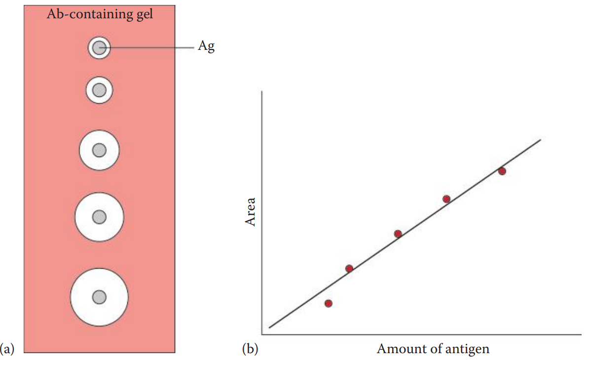
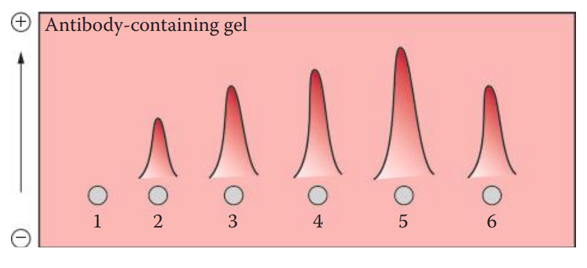
- Rocket Immunoelectrophoresis: An antibody-containing agarose gel is used. The antigen is loaded into the well.
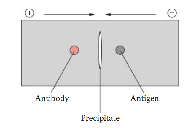
- Crossed-Over Immunoelectrophoresis: This technique is also known as counterimmunoelectrophoresis (CIE).
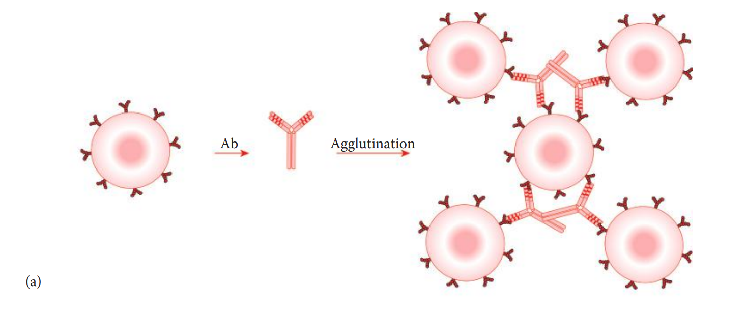
Agglutination-Based Assays
- Agglutination reactions can be used as forensic serological assays such as for blood group typing and menstrual blood identification.
- Agglutination assays are qualitative, indicating the absence or presence of antigens or antibodies.
- Semi-quantitative assays results can be obtained by titration.
- Direct agglutination assays involve reactions in which an antibody interacts with antigens originally located on cell surfaces.
- In a hemagglutination reaction, an antibody binds to the antigens located on erythrocytes. This method is used for the identification of blood types.
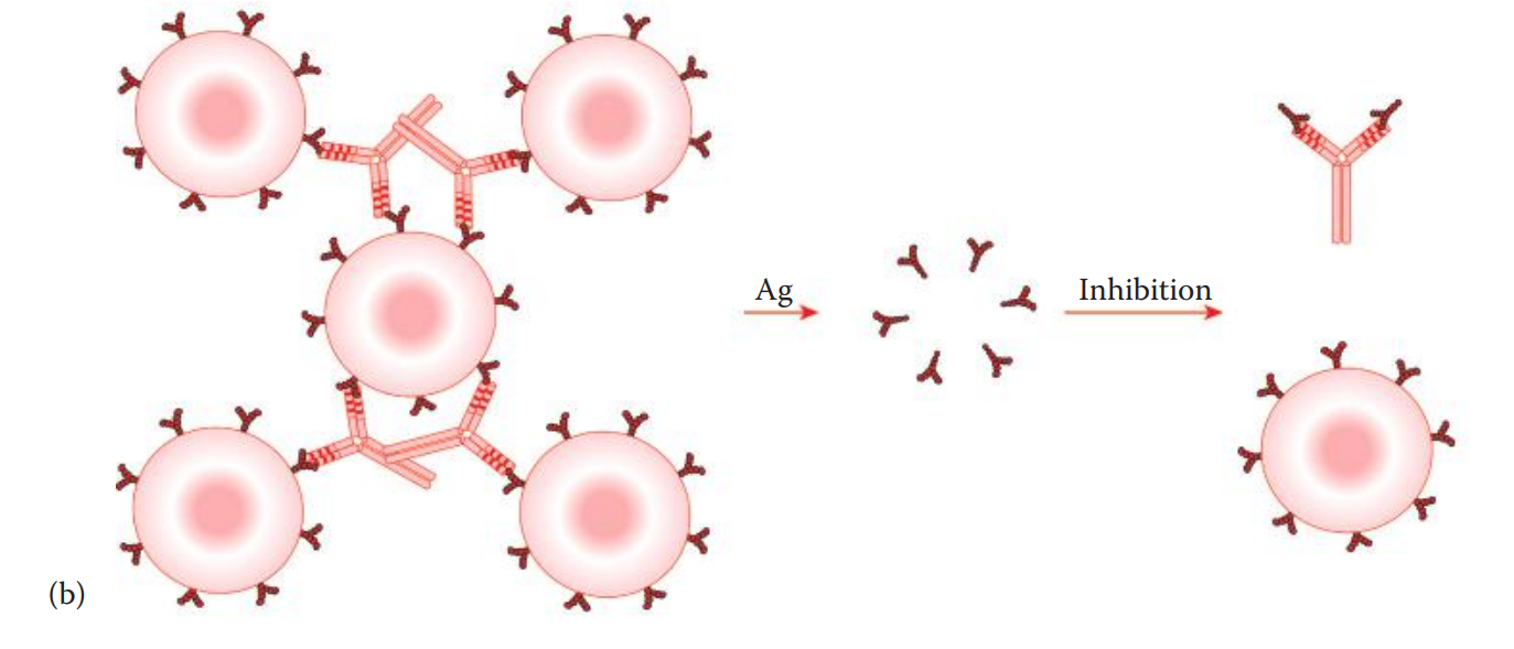
- Agglutination inhibition assays: The presence of an antigen in question is indirectly detected.
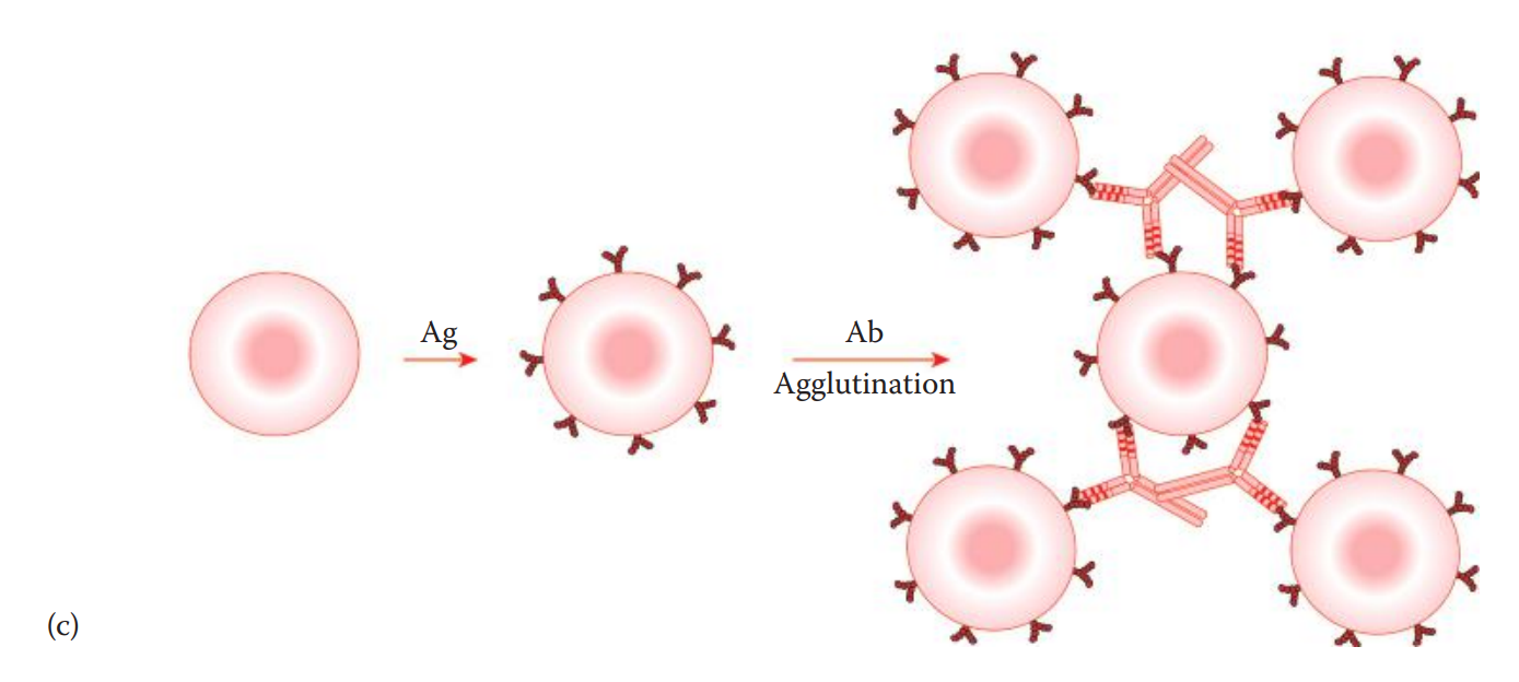
- Passive agglutination assays: The antigen is coated on the surface of carrier cells such as tannic acid–treated sheep erythrocytes.
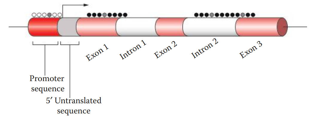
11.4: DNA Methylation Assays for Bodily Fluid Identification
- In the eukaryotic genome, methylation occurs at the cytosine residues commonly in the CpG dinucleotide sequences of both DNA strands.
- The “p” in CpG refers to the phosphodiester bond between cytosine and guanine.
- Cytosine methylation is carried out in vivo by a methyltransferase.
- Genomic Loci: Also known as tissue-specific differentially methylated regions (TDMRs).
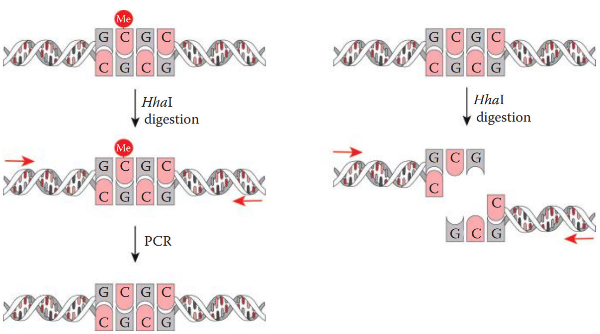
- Methylation-sensitive restriction enzyme digestion polymerase chain reaction (MSRE-PCR) can be used for the rapid detection of DNA methylation.
- This method utilizes the methylation-sensitive restriction enzyme (MSRE).
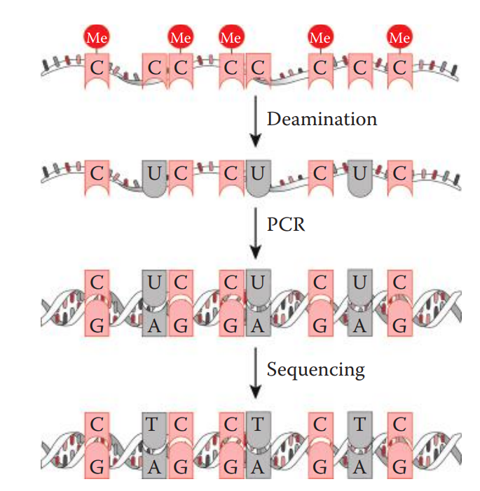
- Bisulfite sequencing: The genomic DNA is treated with sodium bisulfite, which catalyzes the hydrolytic deamination of unmethylated cytosines.
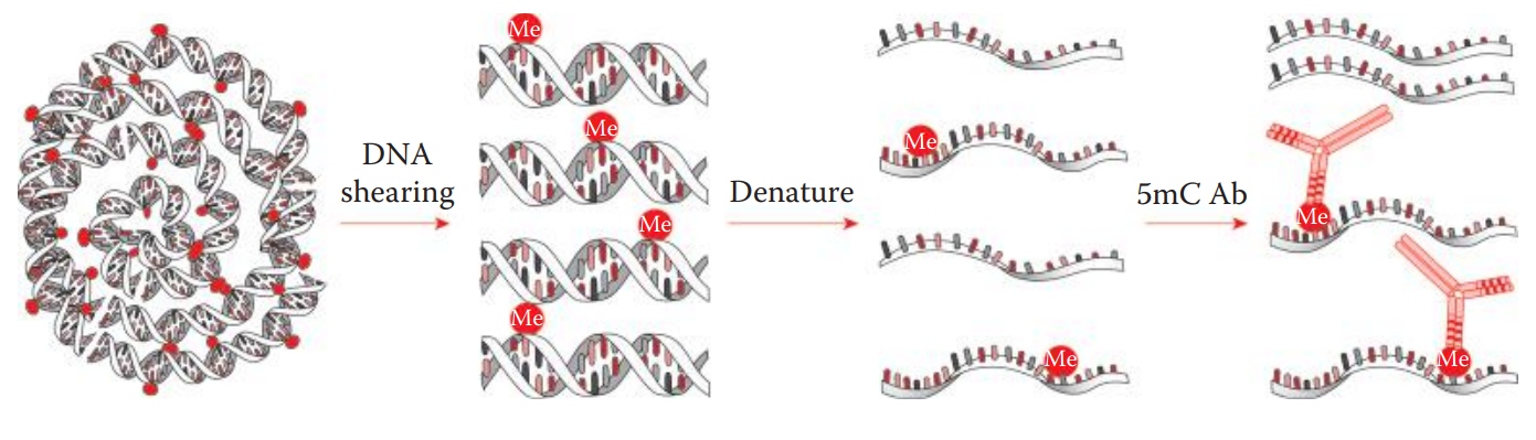
- Methyl-DNA immunoprecipitation (MeDIP) is a useful method for isolating methylated DNA.
11.5: Forensic Applications of RNA-Based Assays and RNA Profiling
Messenger RNA-Based Assays
- mRNA-based Assays can potentially be used for wound age estimation and the age of biological stains.
- The tissue-specific genes that are utilized for bodily fluid identification.
- Reference genes: Constitutively expressed housekeeping genes are utilized as internal controls.
MicroRNA-Based Assays
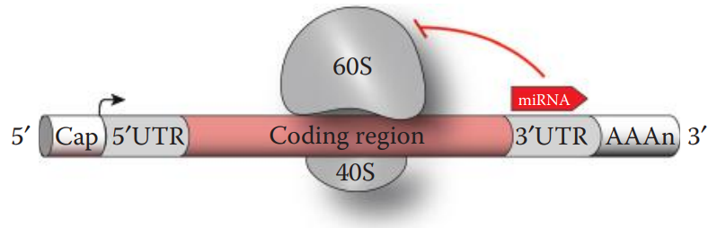
- The biological function of microRNAs (miRNAs) is to regulate gene expression.
- The mature miRNA strand associated with the RNA-induced silencing complex (RISC) binds to its target mRNA.
- A single miRNA can bind different mRNA transcripts encoded by multiple genes.
- Animal miRNAs usually form a base pairing with their target mRNAs through partial complementary sequences, which lead to translation repression to inhibit protein synthesis.
- If the base pairing is exactly complementary to its target mRNA sequence, then the cleavage and the degradation of the target mRNA occur.
11.6: Proteomic Approaches Using Mass Spectrometry for Bodily Fluid Identification
- Mass spectrometry (MS) is a susceptible and rapid technique for protein identification from complex biological samples.
Mass Spectrometric Instrumentation for Protein Analysis
- Mass spectrometer: An analytical technique to identify a molecule by measuring the ratio of the mass (m) to the charge (z) of a charged molecule.
- A typical mass spectrometer instrument analyzes ionized molecules while in their gas phase.
- Ion source: Converts analytes into gaseous phase ions through ionization.
- Ionization: A process used by gaining a positive or a negative charge from a neutral species.
- Mass Analyzer: A part of a mass spectrometer where ions are accelerated under electric fields and separated based on their m/z ratio.
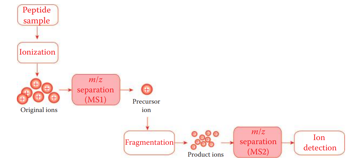
- Biomarker proteins can be identified by peptide sequencing using the tandem mass spectrometry (MS/MS) mode of operation in which a mass spectrometer uses two or more mass analyzers.
- After an initial mass analysis, individual peptide ions in the mass spectrum can be selected. The selected ions are known as precursor ions.
- A precursor ion is then subjected to the second round of fragmentation through collision and is broken into smaller ions known as product ions.
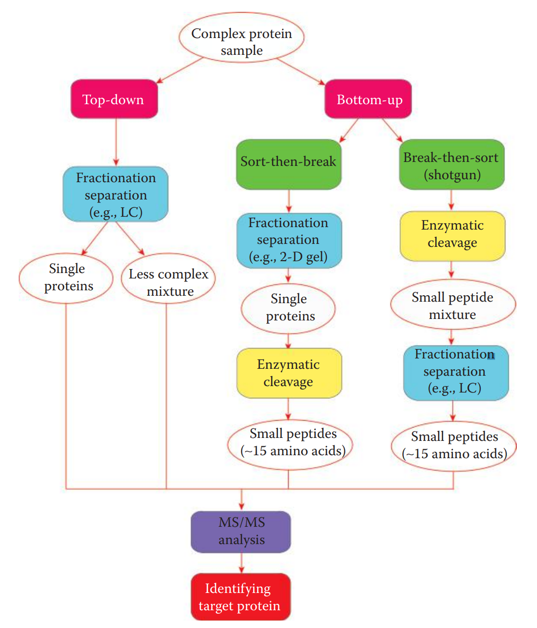
Analysis Strategies for Protein Identification
- In top-down strategy, intact proteins in a complex mixture are fractionated and separated into less complex protein mixtures or single proteins.
- The bottom-up strategy is commonly used for high-throughput analysis of small peptides derived from highly complex samples.
- Sort-then-break approach: Proteins in a complex mixture are first isolated and then enzymatically cleaved.
- Break-then-sort approach: Also known as the shotgun approach, wherein proteins are cleaved first and the resulting peptide mixture is then separated by LC and analyzed by tandem MS.
11.7: Microbial DNA Analysis for Bodily Fluid Identification
- Human Microbiota: Microbial community.
- Metagenomics: Allows the study of the microbial genome of complete microbial communities harvested directly from natural environments.
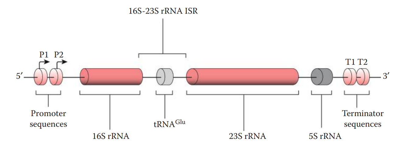
- The bacterial rRNA operon contains three rRNA genes:
- 5S and 23S rRNA genes encode the RNA components of the large subunit of the ribosome.
- 16S rRNA gene encodes the RNA component of the small subunit of the ribosome.
- The intergenic spacer region (ISR) between the 16S rRNA and 23S rRNA genes in the rRNA operon is another commonly used marker.
11.8: Nondestructive Assays for the Identification of Bodily Fluids
- Fluorescence: The emission of light by a fluorophore.
- Fluorophore: A moiety in a molecule that fluoresces on absorbing the energy from an excitation light source or radiation.
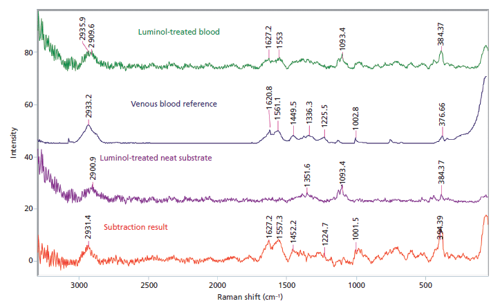
- Raman spectroscopy: Utilizes a near-infrared excitation light source and measures the scattering of laser light caused by the vibrating molecules of a sample.