ANPS 019 Block 2 (Lab)
1/356
There's no tags or description
Looks like no tags are added yet.
Name | Mastery | Learn | Test | Matching | Spaced |
|---|
No study sessions yet.
357 Terms
B
The divisions of the nervous system are the
A. muscle tissue and nervous tissue.
B. central nervous system and peripheral nervous systems.
C. nerves and ganglia.
D. brain and spinal cord.
D
The Peripheral Nervous System or PNS is made up of __________________.
A. spinal cord and ganglia
B. spinal cord and skeletal muscles
C. brain and spinal cord
D. nerves and ganglia
D
Spinal nerves
A. carry only sensory information.
B. carry only motor information.
C. travel from the spinal cord to the brain.
D. are mixed nerves with both sensory and motor nerve fibers.
E
Which structure is indicated by the letter A?
A. Ganglia
B. Spinal Nerves
C. Spinal Cord
D. Cranial Nerves
E. Brain

D
Which structure is indicated by the letter B?
A. Ganglia
B. Spinal Nerves
C. Spinal Cord
D. Cranial Nerves
E. Brain

A
Which structure is indicated by the letter C?
A. Ganglia
B. Spinal Nerves
C. Spinal Cord
D. Cranial Nerves
E. Brain
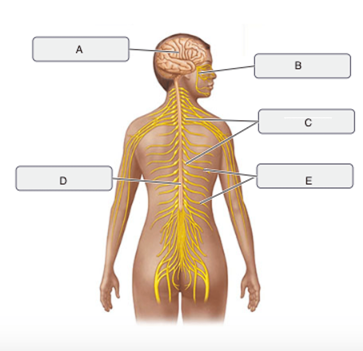
C
Which structure is indicated by the letter D?
A. Ganglia
B. Spinal Nerves
C. Spinal Cord
D. Cranial Nerves
E. Brain

B
Which structure is indicated by the letter E?
A. Ganglia
B. Spinal Nerves
C. Spinal Cord
D. Cranial Nerves
E. Brain

A
Structure: Brain and spinal cord
Function: Integrative and control centers
A. Central Nervous System
B. Peripheral Nervous System
C. Sensory (Afferent) Division
D. Motor (Efferent) Division
E. Somatic Nervous System
F. Autonomic Nervous System
G. Sympathetic Division
H. Parasympathetic Division
B
Structure: Cranial nerves and spinal nerves
Function: communication lines between the CNS and the rest of the body
A. Central Nervous System
B. Peripheral Nervous System
C. Sensory (Afferent) Division
D. Motor (Efferent) Division
E. Somatic Nervous System
F. Autonomic Nervous System
G. Sympathetic Division
H. Parasympathetic Division
C
Structure: Somatic and visceral sensory nerve fibers
Function: Conducts impulses from receptors to the CNS
A. Central Nervous System
B. Peripheral Nervous System
C. Sensory (Afferent) Division
D. Motor (Efferent) Division
E. Somatic Nervous System
F. Autonomic Nervous System
G. Sympathetic Division
H. Parasympathetic Division
D
Structure: Motor nerve fibers
Function: Conducts impulses from the CNS to effectors (muscles and glands)
A. Central Nervous System
B. Peripheral Nervous System
C. Sensory (Afferent) Division
D. Motor (Efferent) Division
E. Somatic Nervous System
F. Autonomic Nervous System
G. Sympathetic Division
H. Parasympathetic Division
E
Structure: Somatic motor (voluntary)
Function: Conducts impulses from the CNS to skeletal muscles
A. Central Nervous System
B. Peripheral Nervous System
C. Sensory (Afferent) Division
D. Motor (Efferent) Division
E. Somatic Nervous System
F. Autonomic Nervous System
G. Sympathetic Division
H. Parasympathetic Division
F
Structure: Visceral motor (involuntary)
Function: Conducts impulses from the CNS to cardiac muscle, smooth muscle and glands
A. Central Nervous System
B. Peripheral Nervous System
C. Sensory (Afferent) Division
D. Motor (Efferent) Division
E. Somatic Nervous System
F. Autonomic Nervous System
G. Sympathetic Division
H. Parasympathetic Division
G
Mobilizes body systems during activity
A. Central Nervous System
B. Peripheral Nervous System
C. Sensory (Afferent) Division
D. Motor (Efferent) Division
E. Somatic Nervous System
F. Autonomic Nervous System
G. Sympathetic Division
H. Parasympathetic Division
H
Conserves energy
Promotes housekeeping functions during rest
A. Central Nervous System
B. Peripheral Nervous System
C. Sensory (Afferent) Division
D. Motor (Efferent) Division
E. Somatic Nervous System
F. Autonomic Nervous System
G. Sympathetic Division
H. Parasympathetic Division
D
In the above image, the visceral motor division of the PNS is labeled _____.
A. A
B. B
C. C
D. D

B
The somatic motor (efferent) division is indicated by which panel in the above image?
A. A
B. B
C. C
D. D

A
The somatic sensory (afferent) division is indicated by which panel in the above image?
A. A
B. B
C. C
D. D

B
Which part of the nervous system can you use to kick a soccer ball?
A. A
B. B
C. C
D. D
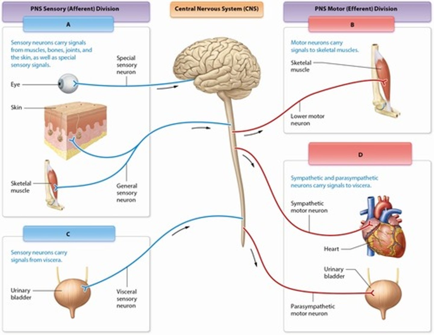
D
Which division can tell the bronchioles (air passages) in your lungs to dilate?
A. A
B. B
C. C
D. D

B
Neurons can communicate with (form synapses on)
A. neurons and muscles.
B. neurons, muscles, and glands.
C. muscles and glands.
D. only other neurons.
A
What are the three main structural components of neurons?
A. dendrites, cell body (soma), axon
B. dendrites, axons, synapses
C. axons, nucleus, cell body (soma)
D. synapses, axons, axon hillock
D
What is the name of the cell that forms the myelin sheath in the peripheral nervous system?
A. Muscle cell
B. Neuron
C. Axon
D. Schwann cell
A
Which is considered the conductive portion of the neuron?
A. Axon
B. Dendrites
C. Cell body (soma)
A
What is considered the receptive portion(s) of the neuron?
A. dendrites and cell body (soma)
B. dendrites only
C. axon
D. axon terminal
G
Which structure is indicated by the letter A?
A. Axon terminals
B. Cell body
C. Axon hillock region of axon
D. Chromatophilic (Nissl) substance
E. Schwann cell
F. Node of Ranvier
G. Dendrites
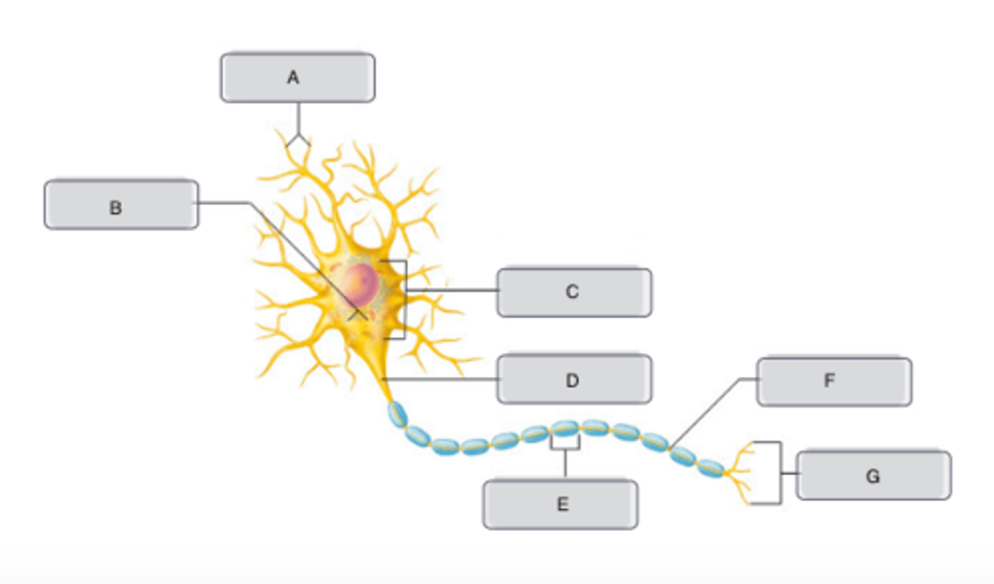
D
Which structure is indicated by the letter B?
A. Axon terminals
B. Cell body
C. Axon hillock region of axon
D. Chromatophilic (Nissl) substance
E. Schwann cell
F. Node of Ranvier
G. Dendrites

B
Which structure is indicated by the letter C?
A. Axon terminals
B. Cell body
C. Axon hillock region of axon
D. Chromatophilic (Nissl) substance
E. Schwann cell
F. Node of Ranvier
G. Dendrites

C
Which structure is indicated by the letter D?
A. Axon terminals
B. Cell body
C. Axon hillock region of axon
D. Chromatophilic (Nissl) substance
E. Schwann cell
F. Node of Ranvier
G. Dendrites

E
Which structure is indicated by the letter E?
A. Axon terminals
B. Cell body
C. Axon hillock region of axon
D. Chromatophilic (Nissl) substance
E. Schwann cell
F. Node of Ranvier
G. Dendrites

F
Which structure is indicated by the letter F?
A. Axon terminals
B. Cell body
C. Axon hillock region of axon
D. Chromatophilic (Nissl) substance
E. Schwann cell
F. Node of Ranvier
G. Dendrites

A
Which structure is indicated by the letter G?
A. Axon terminals
B. Cell body
C. Axon hillock region of axon
D. Chromatophilic (Nissl) substance
E. Schwann cell
F. Node of Ranvier
G. Dendrites

B
Which structure is indicated by the letter A?
A. Receptive region
B. Impulse direction
C. Secretory region
D. Impulse generating and conducting region
E. Biosynthetic center and receptive region

D
Which structure is indicated by the letter B?
A. Receptive region
B. Impulse direction
C. Secretory region
D. Impulse generating and conducting region
E. Biosynthetic center and receptive region

A
Which structure is indicated by the letter C?
A. Receptive region
B. Impulse direction
C. Secretory region
D. Impulse generating and conducting region
E. Biosynthetic center and receptive region
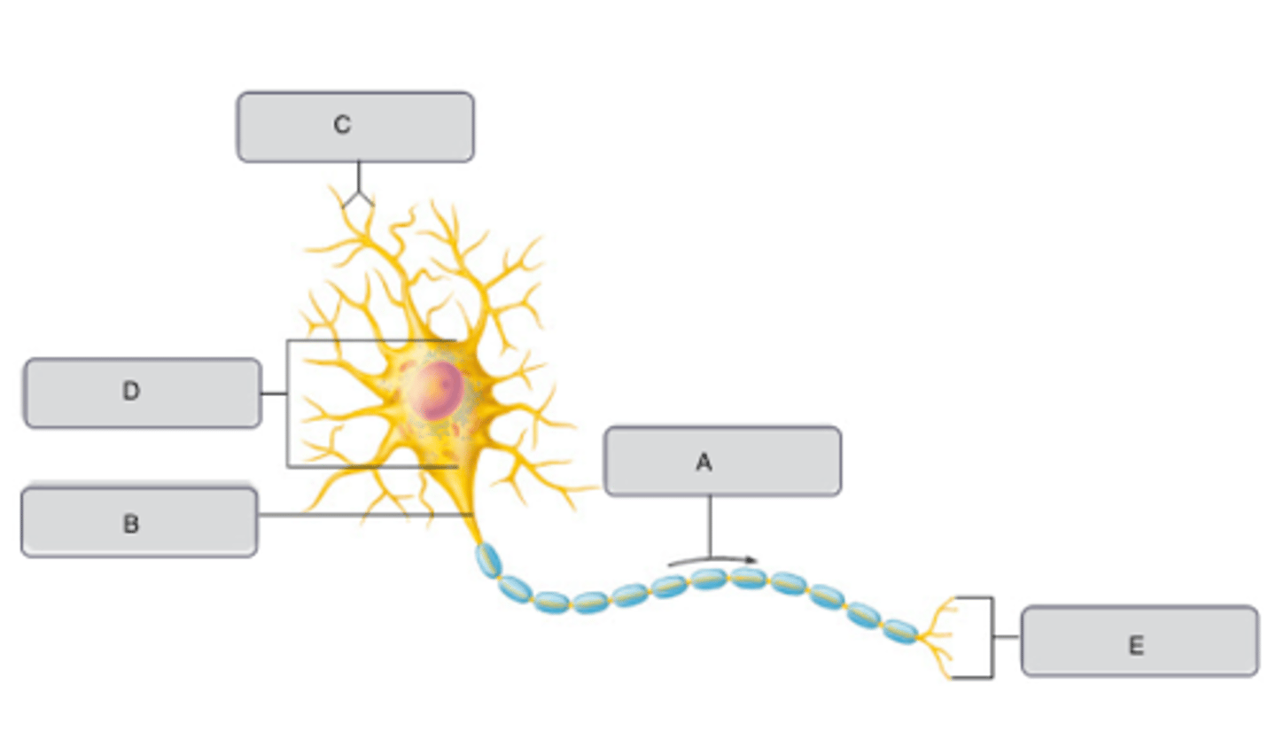
E
Which structure is indicated by the letter D?
A. Receptive region
B. Impulse direction
C. Secretory region
D. Impulse generating and conducting region
E. Biosynthetic center and receptive region

C
Which structure is indicated by the letter E?
A. Receptive region
B. Impulse direction
C. Secretory region
D. Impulse generating and conducting region
E. Biosynthetic center and receptive region

D
What is the structure at A?
A. axon collateral
B. soma
C. axon
D. dendrite

B
What is the structure at B?
A. axon
B. cell body or soma
C. dendrites
D. axon collateral

D
What is the structure at E?
A. axon hillock
B. dendrite
C. Cell body or soma
D. axon terminal
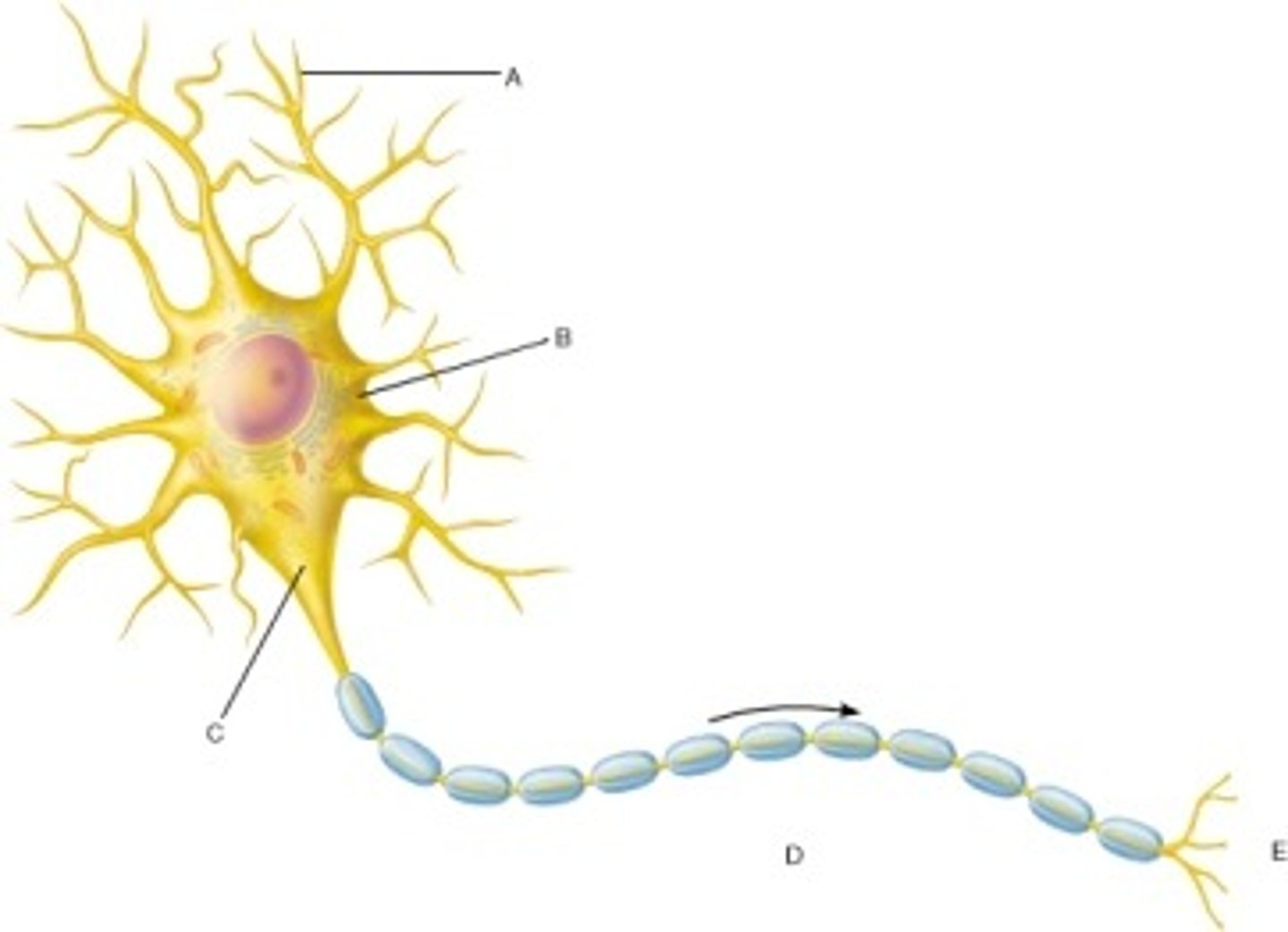
D
This is where synaptic vesicles containing neurotransmitters are released, so it can also be called the synaptic terminal.
A. axon hillock
B. dendrite
C. Cell body or soma
D. axon terminal
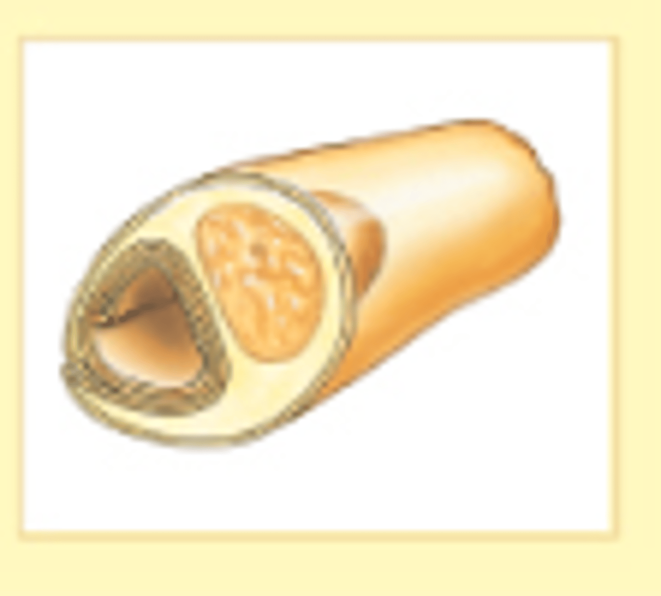
B
Which structure is indicated by the letter A?
A. Axon
B. Endoneurium
C. Perineurium
D. Epineurium
E. Fascicle
F. Myelin sheath

C
Which structure is indicated by the letter B?
A. Axon
B. Endoneurium
C. Perineurium
D. Epineurium
E. Fascicle
F. Myelin sheath

D
Which structure is indicated by the letter C?
A. Axon
B. Endoneurium
C. Perineurium
D. Epineurium
E. Fascicle
F. Myelin sheath

E
Which structure is indicated by the letter D?
A. Axon
B. Endoneurium
C. Perineurium
D. Epineurium
E. Fascicle
F. Myelin sheath

F
Which structure is indicated by the letter E?
A. Axon
B. Endoneurium
C. Perineurium
D. Epineurium
E. Fascicle
F. Myelin sheath

A
Which structure is indicated by the letter F?
A. Axon
B. Endoneurium
C. Perineurium
D. Epineurium
E. Fascicle
F. Myelin sheath

D
Which type of neuron is indicated by the letter A?
A. Pseudounipolar neuron
B. Multipolar neuron
C. Bipolar neuron
D. Anaxonic neuron

C
Which type of neuron is indicated by the letter B?
A. Pseudounipolar neuron
B. Multipolar neuron
C. Bipolar neuron
D. Anaxonic neuron

A
Which type of neuron is indicated by the letter C?
A. Pseudounipolar neuron
B. Multipolar neuron
C. Bipolar neuron
D. Anaxonic neuron

B
Which type of neuron is indicated by the letter D?
A. Pseudounipolar neuron
B. Multipolar neuron
C. Bipolar neuron
D. Anaxonic neuron

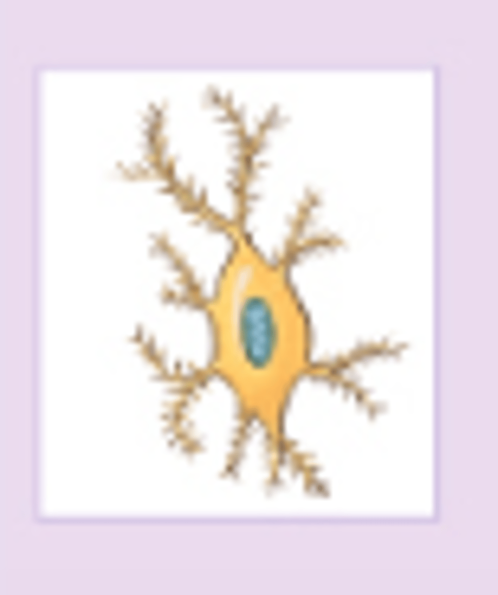
F
Identify the cell type in the picture:
A. Schwann cells
B. Satellite cells
C. Microglia
D. Oligodendrocytes
E. Astrocytes
F. Ependymal cells
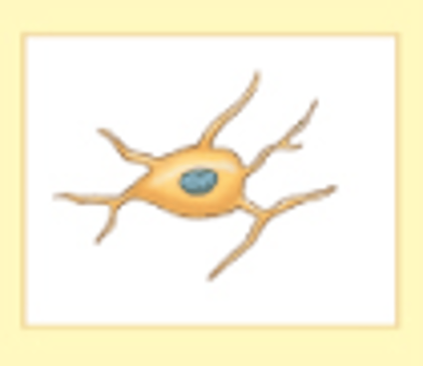
E
Identify the cell type in the picture:
A. Schwann cells
B. Satellite cells
C. Microglia
D. Oligodendrocytes
E. Astrocytes
F. Ependymal cells
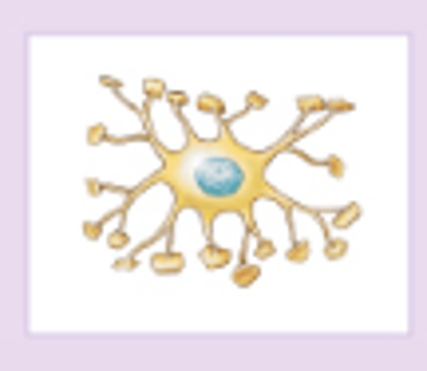
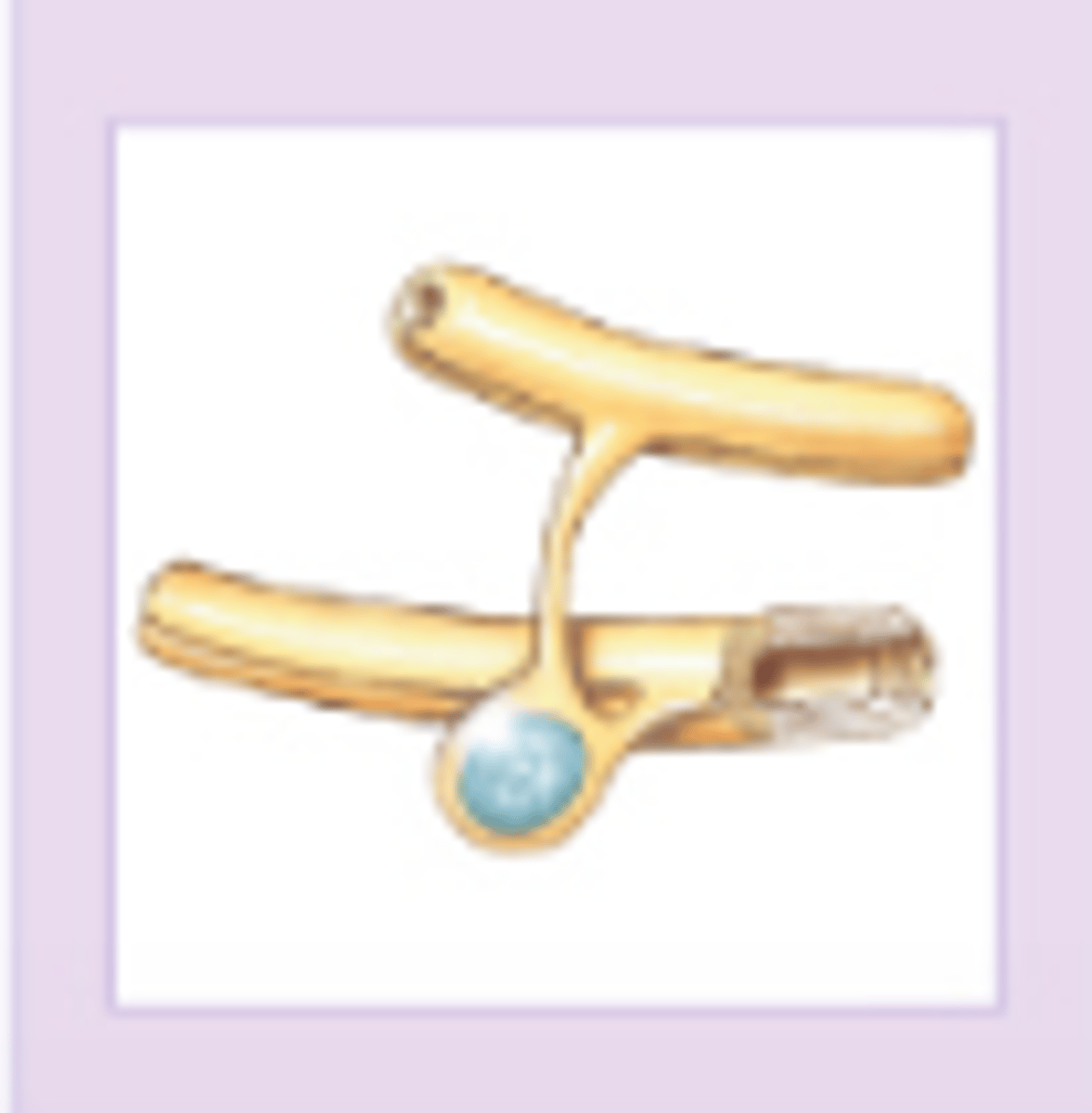
D
Identify the cell type in the picture:
A. Schwann cells
B. Satellite cells
C. Microglia
D. Oligodendrocytes
E. Astrocytes
F. Ependymal cells
C
Identify the cell type in the picture:
A. Schwann cells
B. Satellite cells
C. Microglia
D. Oligodendrocytes
E. Astrocytes
F. Ependymal cells
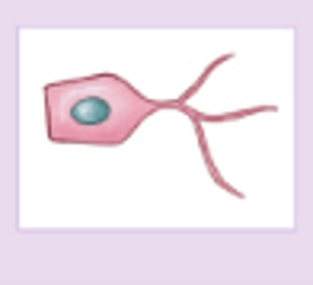
B
Identify the cell type in the picture:
A. Schwann cells
B. Satellite cells
C. Microglia
D. Oligodendrocytes
E. Astrocytes
F. Ependymal cells
A
Identify the cell type in the picture:
A. Schwann cells
B. Satellite cells
C. Microglia
D. Oligodendrocytes
E. Astrocytes
F. Ependymal cells
F
These cells produce and circulate cerebrospinal fluid in the CNS
A. Schwann cells
B. Satellite cells
C. Microglia
D. Oligodendrocytes
E. Astrocytes
F. Ependymal cells
E
These cells provide structural and nutritional support for CNS neurons, and recycle neurotransmitters
A. Schwann cells
B. Satellite cells
C. Microglia
D. Oligodendrocytes
E. Astrocytes
F. Ependymal cells
D
These cells myelinate CNS axons; provide structural framework
A. Schwann cells
B. Satellite cells
C. Microglia
D. Oligodendrocytes
E. Astrocytes
F. Ependymal cells
C
These cells conduct phagocytosis in the CNS
A. Schwann cells
B. Satellite cells
C. Microglia
D. Oligodendrocytes
E. Astrocytes
F. Ependymal cells
B
These cells support neuron cell bodies in PNS ganglia
A. Schwann cells
B. Satellite cells
C. Microglia
D. Oligodendrocytes
E. Astrocytes
F. Ependymal cells
A
These cells produce myelin sheath around PNS axons
A. Schwann cells
B. Satellite cells
C. Microglia
D. Oligodendrocytes
E. Astrocytes
F. Ependymal cells
A
When in resting membrane potential, the outside of the cell is positively charged, and the inside is negatively charged.
A. True
B. False
A
The ________________ contains high concentrations of sodium ions (Na+) and chloride ions (Cl-), while the intracellular fluid (cytosol) of the neuron contains high concentrations of potassium ions (K+) and negatively charged proteins. These large protein anions are not membrane permeable, while K+ are membrane permeable.
A. extracellular fluid (ECF)
B. intracellular fluid (cytosol)
B
The extracellular fluid (ECF) contains high concentrations of sodium ions (Na+) and chloride ions (Cl-), while the ____________________ of the neuron contains high concentrations of potassium ions (K+) and negatively charged proteins. These large protein anions are not membrane permeable, while K+ are membrane permeable.
A. extracellular fluid (ECF)
B. intracellular fluid (cytosol)
A
The concentration of _____ is higher inside than outside the cell
A. K+
B. Na+
A
The resting membrane potential is maintained by Na+-K+ pumps that actively transport _____ into and ______ out of the cell.
A. K+, Na+
B. Na+, K+
B
The concentration of ________ is higher outside than inside the cell.
A. K+
B. Na+
A
The membrane is more permeable to ____.
A. K+
B. Na+
B
Ions are unequally distributed across the plasma membrane of all cells. This ion distribution creates an electrical potential difference (voltage) across the membrane. What is the name given to this potential difference?
A. Positive membrane potential
B. Resting membrane potential (RMP)
C. Threshold potential
D. Action potential
C
Sodium and potassium ions can diffuse across the plasma membranes of all cells and generate a resting membrane potential because of the presence of what type of channel?
A. Sodium-potassium ATPases
B. Chemically-gated channels
C. Leak channels
D. Voltage-gated channels
A
On average, the resting membrane potential is -70 mV. What does the sign and magnitude of this value tell you?
A. The inside surface of the plasma membrane is much more negatively charged than the outside surface.
B. There is no electrical potential difference between the inside and the outside surfaces of the plasma membrane.
C. The inside surface of the plasma membrane is much more positively charged than the inside surface.
D. The outside surface of the plasma membrane is much more negatively charged than the inside surface.
B
The plasma membrane is much more permeable to K+ than to Na+. Why?
A. The Na+-K+ pumps transport more K+ into cells than Na+ out of cells.
B. There are many more K+ leak channels than Na+ leak channels in the plasma membrane.
C. Chemically-gated cation channels favor a greater influx of Na+ than K+.
D. There are many more voltage-gated K+ channels than voltage-gated Na+ channels.
C
The resting membrane potential depends on two factors that influence the magnitude and direction of Na+ and K+ diffusion across the plasma membrane. Identify these two factors.
A. The presence of concentration gradients and Na+-K+ pumps
B. The presence of a resting membrane potential and leak channels
C. The presence of concentration gradients and leak channels
D. The presence of concentration gradients and voltage-gated channels
B
What prevents the Na+ and K+ concentration gradients from dissipating (running down)?
A. Na+ cotransporter
B. Na+-K+ ATPase
C. Na+ and K+ leaks
D. H+-K+ ATPase
A
Also known as the Na+-K+ pump, or simply the pump, this transporter moves_______ Na+ out of the cell and _______ K+ into the cell for every ATP it hydrolyzes. This pumping action prevents the Na+ and K+ concentration gradients from running down as these ions passively move through leak channels.
A. three, two
B. two, three
D
The concentrations of which two ions are highest outside the cell?
A. Na+ and protein anions (A-)
B. K+ and Cl-
C. K+ and protein anions (A-)
D. Na+ and Cl-
A
For each of the following, indicate whether the condition will cause the membrane potential to become more positive, more negative, or largely unchanged when compared to the normal physiological resting membrane potential.
Triple the number of Na+ leak channels
A. more positive
B. more negative
C. largely unchanged
A
For each of the following, indicate whether the condition will cause the membrane potential to become more positive, more negative, or largely unchanged when compared to the normal physiological resting membrane potential.
Double the concentration of K+ outside the cell
A. more positive
B. more negative
C. largely unchanged
B
For each of the following, indicate whether the condition will cause the membrane potential to become more positive, more negative, or largely unchanged when compared to the normal physiological resting membrane potential.
Double the number of K+ leak channels
A. more positive
B. more negative
C. largely unchanged
B
For each of the following, indicate whether the condition will cause the membrane potential to become more positive, more negative, or largely unchanged when compared to the normal physiological resting membrane potential.
Decrease the concentration of Na+ outside the cell by half
A. more positive
B. more negative
C. largely unchanged
C
For each of the following, indicate whether the condition will cause the membrane potential to become more positive, more negative, or largely unchanged when compared to the normal physiological resting membrane potential.
Double the size of the cell, without adding channels
A. more positive
B. more negative
C. largely unchanged
C
For each of the following, indicate whether the condition will cause the membrane potential to become more positive, more negative, or largely unchanged when compared to the normal physiological resting membrane potential.
Double the number of closed channels for K+
A. more positive
B. more negative
C. largely unchanged
A
__________: The resting membrane potential of the cell.
A. Membrane potential of -70 mV
B. Membrane potential of -90 mV
C. Na+ permeability
D. K+ permeability
B
________: There is no net movement of K+ into or out of the cell because electrical gradient equals chemical gradient.
A. Membrane potential of -70 mV
B. Membrane potential of -90 mV
C. Na+ permeability
D. K+ permeability
C
_______: Although it plays a role, it is not a primary determinant of the resting membrane potential
A. Membrane potential of -70 mV
B. Membrane potential of -90 mV
C. Na+ permeability
D. K+ permeability
D
__________: The primary determinant of the resting membrane potential.
A. Membrane potential of -70 mV
B. Membrane potential of -90 mV
C. Na+ permeability
D. K+ permeability
A
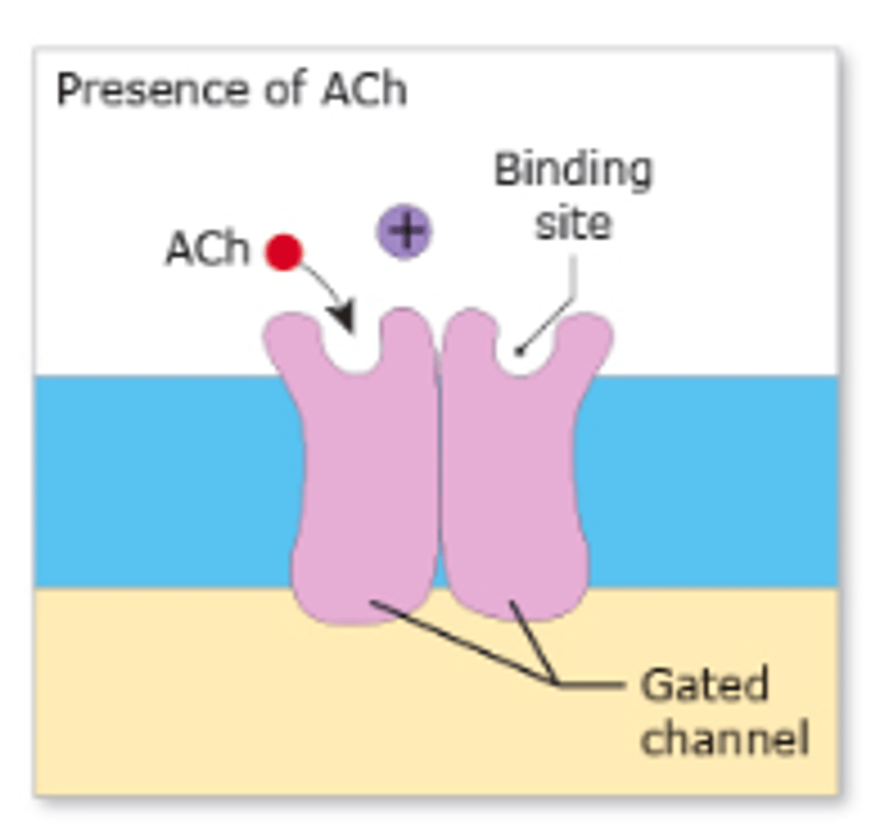
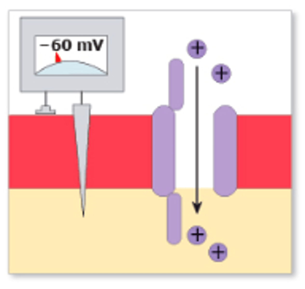
Identify the gated channel in the picture:
A. Chemically gated channel
B. Voltage-gated channel
C. Mechanically gated channel
B
Identify the gated channel in the picture:
A. Chemically gated channel
B. Voltage-gated channel
C. Mechanically gated channel
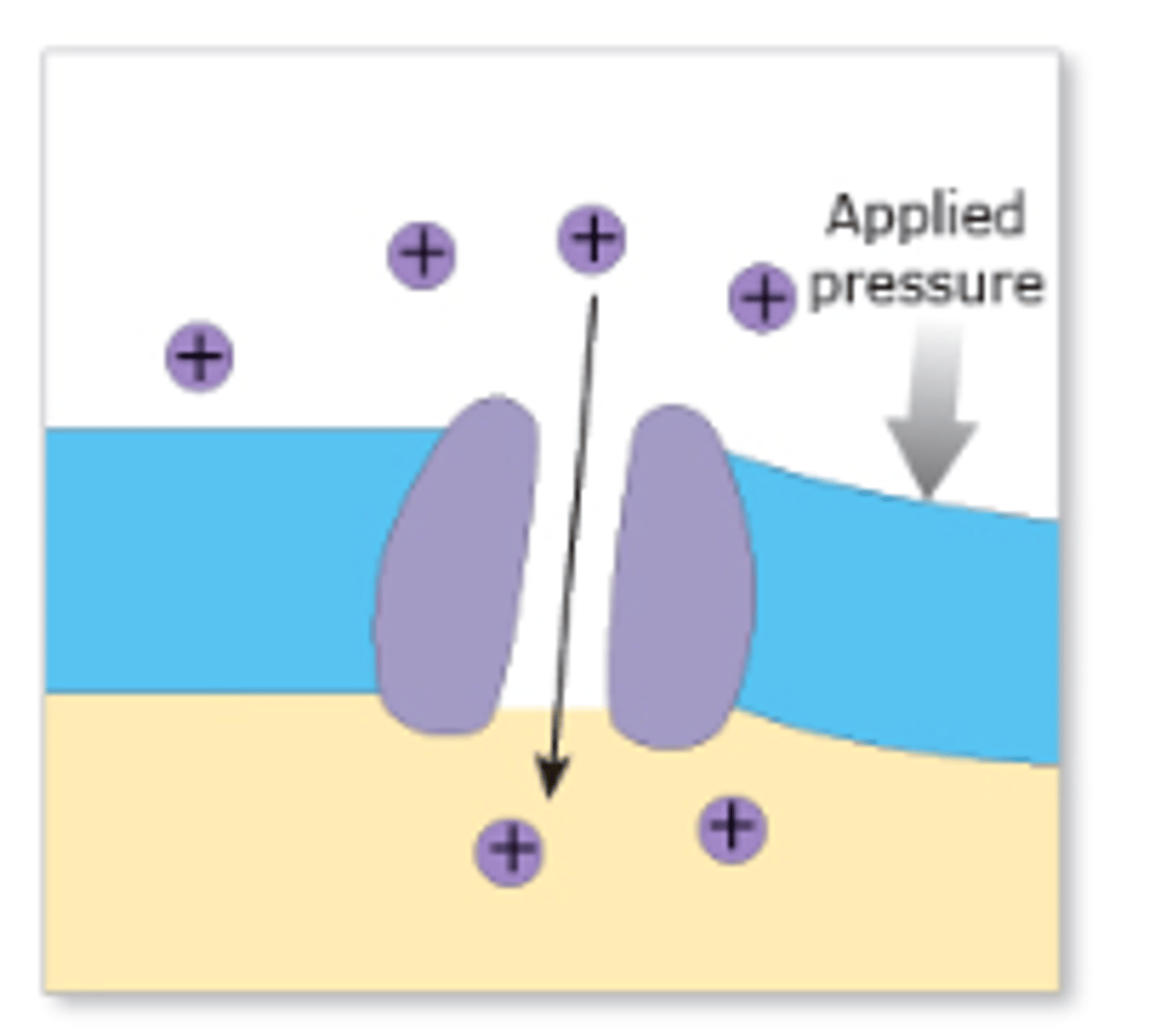
C
Identify the gated channel in the picture:
A. Chemically gated channel
B. Voltage-gated channel
C. Mechanically gated channel
A
________: The membrane potential is becoming more positive than the resting membrane potential
A. Depolarize
B. Repolarize
C. Hyperpolarize
B
_____: The membrane potential is moving from a more positive value toward resting membrane potential
A. Depolarize
B. Repolarize
C. Hyperpolarize
C
_____: The membrane potential is more negative than the resting membrane potential
A. Depolarize
B. Repolarize
C. Hyperpolarize
E
Identify the phases of the action potential: (Letter A)
A. Hyperpolarization
B. Repolarization
C. Peak
D. Depolarization
E. Rest

D
Identify the phases of the action potential: (Letter B)
A. Hyperpolarization
B. Repolarization
C. Peak
D. Depolarization
E. Rest
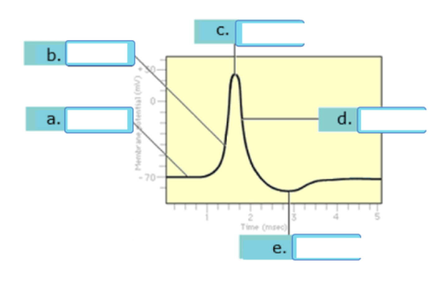
C
Identify the phases of the action potential: (Letter C)
A. Hyperpolarization
B. Repolarization
C. Peak
D. Depolarization
E. Rest

B
Identify the phases of the action potential: (Letter D)
A. Hyperpolarization
B. Repolarization
C. Peak
D. Depolarization
E. Rest

A
Identify the phases of the action potential: (Letter E)
A. Hyperpolarization
B. Repolarization
C. Peak
D. Depolarization
E. Rest
