lecture 6: epithelial cell surface specialisations
1/6
There's no tags or description
Looks like no tags are added yet.
Name | Mastery | Learn | Test | Matching | Spaced |
|---|
No study sessions yet.
7 Terms
cytoskeleton
Actin/microfilaments
Found in the periphery of the cell
At the plasma membrane
When 2 cells divide (undergoing mitosis) --> cytokinesis
Found in the surface specialisations
Found in some cells e.g. muscle cells; skeletal and caridean muscles cells actin and myosin come together to form sarcomeres(contractile units)
Microtubules
Surface specialisations
Cells undergoing mitosis (forms the mitotic spindle and at the centromeres)
Intermediate filaments
Around the nucleus
Skin cells
microtubules
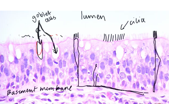
cilia
Surface specialisation on eppithelial cells
Ovaduct
Fallopian tube
Trachea
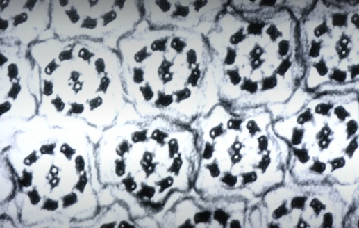
One cilium is composed of cytoskeleton (inside the epithial lians)
Cilia is made up of microtubules
Image above shows transverse cross sections
About 10 microns in length
Microtubles/cilia come up through the plasma membrane to create the increase SA to V ratio
Point; movement
They sweep
9 + 2 microtubule doublet arrangement
electron micrograph
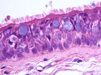
cilia light micrograph
Pseudo stratified as all the epithelial cells touch the basement membrane
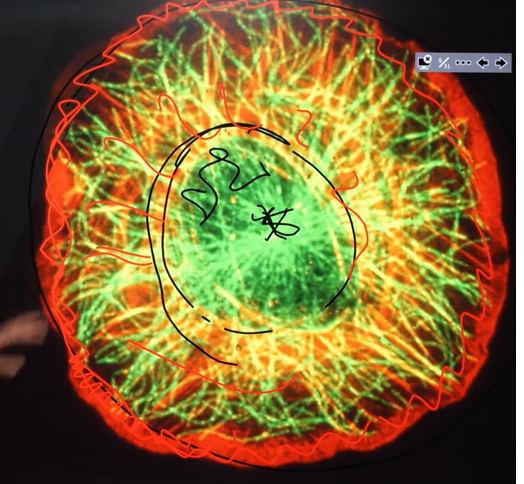
cilia florescence microscopy image
Actin is on the periphery of the cell therefore actin is the red in the image; keeps the integrity of the cell
Microtubules present as well
Nucleus is in the middle
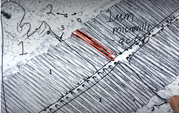
actin microfilaments; electron micrograph
Shows a small part of one cell
4 cells in this electron micrograph
Cytoskeleton is highly organised to the periphery of the cell
1 micron in length
Forms little fingers at the edge of the cell
Actin microfilaments stick into the plasma membrane of the cell; forming microvilli
Microvilli create large surface area to volume ratio
Places where you would find lots of microvilli;
Small intestine; simple columnar epithelial cell with microvilli allowing for the absorption of food
actin microfilaments
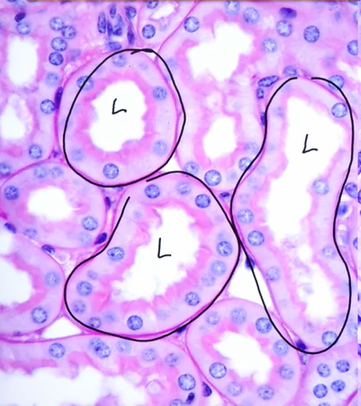
Proximal convolute tubule of the kidney
Needs lots and lots of absorption
Needs to reabsorb 70% of the plasma ultrafiltrate that gets filtered through the glomerulus back to the body
If the microvilli didn’t work we would loose all of our blood and filtrate and water to our urine
We can see the microvilli in the 'brush border'
The carbohydrates that are studded into the membrane are picking up the colour on the stain (pink circular area surrounding the lumen)
trachea
Sits posterior to the esophagus
Purple is the thick band of purple
Epithelium is the cells lining the trachea
Simple, columnar cells with cilia
Pseudostratified epithelium
cilia sweeps the mucus up