PSY 246 Occipital lobes and networks
1/22
There's no tags or description
Looks like no tags are added yet.
Name | Mastery | Learn | Test | Matching | Spaced | Call with Kai |
|---|
No analytics yet
Send a link to your students to track their progress
23 Terms
What is the retina
Light-senstive layer at the back of the eye that converts light into neural signals
Light passes through several layers of cells before reaching rods and cones
What forms the optic nerve?
Ganglion cells have axons that leave the retina through the optic disc that form the optic nerve which creates the blindspot
Optic Nerve vs Optic Chaism
Optic nerve = ganglion cell axons. Leave eye at optic disc
Optic Chiasm = point of crossover for half the visual projections
What is our blind spot
Where optic nerve passes through optic disc
Since there are no cells to detect light an optic disc, that part of retina is blind
We dont notice it because our brain “fills in” this spot by taking in the best guess of whats actually there based on our surroundings
What is the pathway from eye to the brian
Eye - Optic Nerve - optic chiasm - LGN - Visual Cortex
What are the two visual pathways
Retino-geniculate-striate pathway = Eye → optic chiasm → LGN of thalamus → striate (primary visual cortex) [solid line of diagram]
when talking about vision, this is what its talking about
Tectopulvinar Pathway = eye → optic chiasm → superior colliculi → lateral posterior and pulvinar nuclei of thalamus
Name the parts of the occipital lobe and their functions
Primary Visual cortex (V1) = basic features (edges, orientation, spatial frequency)
Secondary visual cortex (V2/V3) = form depth, binocular vision
Association cortex (V4) = COLOUR
Middle temporal region (V5) = Motion
What are the consequences of damage to primary visual cortex
Hemanopia = loss of vision in HALF the visual field
Scotoma = loss of vision in one point
Quadrantanopia = loss of vision to a quarter of the visual field
Compare and contrast the ventral and dorsal visual streams
Ventral Stream = “what” pathway
Recognizes objects
Names and functions of objects regardless of location
Dorsal stream = “Where”/”How” pathway
Locations of objects, but not their names or functions
How to interact with objects
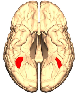
Inferotemporal cortex (IT); what visual stream is it apart of, part in the brain; what is it important for
Part of the cerebral cortex in the lower portion of temporal lobe, important for object recognitionW
What are the consequecnes of damaging inferotemporal cortex (IT)
Agnosias = inability to recognize objects
Prosopagnosia = inability to recognize faces
What do neurons in IT cortex respond and dont respond to
Dont respond well to spots or lines
Respons well to stimulus such as hands, faces, or objects
Some areas of the cortex are specialized to proccess certain types of stimuli, name the two areas mentioned in class
Parahippocampal Place Area (PPA = responds to places (pictures of houses, landmarks)
Fusiform Face Area (FFA) = respond to faces more than objects
What is Pareidolia
Seeing faces in a mbigious stimuli
(brunt toast, front of vehicles, wall plug ins)
What are the visual perceptual disorders
Damages in higher visual areas and not primary visual cortex
Provlems with interpreting or recognizing visual information despite normal vision
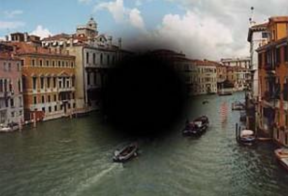
Apperceptive Visual Agnosia
Cant combine visual details into a complete image
Cant recognize objects because they cant assemble visual features into a whole
Basic sensory abilities (acuity, color, shape detection) are intact
“Can see that something is there but cant perceive it (naming it, saying what it is or copying it)”
Assiociative Visual Agnosia
Impairment in linking visual perception to meaning
objects can be seen but not recognized
Can copy and describe objects accurately
Cannot identify or name objects despire normal vision and perception
What are tests for visual agnosias? AND compare how apperceptive and associative individuals differ in these tests
Tests include copying drawings, “real or not” images, or impossible objects
Apperceptive: struggle to tell real vs impossible because they cant form percept
Associative: Can tell real vs impossible but cannot recognize or name a real object
Prosopagnosia; what is it, and where is the damage located that causes this
Impairment in recognizing familiar faces, despite normal vision
Cannot recognize photos of their own face
Damage to Fusiform face area in the cortex (most likely in the right)
Individuals with prosagnosia are able to identify people by
Voice
Clothing
Body image
Context
What is blindsight; where is the damage located?
Ability to respond to visual stimuli without conscious awareness of seeing them
Often due to damage in V1
For invidiuals affected with blindsight, how do they still see
Tecto-Pulvinar Pathway
Not all visual information relies on geniculostriate pathway
Supports unconsious vision: Explains blindsight
What is akinetopsia
Inability to perceive motion, even though static objects are seen normally
Patients sees the world in a series of static “frames”
Caused by damage to visual area V5/MT