Operator controls
1/23
There's no tags or description
Looks like no tags are added yet.
Name | Mastery | Learn | Test | Matching | Spaced |
|---|
No study sessions yet.
24 Terms
Monitor
displays the ultrasound image with 2D, color and spectral doppler
duplex
2D and color
Triflux
2D, color, spectral
Near Field
closest to the transducer
Far field
deeper to the tissue
Power
controls the amount of acoustical energy sent into the human body, controls brightness, decibels
ALARA
as low as reasonably achievable
data field on the monitor
top right, displays image processing information
control panel
has transducer button
phrased array
used for cardiac

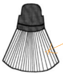
linear array
used for superficial structures

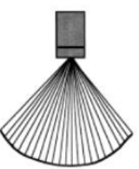
Curved array
powerful, penetrates

transducers
thick crystal: low frequency transducer, more penetration
Thin crystal: high frequency transducer, less penetration
depth
properly display body part being imaged
harmonic imaging
uses frequencies created by the tissues, rather than the fundamental frequency, to create an image
harmonic imaging helps
brings out details in second harmonic
overall gain
adjusts brightness of entire image
TGC