Pediatric Cardiology
1/18
There's no tags or description
Looks like no tags are added yet.
Name | Mastery | Learn | Test | Matching | Spaced | Call with Kai |
|---|
No analytics yet
Send a link to your students to track their progress
19 Terms
Characteristics of Cyanotic Congenital Heart Defects
Profound cyanosis
Not relieved with hyperoxia test
Dyspnea
2D Echocardiogram makes definitive diagnosis
Tetralogy of Fallot
4 defects:
Ventricular septal defect (VSD)
Pulmonary stenosis
Overriding aorta
Right ventricular hypertrophy
Pulmonary stenosis murmur
Crescendo-decrescendo
Hypoxic (Tet) spells with crying or feeding, put knees to chest
Classic chest x-ray finding is a boot-shaped heart, with an upturned cardiac apex
Cyanotic
Transposition of the great vessels
Cyanosis-profound
No murmurs
Chest x-ray classically an "egg on a string" created by a narrow superior mediastinum
Tricuspid Atresia
The absence of the tricuspid valve results in a hypoplastic right ventricle
Severely cyanotic with single S2
Truncus Arteriosus
A single S2 is due to the single valve
Minimal Cyanosis
Total anomalous pulmonary venous return (TAPVR)
All of the pulmonary veins fail to connect to the left atrium and return abnormally via the right side of the heart
May see supracardiac, infracardiac, cardiac, or mixed drainage
Cyanosis
Characteristics of Acyanotic Heart disease
Lesions result in increase volume load
Shunting of fully oxygenated blood back to the lungs
Pulmonary edema → Tachypnea, chest retractions, nasal flaring, wheezing
Ventricular Septal Defect
Loud, harsh, systolic murmur, usually heard best at the left lower sternal border
No cyanosis
Pulmonary HTN, poor feeding, dyspnea
Atrial Septal defect
Systolic ejection murmur at the left upper sternal border
Eisenmenger syndrome
Uncorrected left to right shunts →
Severe irreversible pulmonary hypertension
Right ventricular failure resulting in right-to-left shunting
Cyanosis, dyspnea with exertion, hepatomegaly, and clubbing of the fingers and toes
Patent Ductus Arteriosus (PDA)
Continuous, machinery-like murmur can be heard at the left infraclavicular area
Coarctation of the Aorta
Narrowing of descending Aorta
Disparity of pulse and BP in arms and legs
Normal BP: higher in leg
In CoA, BP much lower in legs
Check BP in all 4 extremities
Check ipsilateral pulses of radial and femoral
Severe CoA will have absent femoral pulses
Rib notching may be seen in older children (>8 years of age) with large collaterals (commonly from subclavian artery)
Most common heart defects in Down Syndrome
Complete atrioventricular septal defect
Ventricular septal defect
Atrial septal defect
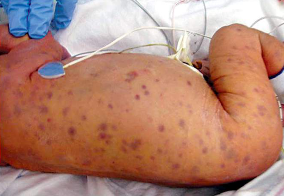
Congenital Rubella Syndrome
In utero infection
Blueberry muffin rash
Deafness, cataracts, and cardiac disease (PDA)
Most common cardiac defect in Turner Syndrome
Coarctation of the aorta
Bicuspid aortic valve leading to aortic stenosis
Hypertension
Prolonged QT
Aortic dissection
Marfan Syndrome
Aortic root dilatation/dissection
Ectopia lentis: lens dislocation
Above average height
Arachnodactyly
Dolichostenomelia (elongated limbs)
Scoliosis is common
Pectus excavatum
Pectus carinatum
Major Jones criteria for diagnosis of rheumatic fever
Migratory arthritis
Carditis and valvulitis
Central nervous system involvement
Erythema marginatum
Subcutaneous nodules- located over extensor surface of elbows, knees, knuckles, and ankles or scalp and spine; firm, nontender
Kawasaki Disease
Acute febrile vasculitis
If untreated, can develop coronary artery abnormalities (aneurysms, dilatation, stenosis...)
Most often proceeded by viral infection
Most younger than 5y.o.
Causes severe vasculitis, mostly medium-sized arteries (coronary)
ECHO: coronary abnormalities, repeat at 2-3 wks, then again at 6-8wks
Treatment of Kawasaki Disease
Treatment should not be delayed
First line:
Aspirin + IVIG within 7-10 days of onset
If IVIG resistance (fever persist > 36hrs after initial dose) then give a 2nd dose
If still not improving- add infliximab or methylprednisone
High risk patients would receive Aspirin/IVIG + methylprednisone
< 6mo, >9yo, Asian race, CRP > 13mg/dL, and Zscore >2 in LAD or right coronary artery