Psych 120A Midterm Study Guide: Key Terms & Definitions
1/293
There's no tags or description
Looks like no tags are added yet.
Name | Mastery | Learn | Test | Matching | Spaced |
|---|
No study sessions yet.
294 Terms
scientific study of knowledge and thought
What is cognitive psychology?
examples of cognitive psychology
Memories, Problem Solving, Decision Making, Attention, Reasoning
cognitive psychology
How is our knowledge of the world represented?
What mental processes are involved in acquiring,accessing, and using this knowledge?
How can we measure behavior to make inferences about mental representations and processes?
representation
A set of objects that stand for another set of objects by virtue of having the same causal relational structure.
External Objects
we believe are out there in the world, but we don't have access to it
Internal Representation
bouncing off our retina, and we create a representation and make assumptions of that
Processes
operations that transform one representation into another
Mental representations:
1) are formed in the mind
2) are activated by objects/events in world
3) make it possible to think about objects/events in their absence
Mental processes
operations performed on mental representations
Cognitive offloading
We offload information to the cloud and our devices
We allow ourselves not to remember things we dont need to remember
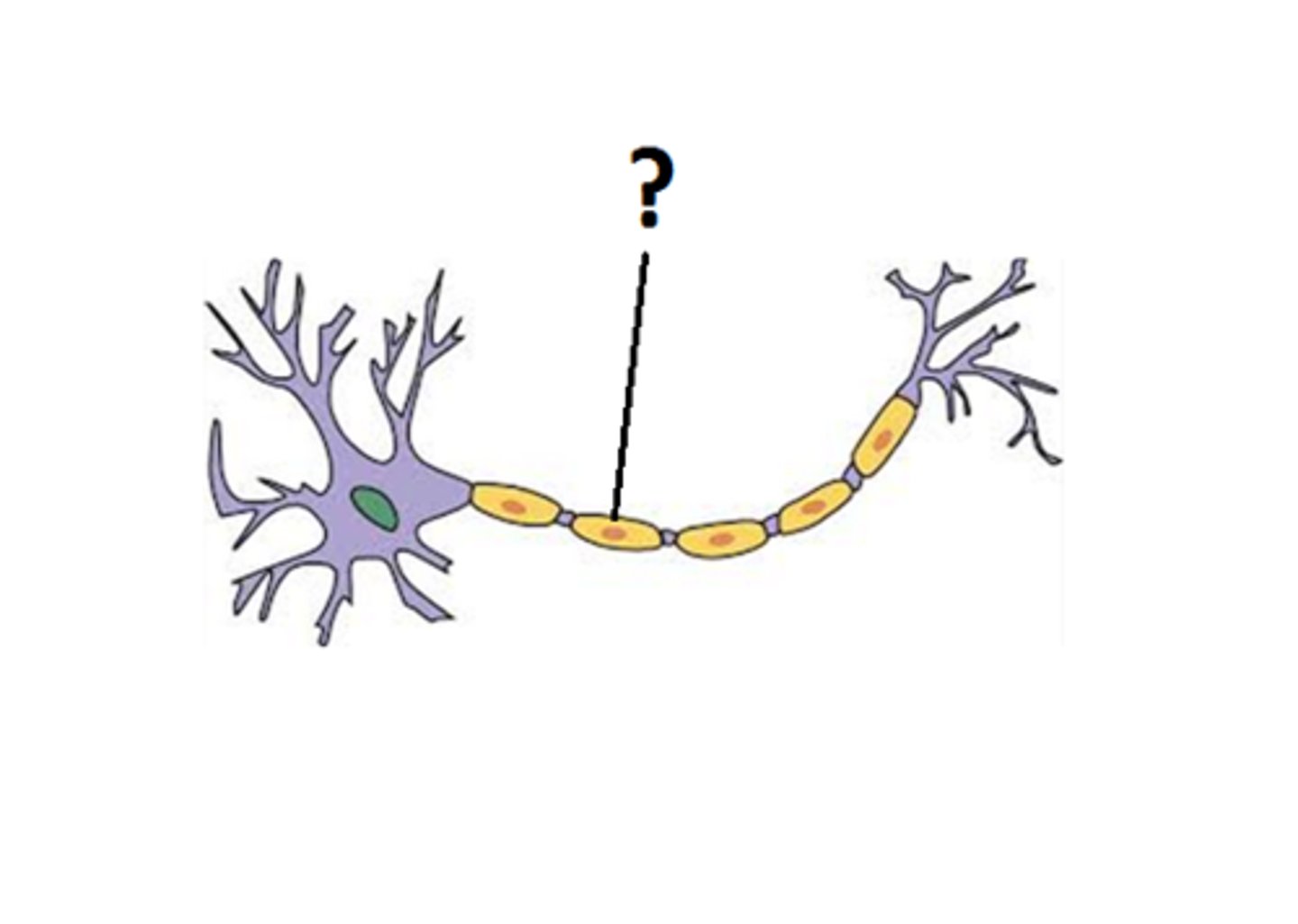
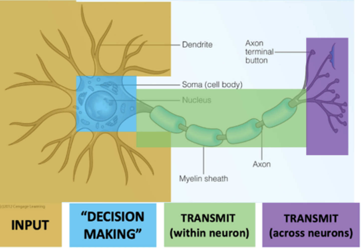
Dendrites
detect incoming signals
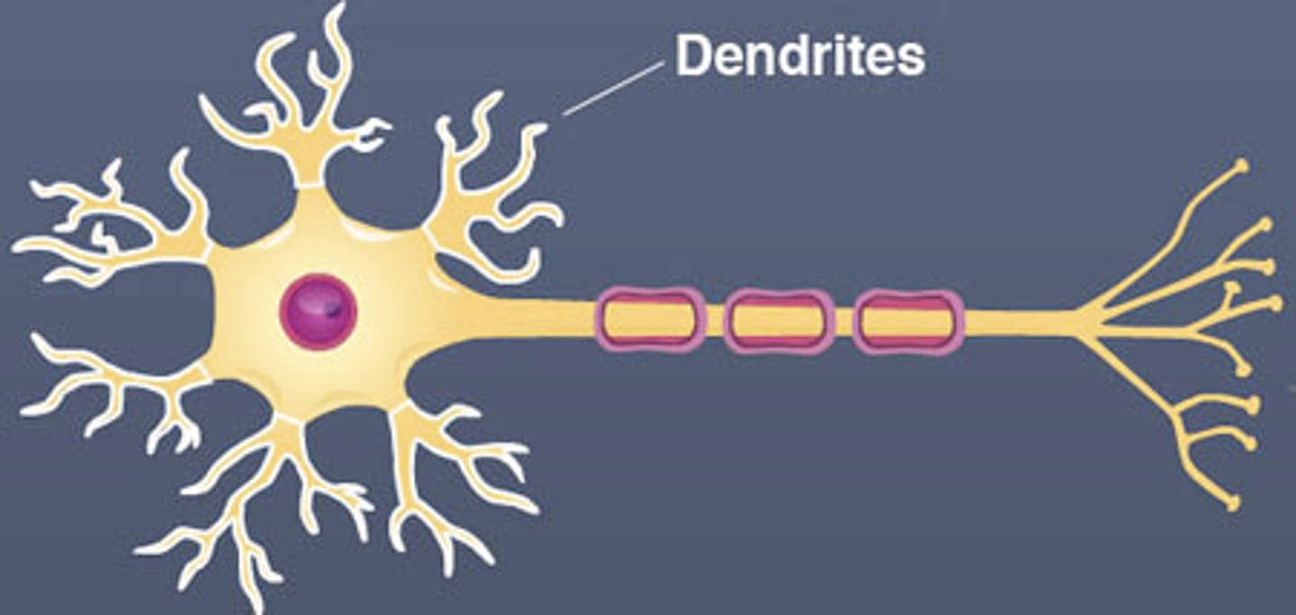
The cell body
contains the nucleus and cellular machinery
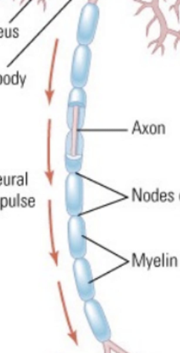
Axons
transmits signals to other neurons
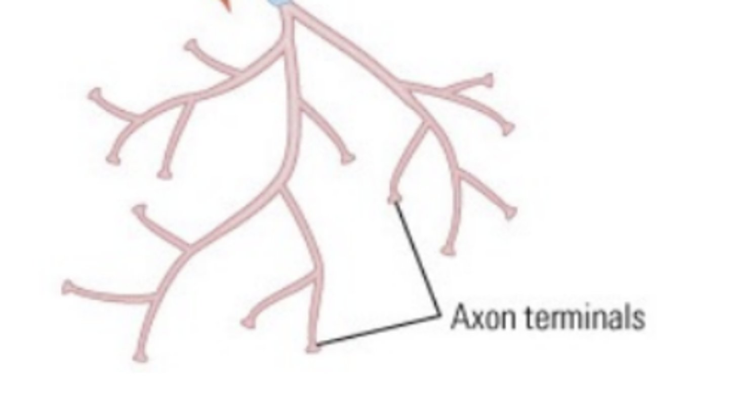
axon terminal
The endpoint of a neuron where neurotransmitters are stored
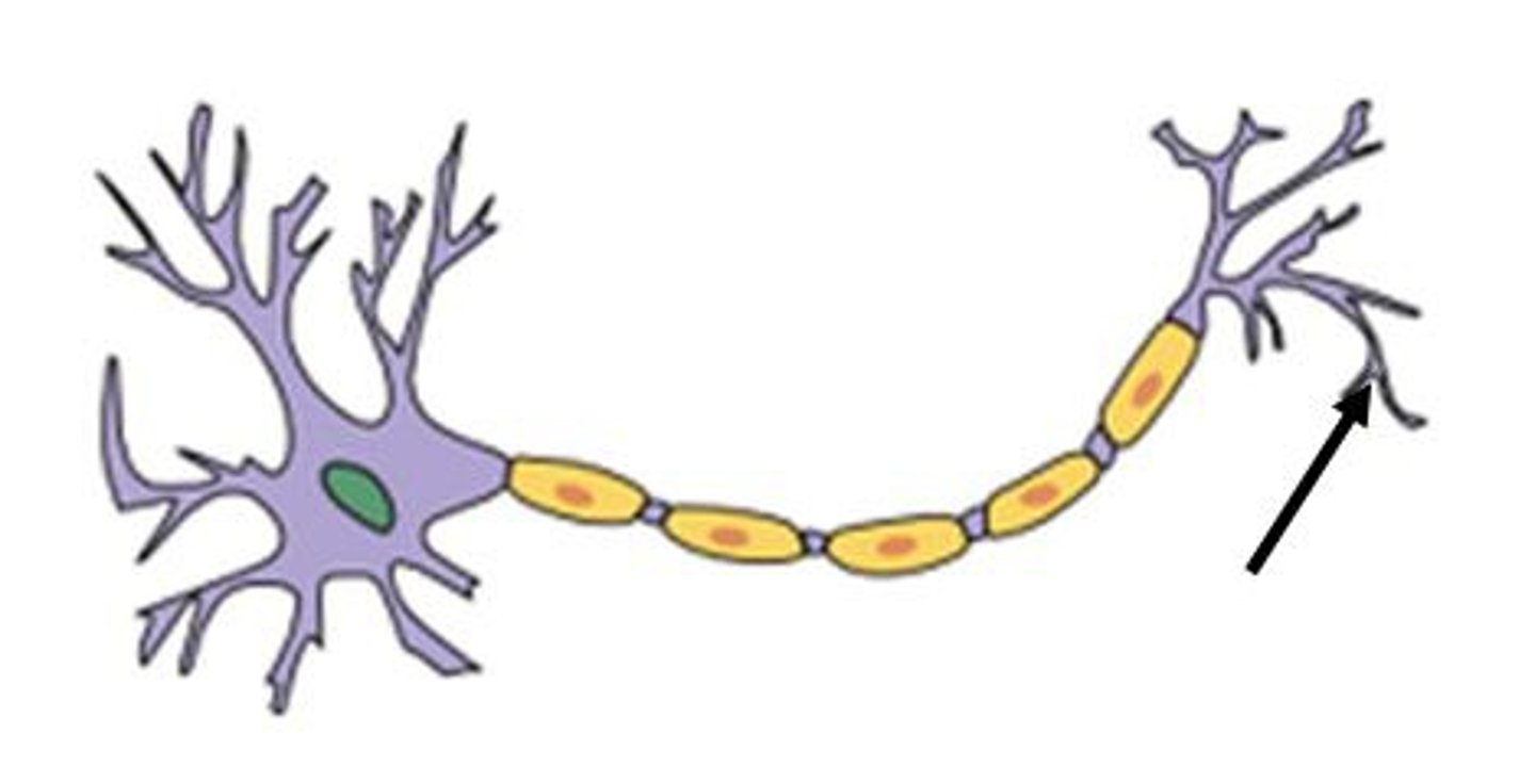
myelin sheath
A layer of fatty tissue segmentally encasing the fibers of many neurons; enables vastly greater transmission speed of neural impulses as the impulse hops from one node to the next.
Neural Impulses
Neurons process information by electric impulses that travel down the axon
Action Potential
Sends an electrical impulse known as an
86 million
How many neurons are in the human brain?
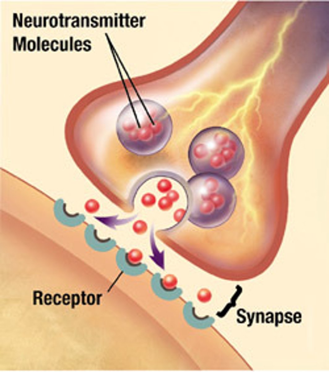
Synaptic Neurotransmission
When the action potential reaches the axon terminal, it causes the release of neurotransmitters into the synapse
100,000,000,000,000
To process information neurons must communicate with one another. How many total connections are there in the human brain?
360 MPH (faster than the fastest car)
Neurons communicate by sending electrical signal. How fast can these signals travel?
Gray matter of the brain
contain the cell bodies of the neurons
White matter of the brain
contains the axons. Axons are covered with myelin, which insulates and increases the speed of conduction
Diffusion Tensor Imaging (DTI)
an MRI-Based technique to image white matter pathways
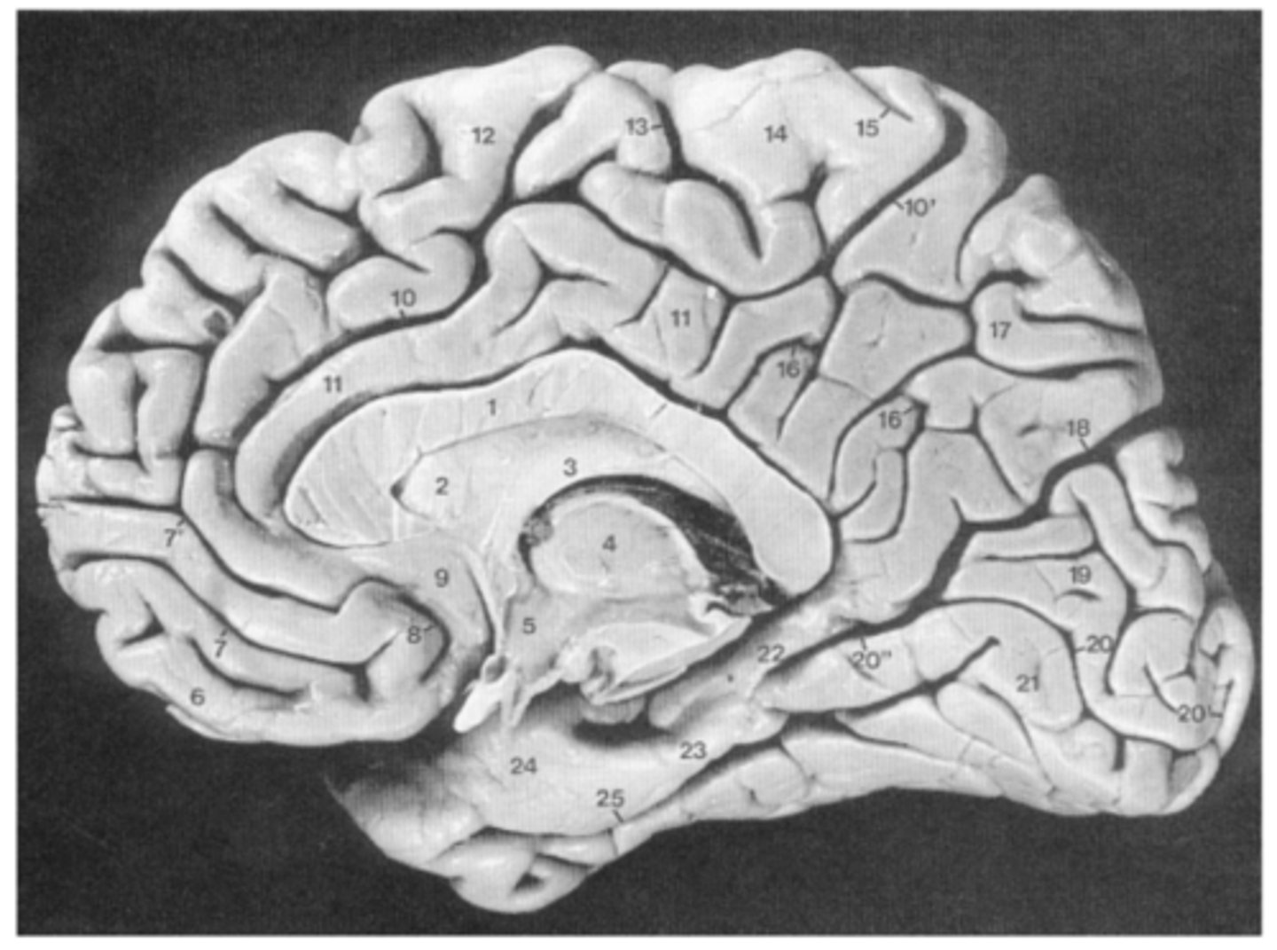
Medial View
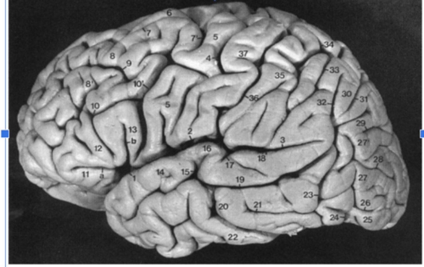
Lateral View
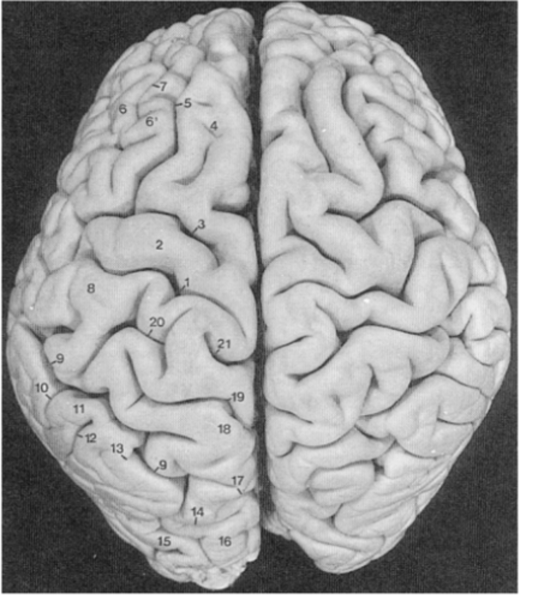
Top-Down View
electric impulses that travel down the axon
How do neurons process information?
Synapse
is the gap between two neurons
The Connectome of the Human Brain
Map out how each area of the brain transmits information to other areas
Synaptic Neurotransmission
communication bw neurons occurs at synapse, neurotransmitters released from axon of presynaptic neuron and picked up by receptors in dendrites or soma of postsynaptic neuron which have receptors that bind specific neurotransmitters, binding triggers electrochemical change in postsynaptic neuron
Brain Tissue Issue Types
Gray and White Matter
Axons are covered with Myelin
Why is the white matter white?
Conduction
The speed at which these electrical signals travel along axons
to track how one area of the brain is connected to one another
How can we Diffusion Tensor Imaging (DTI)?
Anterior
front of the brain
Posterior
back of the brain
Superior
top of the brain
Inferior
bottom of the brain
Left
left hemisphere of the brain
Right
right hemisphere of the brain
Dorsal
superior
Ventral
inferior
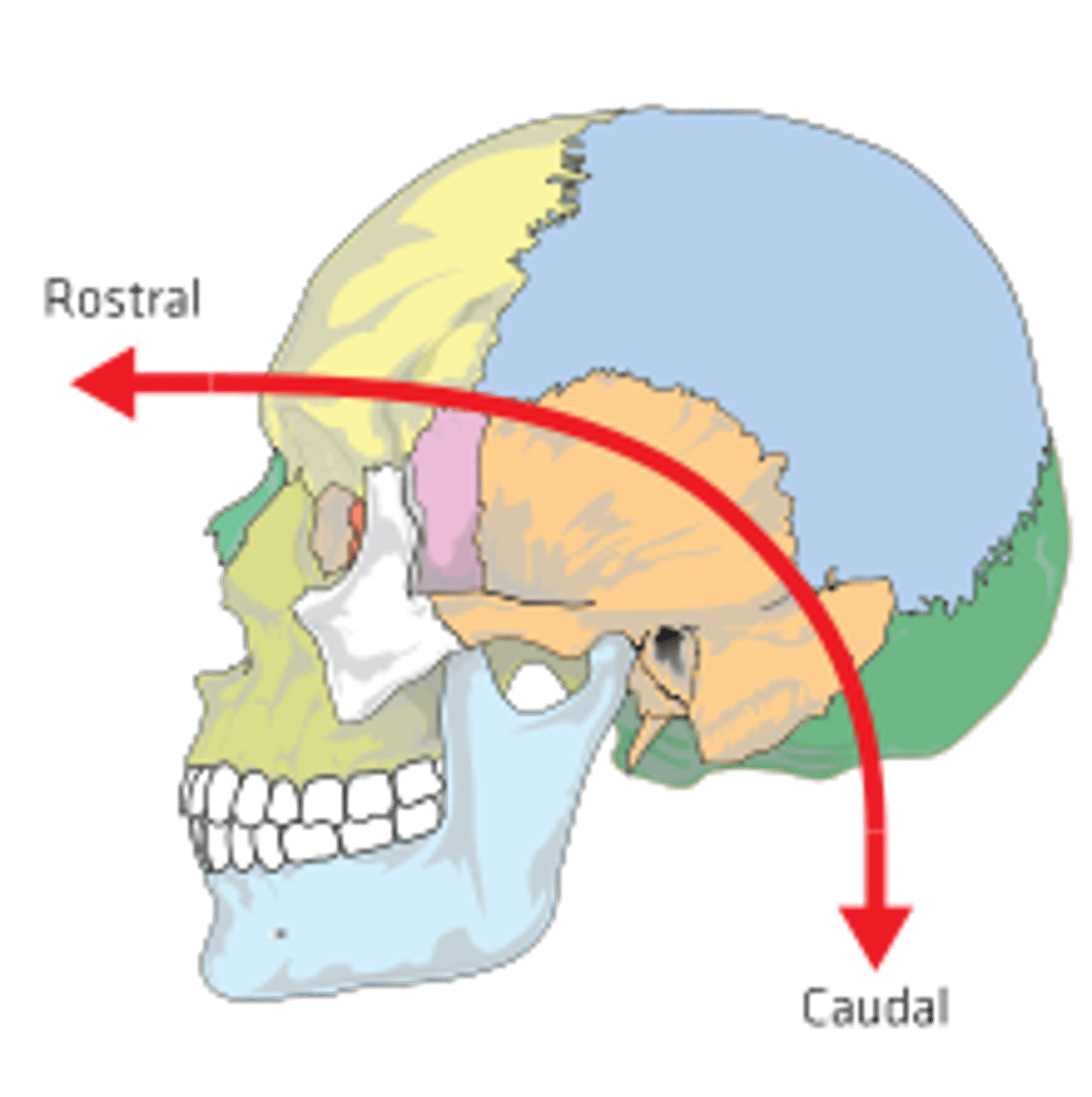
Rostral
toward the nose

Caudal
toward the tail or inferior end
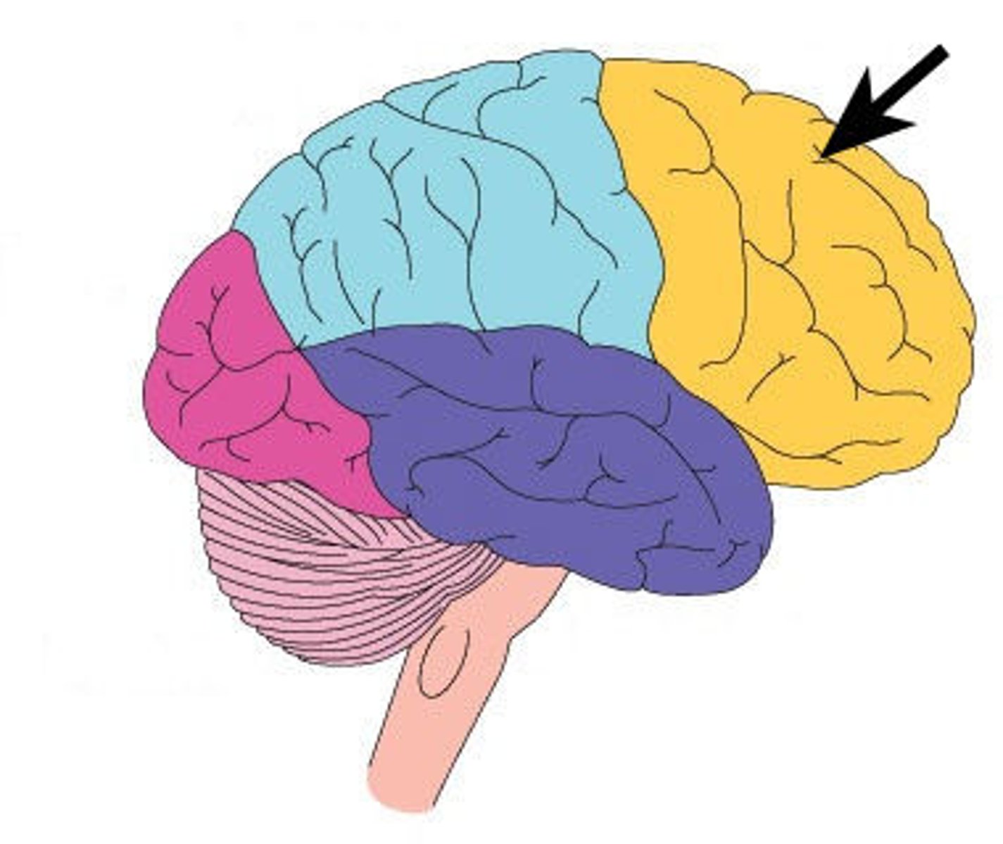
frontal lobe
central fissure
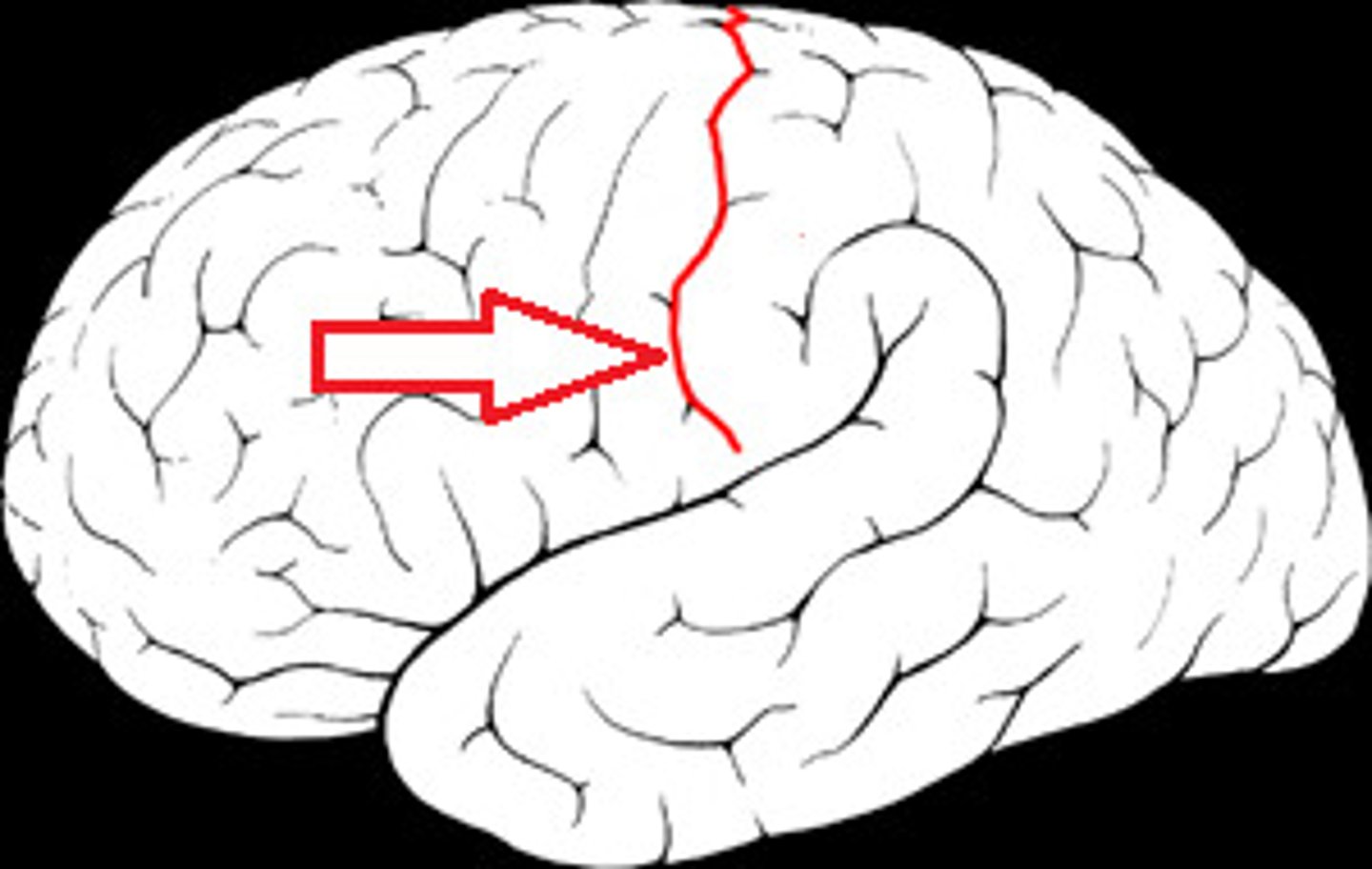
Parietal Lobe
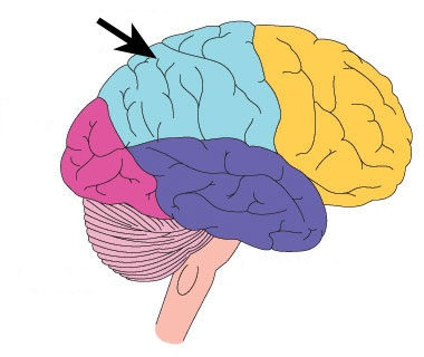
Lateral fissure
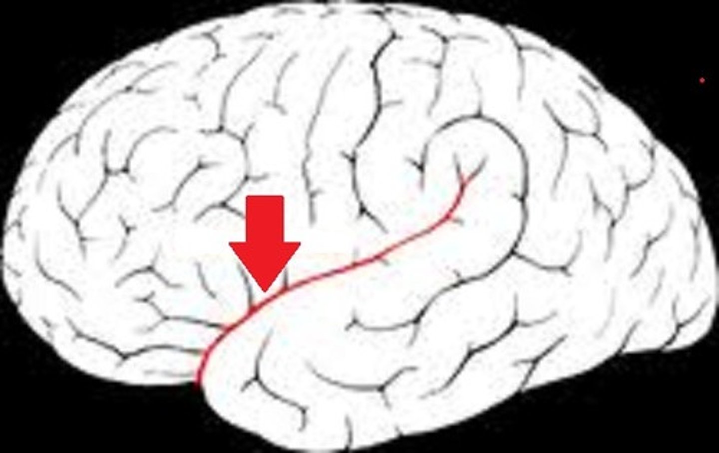
temporal lobe
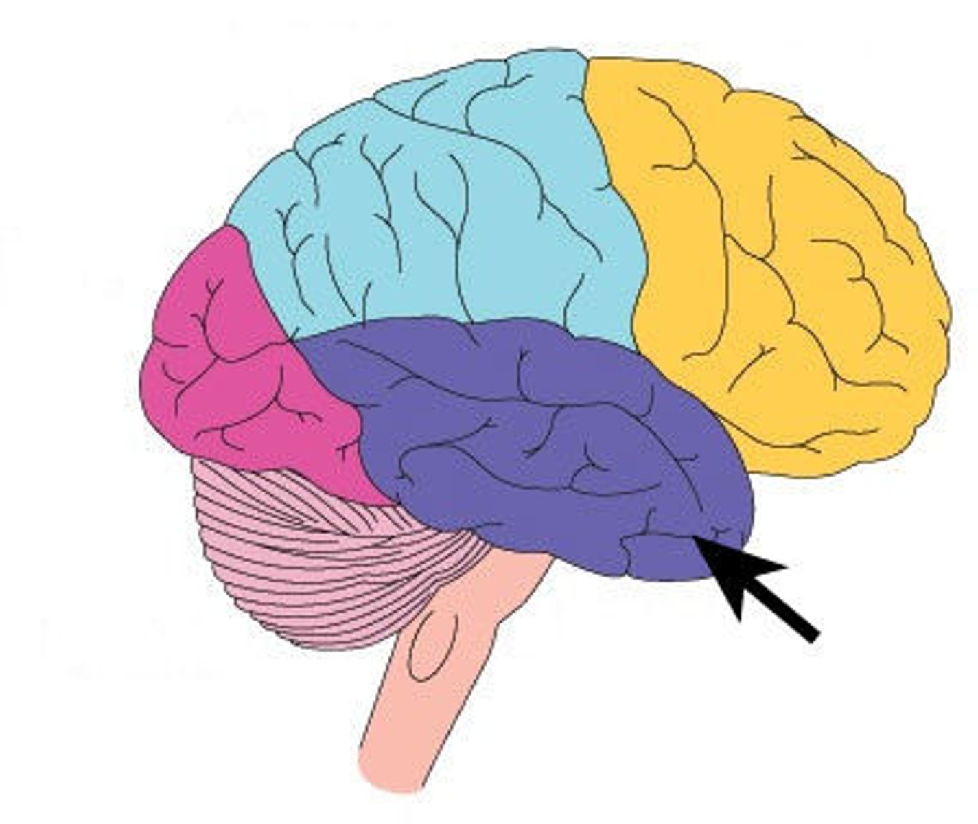
occupital lobe
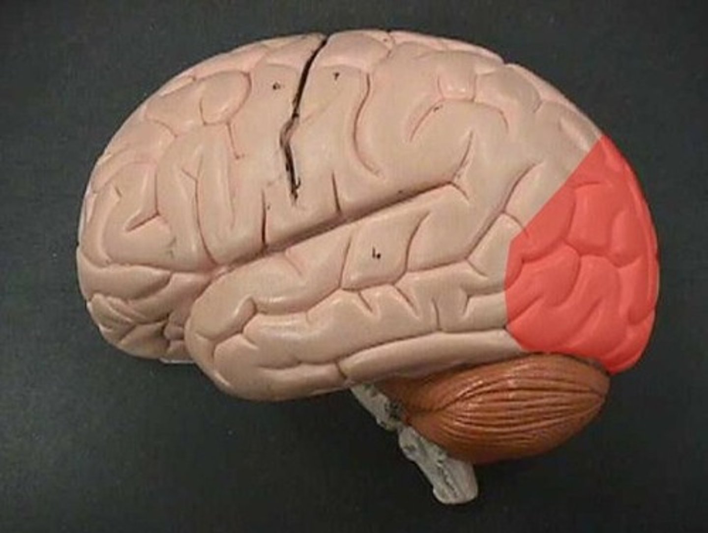
Cerebellum
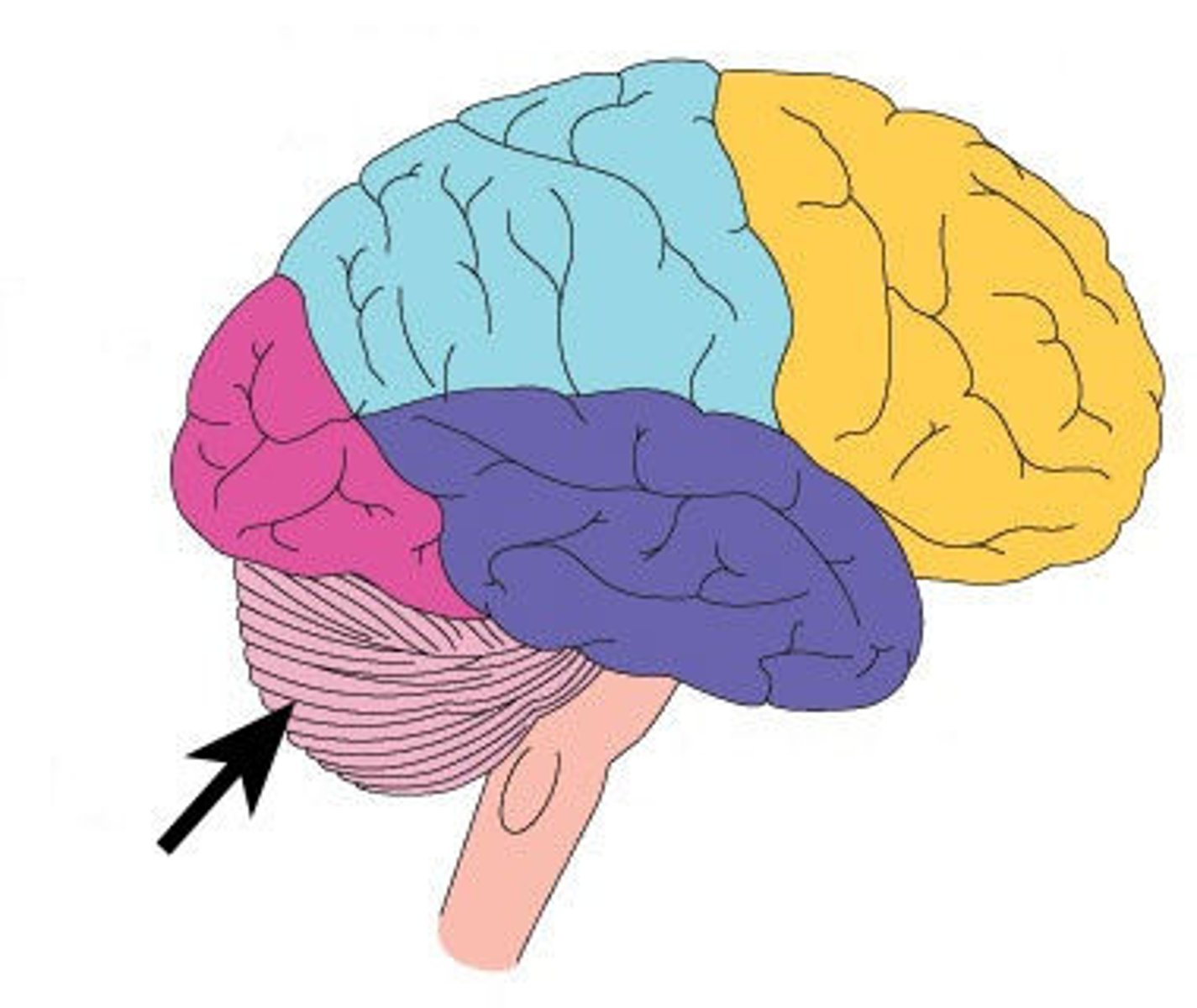
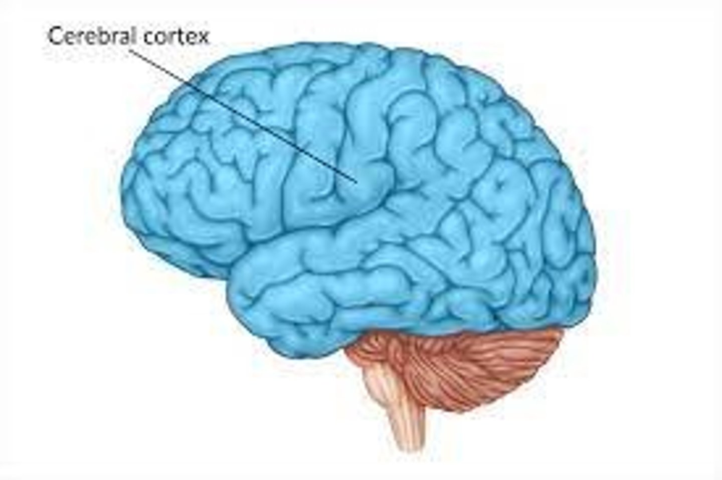
cebrebral cortex
auditory context
strip of cortex in the temporal lobe that is responsible for processing auditory information
visual cortex
The visual processing areas of cortex in the occipital and temporal lobes.
primary sensory cortex
regions of the cerebral cortex that initially process information from the senses
association cortices
complex processing that goes on between the arrival of input in the primary sensory cortices and the generation of behavior.
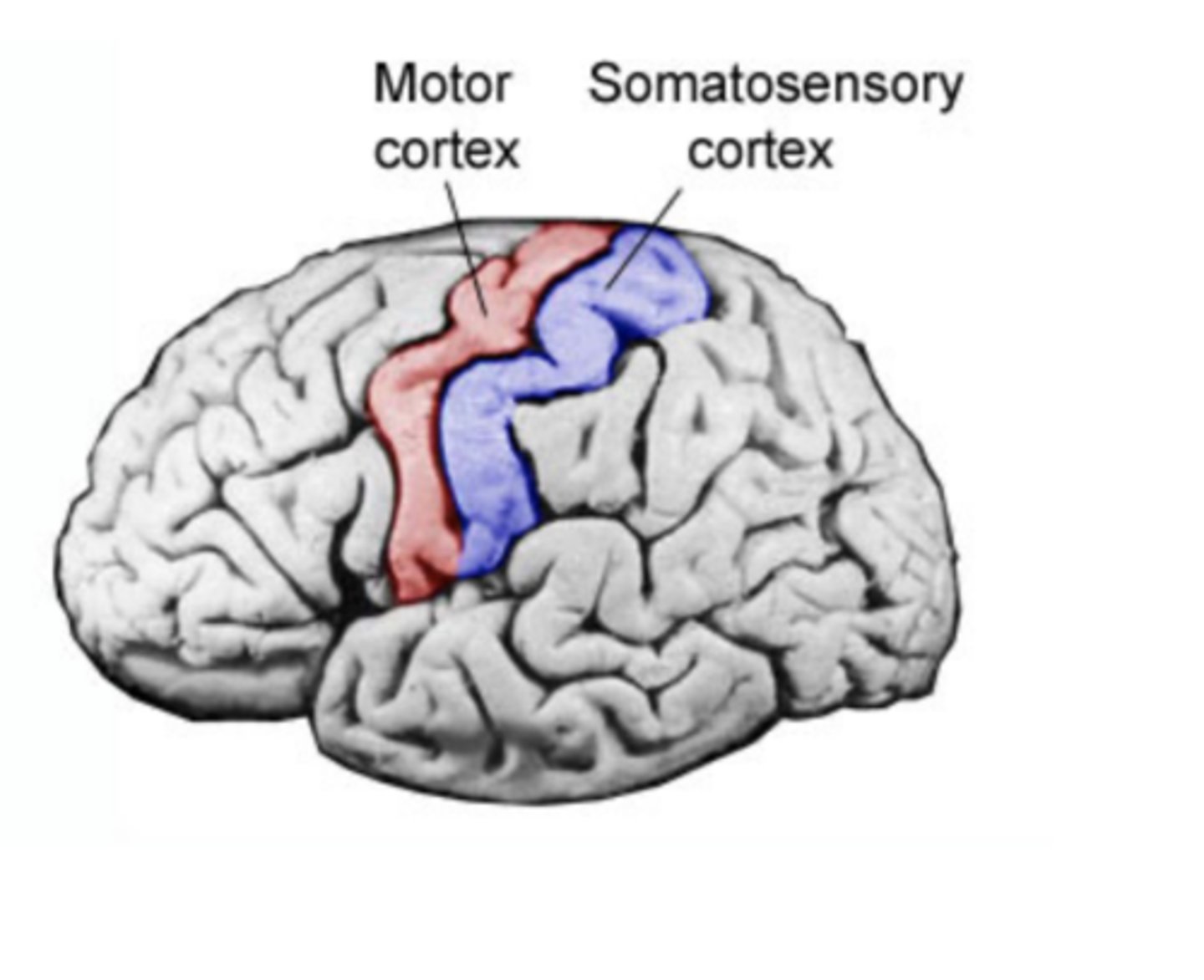
Touch cortex
somatosensory cortex
somatosensory cortex
area at the front of the parietal lobes that registers and processes body touch and movement sensations (arms, legs, fingers)
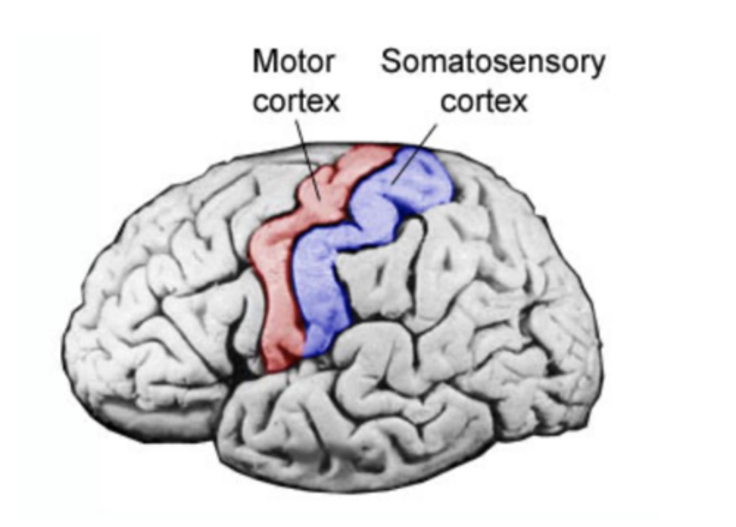
motor cortex
an area at the rear of the frontal lobes that controls voluntary movements (motion of legs, arms, fingers)
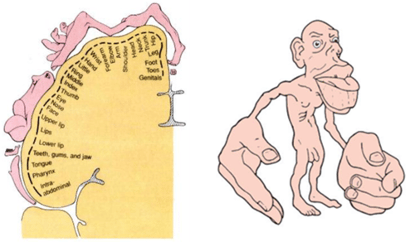
Homumculus
a depiction of what we would look like if each of our parts grew in proportion to how much we sense with them
Integrate Isolate model
involved in integration, adding regions connected to brain areas responsible for coordinating complex movements
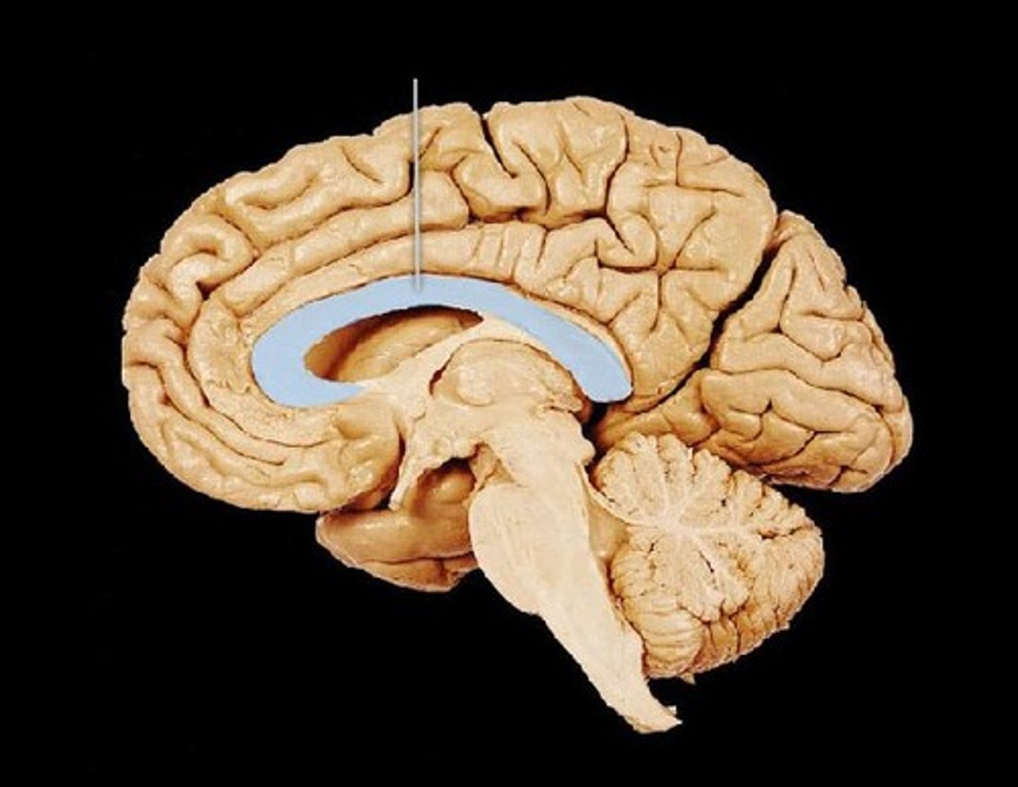
Corpus Callosum
Major white matter pathway that connects the two hemispheres of the brain
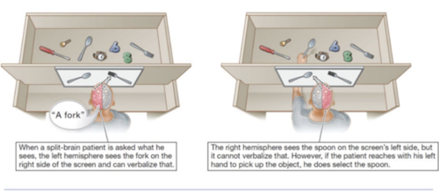
split brain syndrome
A condition in which an individual's corpus callosum is severed such that the two hemispheres of the brain cannot communicate with each other.
left visual field
right hemisphere
right visual field
left hemisphere
left hemisphere
language, speech
right hemisphere
controls the left side of the body; creative, intuitive, spacial
contralateral organization of vision
Information from left visual field is processed in the right hemisphere, and vice versa
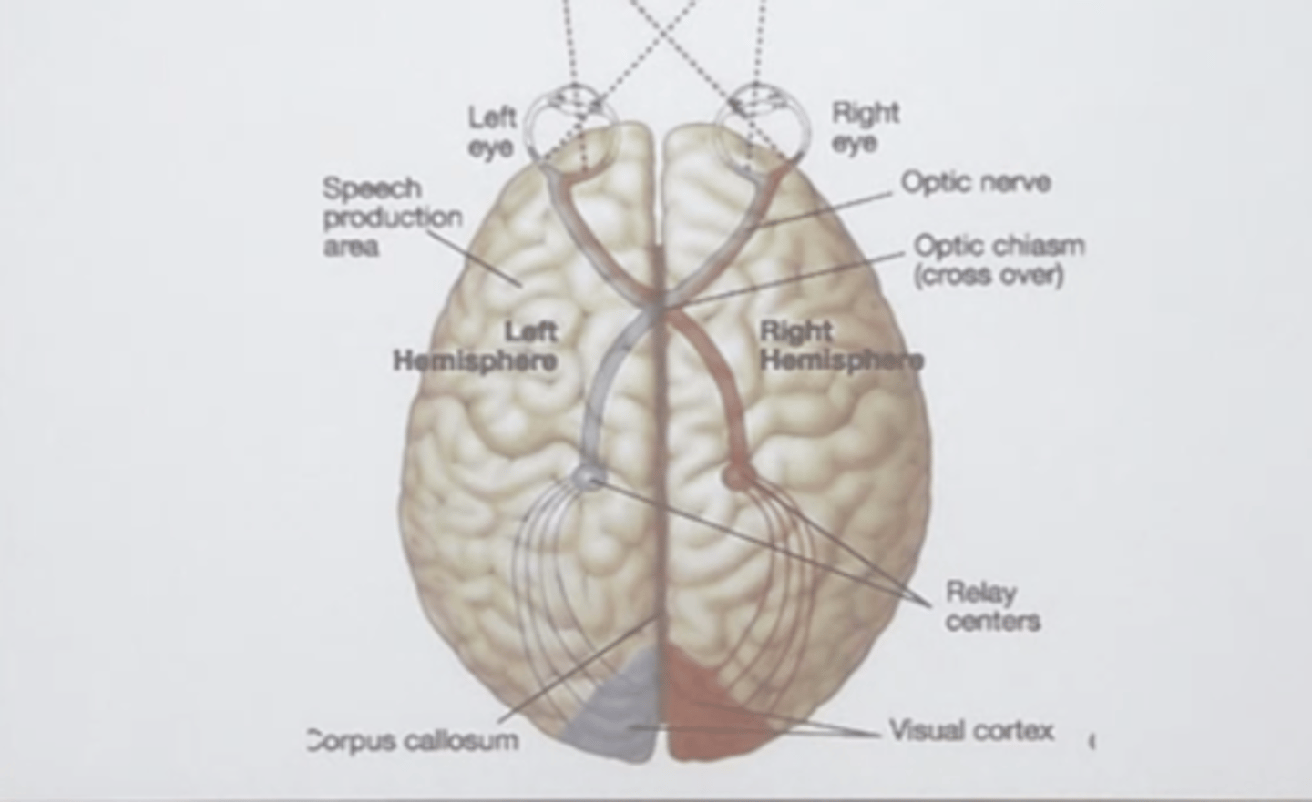
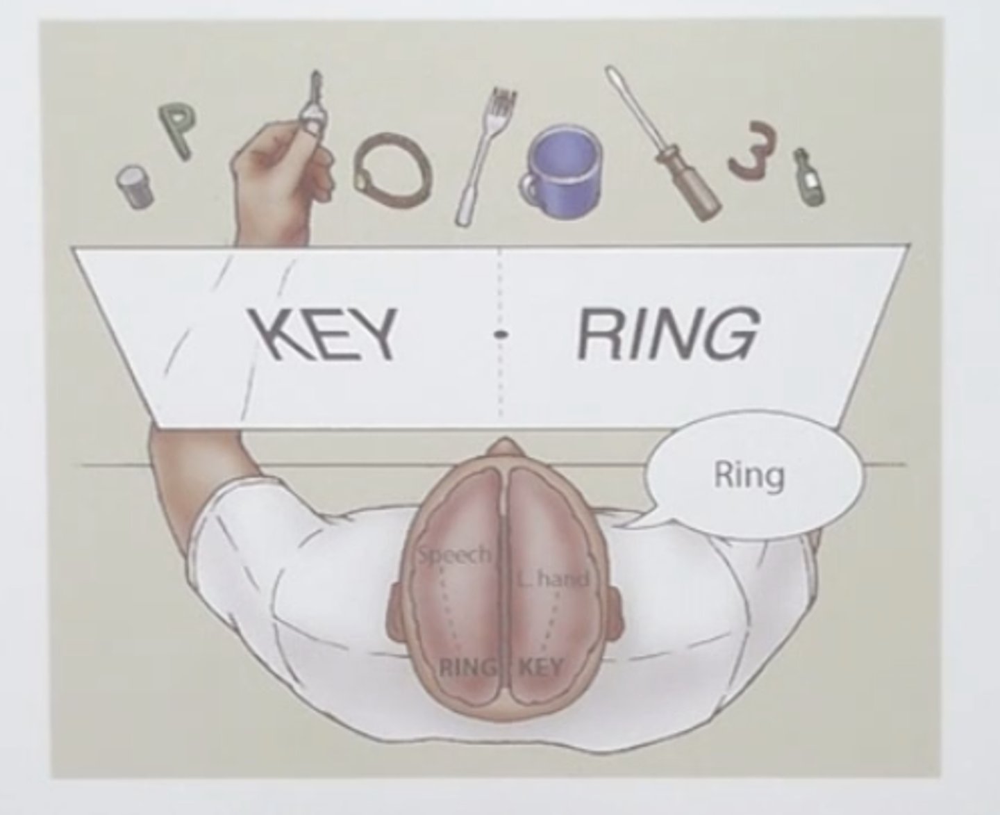
Split Brain Experiment
When a person with a split brain is presented with the picture of a ring on the right and a key on the left side of a screen, she can verbalize ring but not key because the left hemisphere "sees" the ring and language is usually located in the left hemisphere. She would be able to choose a key with her left hand from a set of objects behind a screen. She would not, however, be able to pick out a ring with her left hand because what the left hemisphere "sees" is not communicated to the left side of her body.
Studying split brain patients
History of Functional Brain Mapping
Was right about that certain areas of the brain do certain things, however, was off on the functions
The First "Brain Imaging Experiment"
Angelo Mosso
When the brain is engaged there is more blood flow, the patient would tip down due to redistribution of blood in his system
So it was like a seesaw that was evenly distributed allowing the individual to stay straight. Once they solved math problems or reading, they would tilt downwards representing an increase of blood flow to the brain.
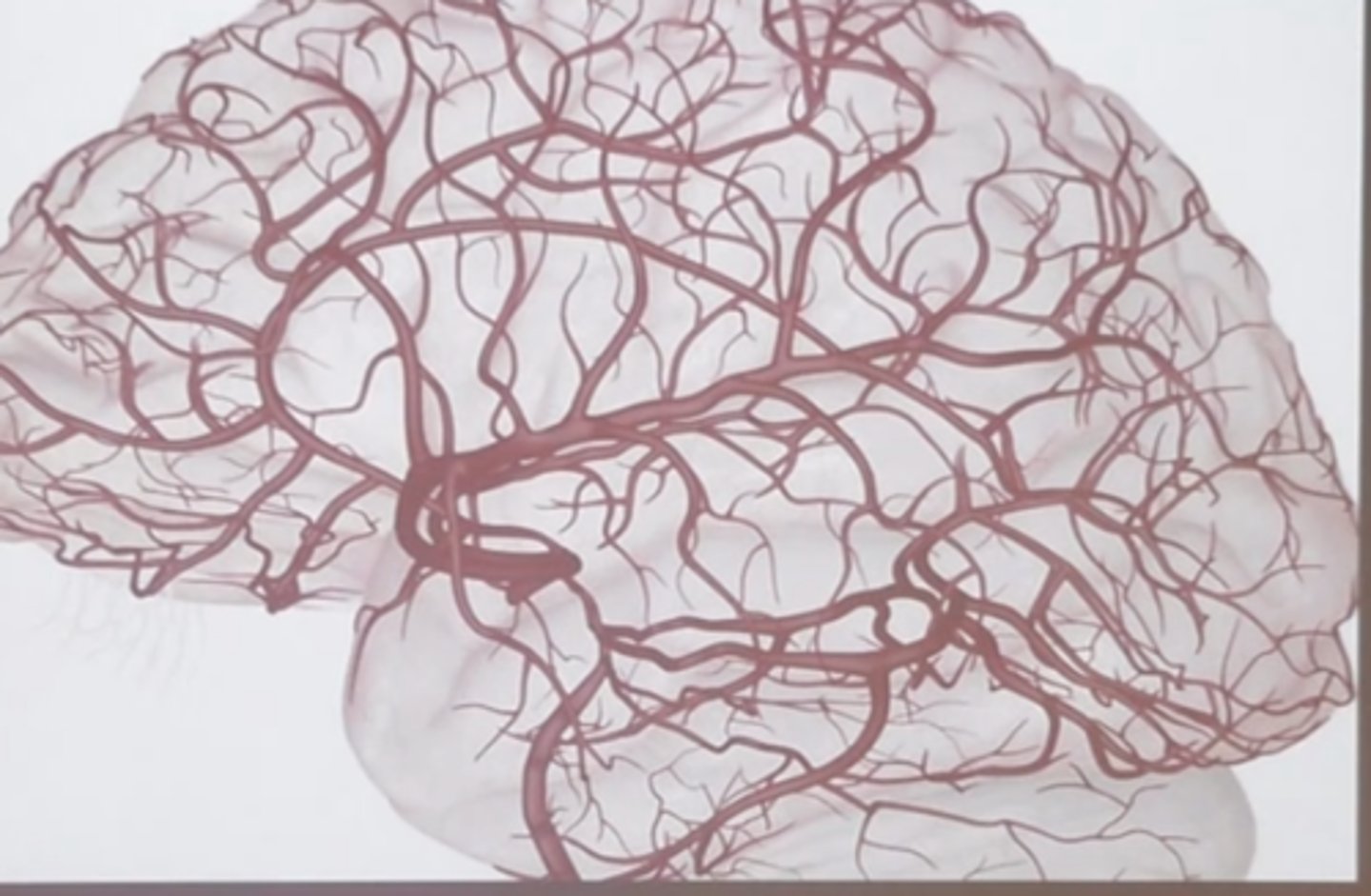
The Brains Blood Supply
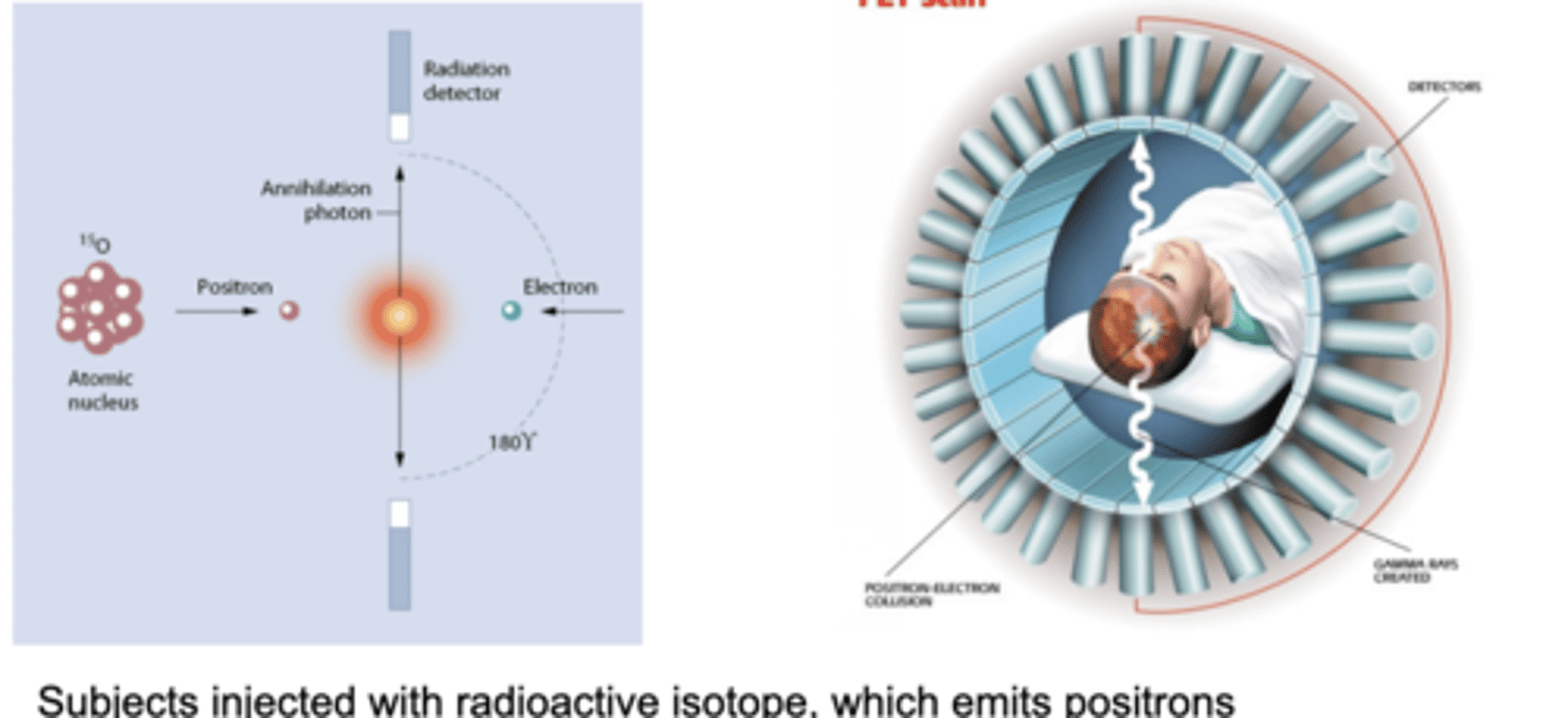
Positron Emission Tomography (PET)
a method of brain imaging that assesses metabolic activity by using a radioactive substance injected into the bloodstream
Neural activity → increased metabolic demand → local increase in blood flow the active region
it’s describing a physiological response called neurovascular coupling. Here's how it unfoldsNeurovascular coupling refers to the relationship between neural activity and blood flow in the brain, where increased brain activation leads to heightened blood supply to those areas, supporting their metabolic needs.
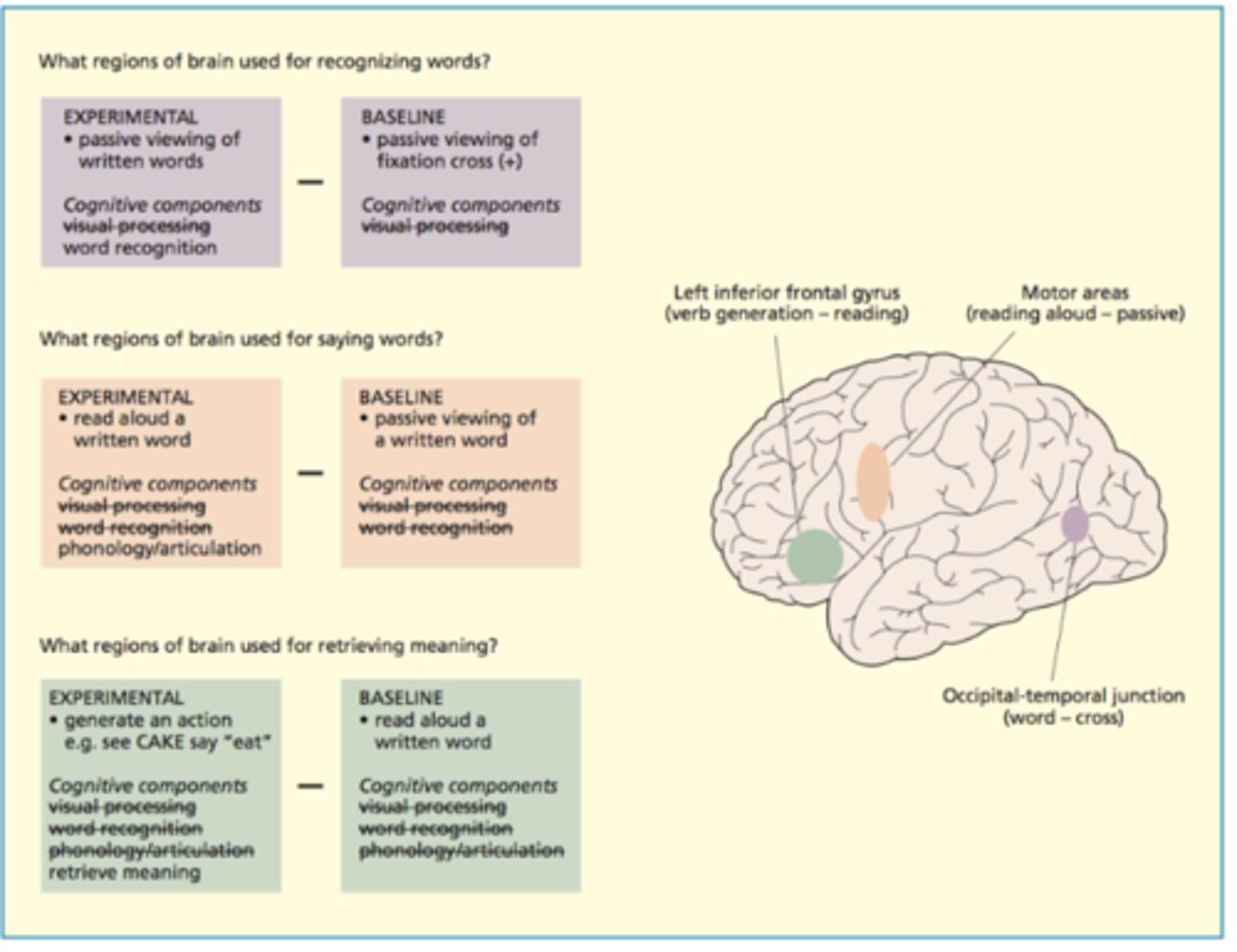
Single word processing with PET
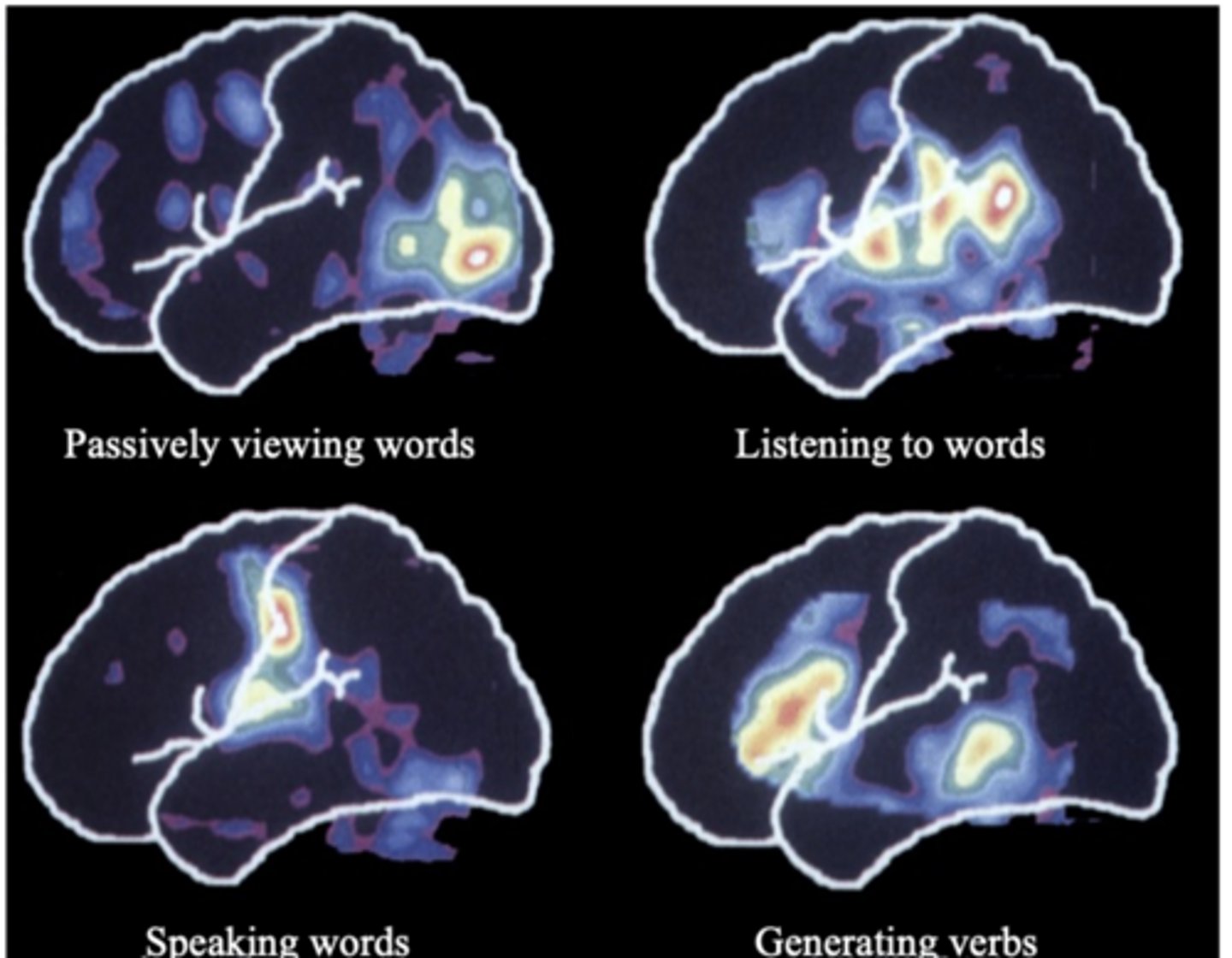
cognitive subtraction
a type of experimental design in functional imaging in which activity in a control task is subtracted from activity in an experimental task
Pros of PET Scan
1. Decent spatial resolution (the quality)
2. Can be used for diagnostic purposes
3. Can also measure neurotransmitter metabolism (e.g., dopamine)
4. Very quiet (good for auditory experiments
Cons of PET
1. invasive
(radioactive injection)
2. Very expensive ($3-5k per scan)
3. Poor temporal resolution (cannot detect the neural response to discrete cognitive effects) (change over tiime is slow and not good at measuring quick changes in brain activity. )
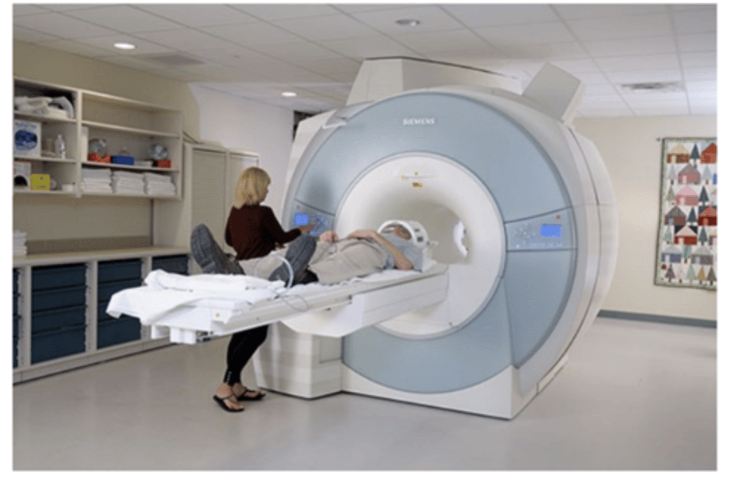
Magnetic Resonance Imaging (MRI)
brain-imaging method using radio waves and magnetic fields of the body to produce detailed images of the brain
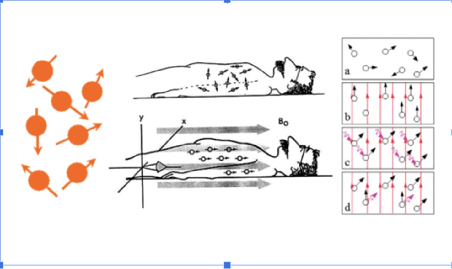
Strong Magnetic Field
You lie inside a large magnet. This magnetic field causes hydrogen protons (found abundantly in water and fat) in your body to align like tiny compass needles.
Radiofrequency Pulse
A burst of radio waves is sent into your body, knocking those aligned protons out of position.
Relaxation & Signal Emission
When the pulse stops, the protons relax back to their original alignment. As they do, they emit radio signals.
Signal Detection & Image Formation
These signals are picked up by receiver coils and processed by a computer to create detailed cross-sectional images of your body.
MRI physics
Atoms with an odd number of protons and/or neutrons are capable of acting as magnets
-A strong magnetic field causes hydrogen atoms to align in the same orientation.
-When a radio frequency wave is passed through the head, atomic nuclei emit electromagnetic energy.
-The MRI scanner is tuned to detect radiation emitted from the hydrogen molecules.
-Computer reconstructs image.
MRI
studies brain anatomy
Functional MRI (fMRI)
studies brain function
Blood Oxygenation Level Dependent (BOLD) signal indirect measure of neural activity
neural activity --> blood oxygen --> FMRI signal
Blood Oxygen Level Dependent (BOLD) Signal
When neurons in a brain region become active, they consume more oxygen.
The body responds by increasing blood flow to that area.
fMRI detects these changes in blood oxygenation, which indirectly reflects neural activity.
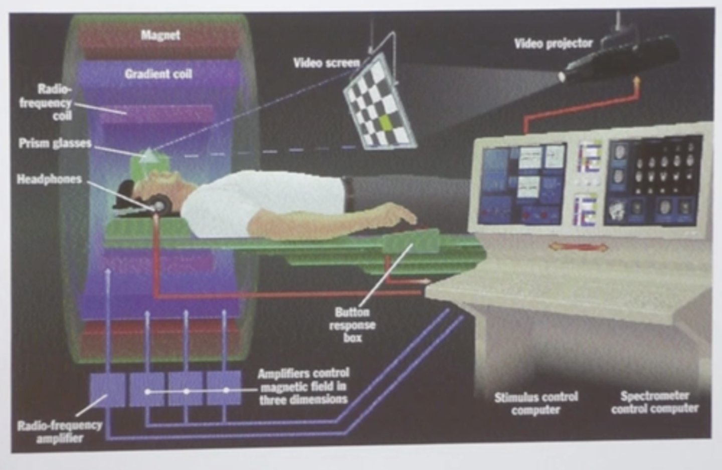
Magnetic Field & Signal Detection
Like a regular MRI, fMRI uses a powerful magnet to align hydrogen atoms.
As blood oxygen levels shift, the magnetic properties of hemoglobin change—this alters the signal picked up by the scanner.
Task-Based or Resting-State Scans
You might be asked to perform a task (e.g., tap your fingers, recall a word) during the scan.
The fMRI maps which brain regions light up during that activity. And is used to visualize brain activity through changes in blood flow.
BOLD
Blood Oxygenation Level Dependent
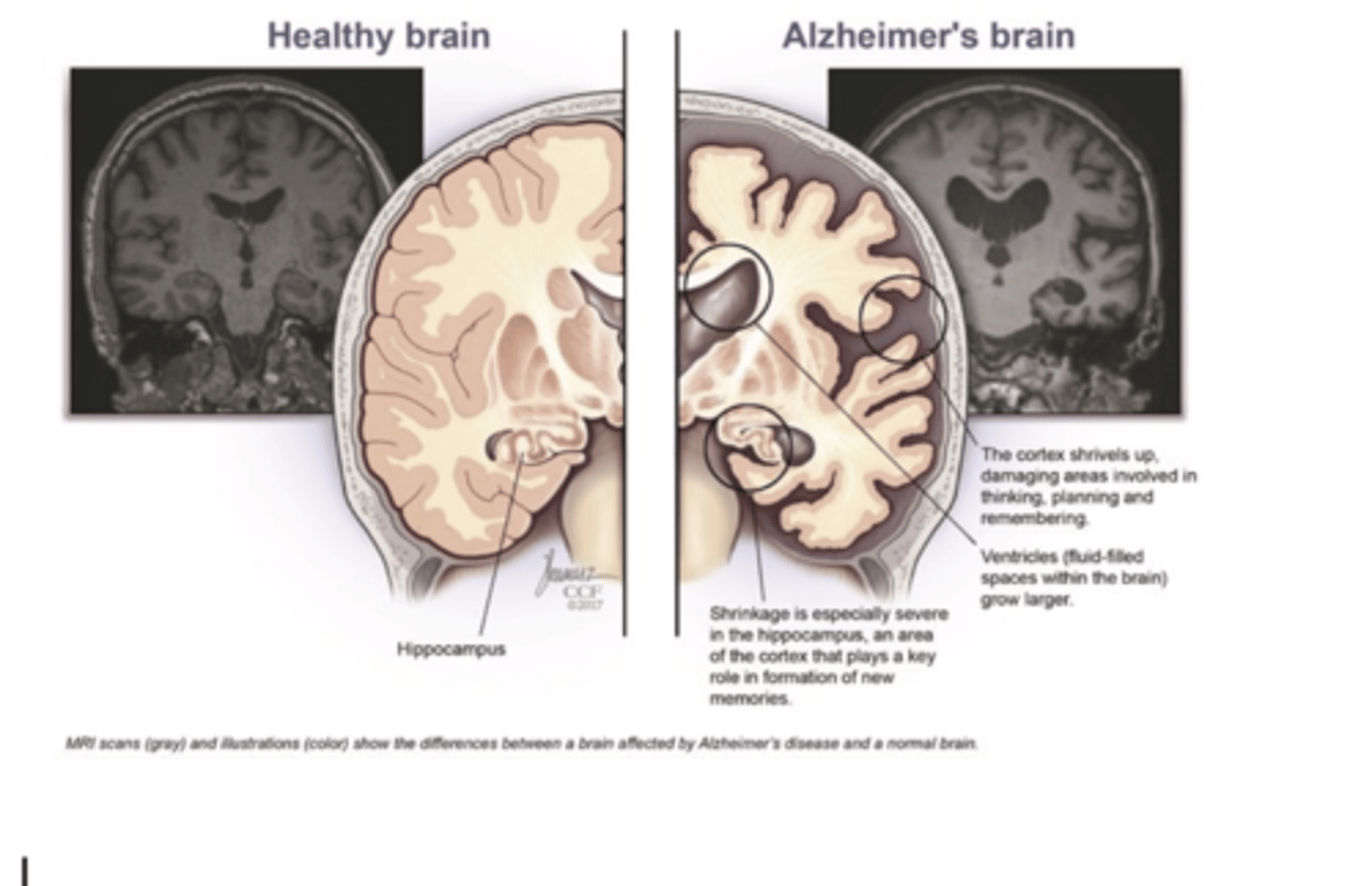
Structural MRI scans
are useful for clinical imaging (diagnosis)
MRI vs. fMRI
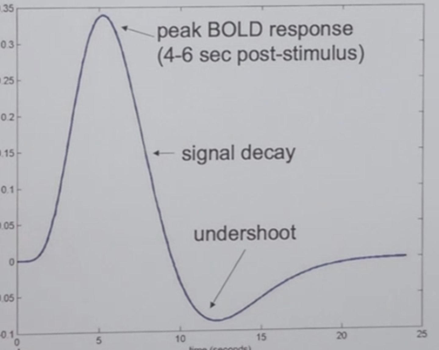
Hemodynamic response function (HRF)
changes in the BOLD signal over time
FMRI setup
Contrasting BOLD activation across conditions
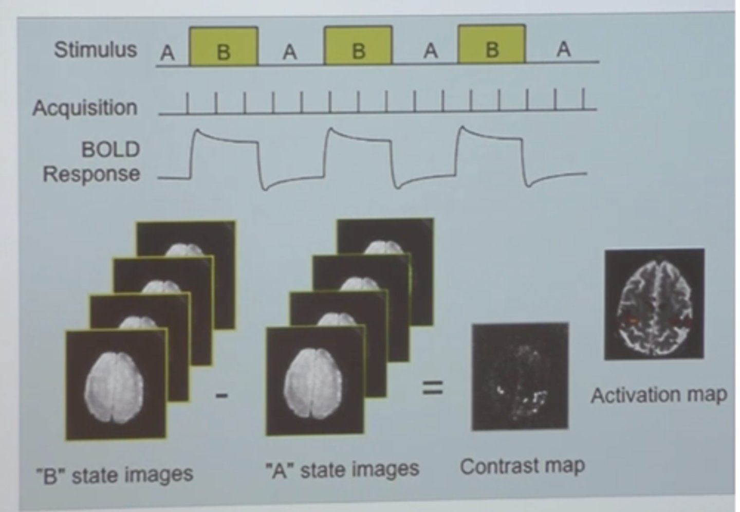
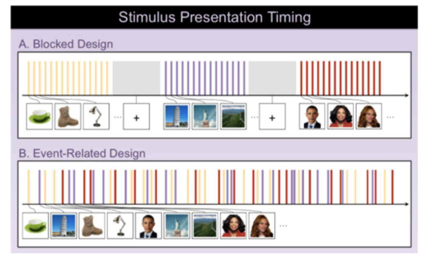
blocked design
experimental units or subjects are placed into groups based on certain properties identified for control purposes. Then blocks are randomly assigned all treatments within each block. Comparisons are drawn within blocks.
event-related design
stimuli from two or more conditions are presented randomly or interleaved
Advantages of Event Related Design
Characterize individual trails after-the-fact, based on the subject's behavioral response
Compare incorrect and correct trials
With FMRI, every two seconds there is a measure level
Group-level statistical analysis
crucial for inference that effects generalize across the population
functional segregation
Organization into different areas, each of which performs a different function; for example, in sensory systems, different areas of secondary and association cortex analyze different aspects of the same sensory stimulus.
functional integration
the way in which different regions communicate with each other
fMRI pros
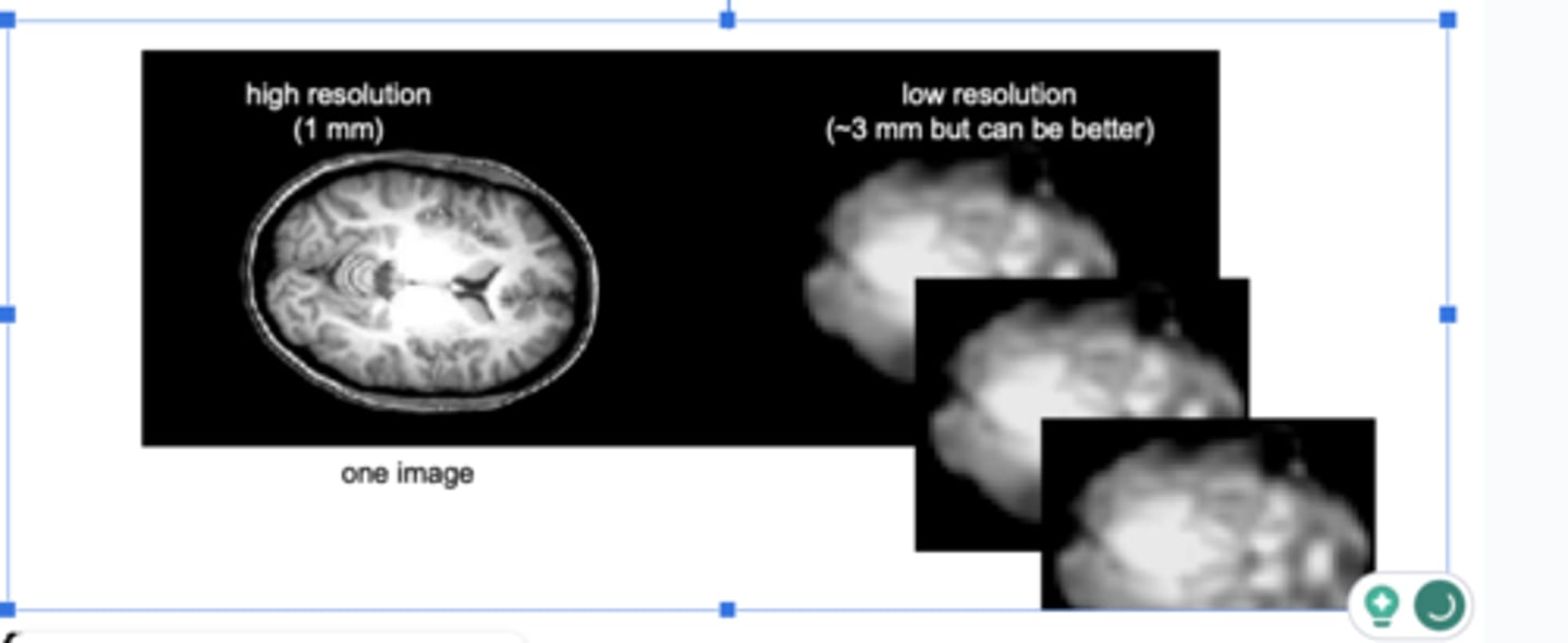
1. Good spatial resolution (1-3mm3) allows precise localization of brain activation
2. Non-invasive (no known harms to subjects)
3. scanners widely available at medical centers and research universities.
fMRI cons
1. Although temporal resolution (1-4 sec) is far better than PET scans, still much slower than measuring actual neural activity
2. Expensive (~ $600/hour)
3. Scanner noise is very loud
4. only measures blood flow, it does NOT provide precise info about the timing of neural activity.
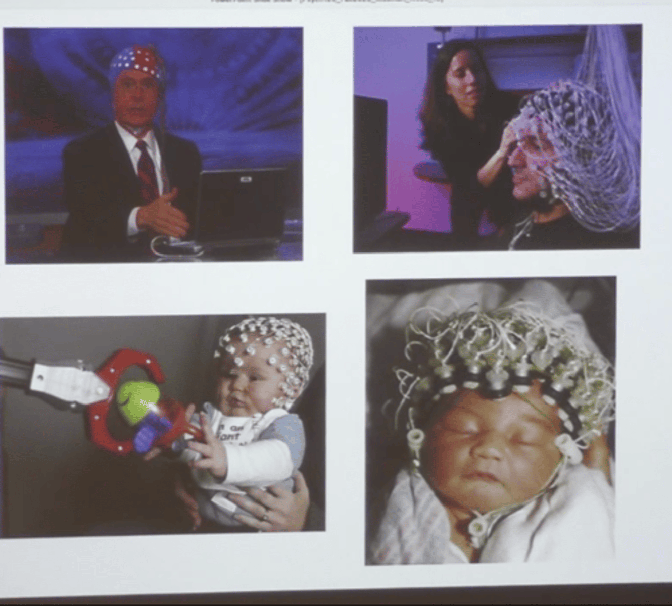
Electroencephalogram (EEG)
an record electrical activity from large populations of simultaneously active neurons at the scalp with millisecond resolution.
is a direct measure of neural activity.
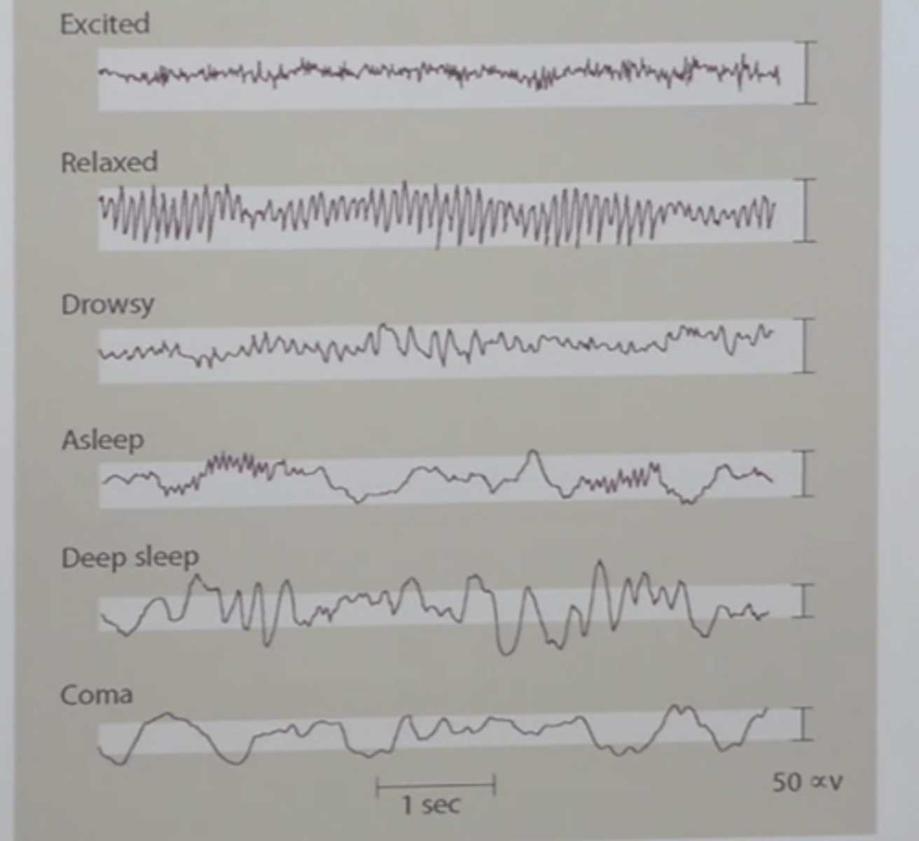
Arousal states of the brain: characteristic waveform patterns
ERP (event-related potential)
Characterizing electrical activity changes
evoked during specific cognitive events
a measure of electrical activity from a subpopulation of neurons in response to particular stimuli that requires averaging many EEG recordings