Sensory Systems: Tactile and Proprioceptive Sensation
1/10
There's no tags or description
Looks like no tags are added yet.
Name | Mastery | Learn | Test | Matching | Spaced |
|---|
No study sessions yet.
11 Terms
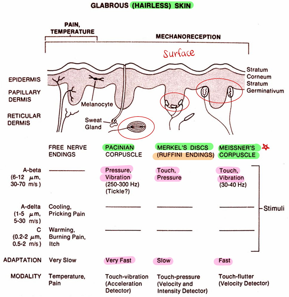
Modalities and Nerve Endings- Skin sense modalities
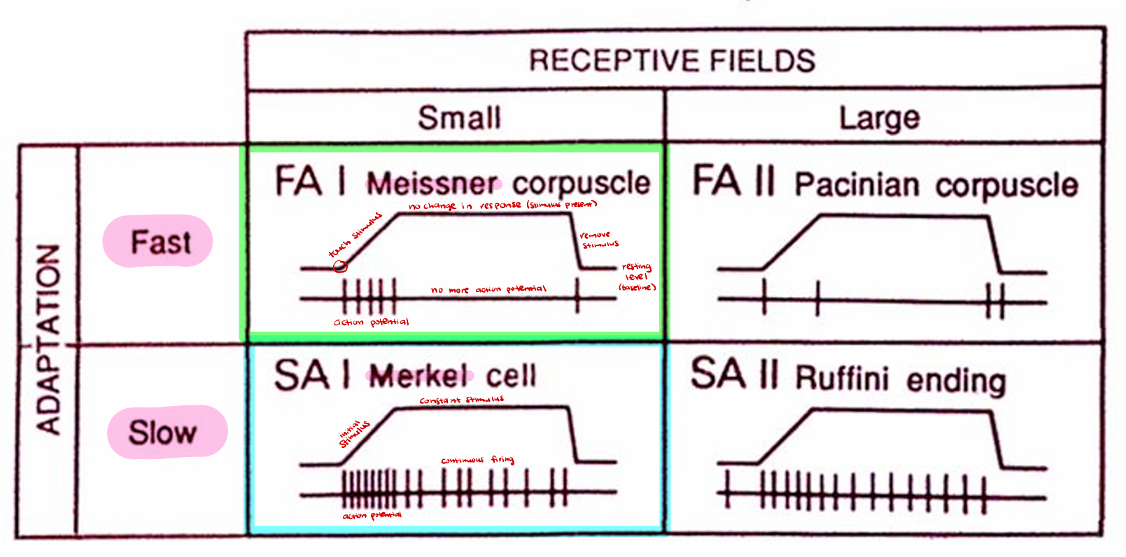
Fine, discriminative touch (2 point discrimination): Require small receptor near surface ( epidermis)
Merkel (II), Pinkus (hairy; = Merkel), Meissner (II, hairless skin), Ruffini organs (deeper in dermis)
Vibration sense: Used to appreciate textures, tested with tuning fork
Pacinian (“cut onion”, very fast), Meissner (fast)
Pressure: Slower adaptation
Pacinian (very fast: good for fast changing but not ideal for slow changing pressure), Merkel (slow)
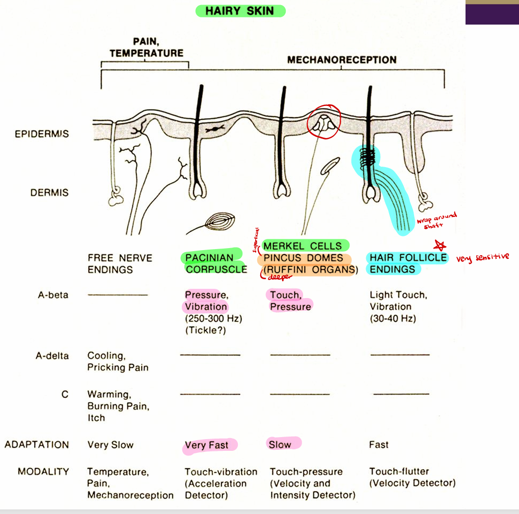
Specialized Sensory Endings (all have group II nerve fibers)
Hair shaft receptors
Merkel cell receptors
Meissner's corpuscles
Pacinian corpuscles
Ruffini endings
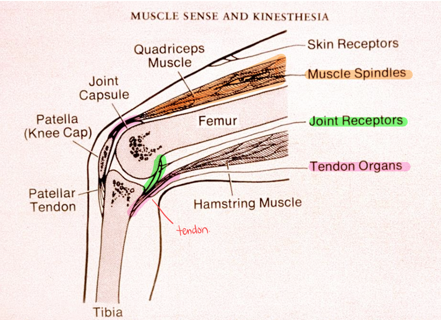
Proprioceptors (muscle and joint senses)
Muscle spindles: muscle stretch (length), primary (Ia) and secondary (II) endings
primary have larger (faster) axons
Golgi tendon organs: muscle & tendon tension, group Ib
almost as fast as primary spindles
Joint receptors: position sense, groups Ib, II
several types
Physiological Properties
Modality-specific - each is tuned to type of mechanical stimulus
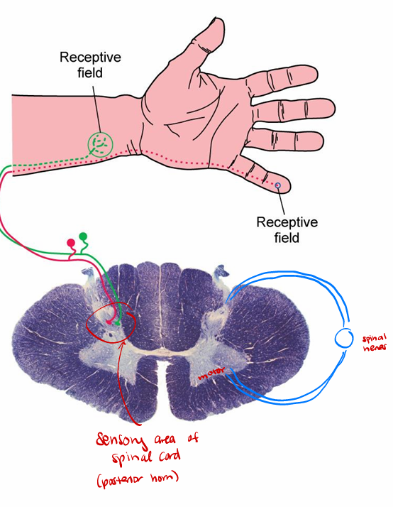
Receptive field: defined surface area of skin- can be mapped out on body surface as dermatomes (area of skin where the particular receptor receives stimulus/ differ by receptor and location of skin)
*photoreceptor cells: exact location in visual field
Smaller receptive field = greater sensitivity (fingertips)
Wider receptive field = lower sensitivity (back)
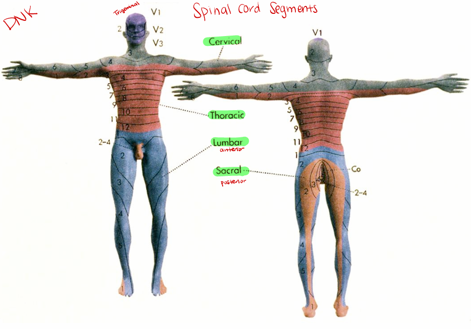
Spinal Cord Segments
Cervical
Thoracic
Lumbar (anterior)
Sacral (posterior)
Adaptation and Transduction
Adaptation determined by structure and electrical properties of ending
changing stimulus- want fast adaptation
slow adaptation = “true representation of stimulus”; keeps firing even after stimulation stops
Transduction thought to be by mechanical deformation of ion channel - differs with receptor type
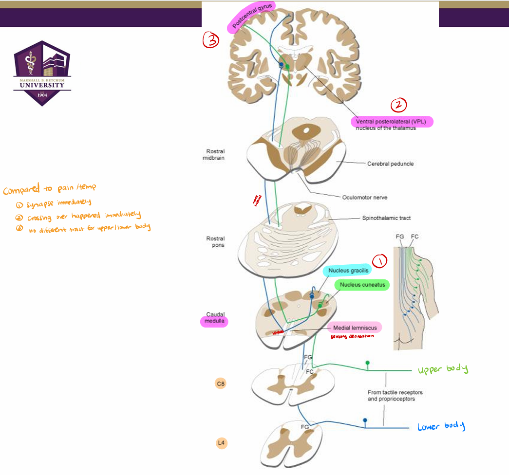
Central Connections (body) (receptor → medulla)
Name of tracts: “medial lemniscus pathway” (medulla) or “dorsal column pathway” (spinal cord”
1o afferents have cell bodies in DRG
First synapse in spinal cord (posterior horn) for reflexes only (different from pain & temperature)
if reflexes not needed, info will continue tract w/o synapses
1o afferents from body ascend in ipsilateral dorsal columns (gracile MEDIAL(receive sensory info from lower body) and cuneate LATERAL (upper body) fasciculus) to medulla (final destination of axon- first synapse if no reflex)
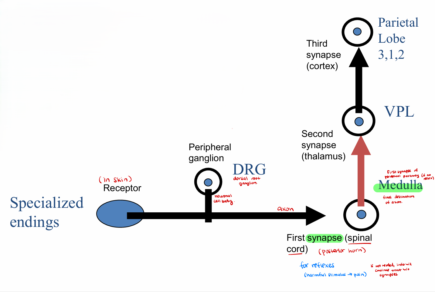
Central Connections (body) (medulla → VPL)
Synapses in medulla (dorsal column nuclei)
nucleus gracilis (lower body) and nucleus cuneatus (upper body) different
2nd order fibers cross midline in medulla (sensory decussation) to form medial lemniscus
lower body (gracile) more ventral in tract
Central Connections (body) (VPL → parietal lobe)
Medial lemniscus ascends to ventral posterior lateral thalamus (VPL)
in pons, moves dorsally and laterally; lower body is most lateral (rearrange)
Third order fibers from VPL ascend to postcentral gyrus in somatotopic order
same locations as pain & temperature: leg medial; arm dorsal; face ventral (homonculous)
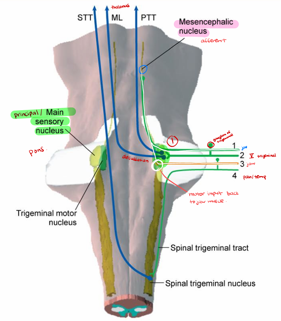
Tactile and Proprioception Pathways for the Face
Primary afferents (CN V) synapse in principal sensory nucleus (of trigeminal) in pons - cell body in trigeminal ganglion
Local reflexes to facial motor nucleus (e.g., blink - V1 afferent)
Second order fibers cross midline (some ipsilateral), join trigeminothalamic tract
more dorsal than pain & temperature fibers
Thalamic relay in ventral posterior medial (VPM) nucleus - same as pain & temp
Third order fibers ascend to inferior aspect of postcentral gyrus
Some primary afferents have cell bodies in mesencephalic nucleus (of trigeminal), participate in jaw reflexes
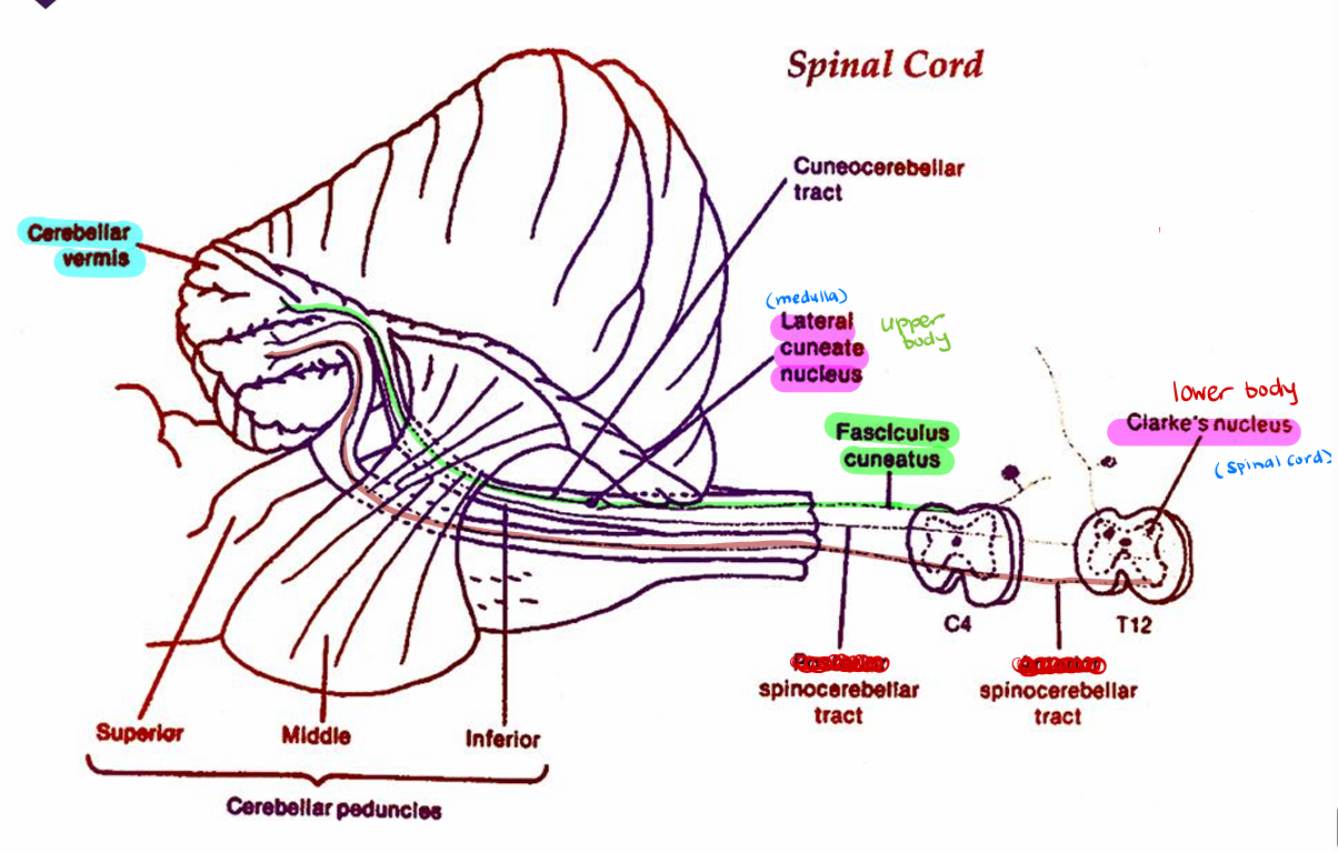
Muscle and Joint Sense Input to Cerebellum
All muscle and joint receptors, some skin receptors (proprioceptors)
Primary afferent may travel in dorsal column
First synapses in spinal cord (Clarke's column- lower) or medulla (lateral cuneate nucleus- upper)
Clarke’s: relays info from lower body (leg)
Lateral cuneate: relays info from upper body (arm)
Destination in ipsilateral cerebellar vermis (most fibers do not cross midline- no decussation)
Spinocerebellar tract from lower body (leg)
Cuneocerebellar tract from upper body (arm)
Does not participate in perception: info used for muscular coordination