Vision 3
1/35
There's no tags or description
Looks like no tags are added yet.
Name | Mastery | Learn | Test | Matching | Spaced | Call with Kai |
|---|
No study sessions yet.
36 Terms
What are 3 features of the retinotopic map within V1?
inverted
horizontally flipped (right VF in LH)
distorted (cortical magnification)
What is cortical magnification?
there are more cells representing the fovea than the visual periphery
Why does fovea take up only 1% of retina but 50% of V1?
only cones which have higher visual acuity as not many to one so good detail at the fovea
at fovea only photoreceptors minimises bluriness so again higher visual acuity
all good for evolution
Who mapped out the retina, focusing on peaks and declines of photoreceptors distribution in retina?
Curcio et al (1990)
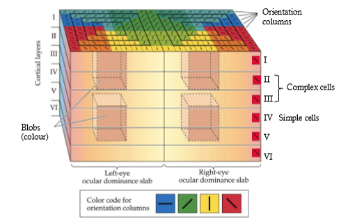
How many layers are in the striate cortex (V1)?
6
Where do most inputs from the thalamus go to?
layers IV and V
What happens in other layers?
higher processing in layers 2/3
outputs mostly from layers 2/3 corticortical
ouputs also from layers 5/6 subcortical
Whats one attribute V1 neurons show?
orientation tuning
Who investigated orientation tuning and how?
Hubel and Wiesel (1959)
microelectrode recordings in cat V1 while presenting bars of light against dark background
cells in V1 layer IV show orientation selectivity respond most strongly to bars in a particular direction
most be located in particular location within neurons spatial receptive field to elecit response (simple cells)
How is simple cell orientation tuning computed?
combining four centrre/surround fields produces an orientated bar receptive field in V1 simple cells
What is the difference in complex cells orientation tuning?
they will respond to orientation anywhere when presented within neurons spatial receptive field
no inhibitory surround - don’t have to be presented in middle of receptive field
What else are complex cells attuned for?
motion direction selectivity
How is orientation tuning organised within V1? (take sulcus and drive electrode perpendicular through)
cells throughout cortical layers prefer the same orientation
clusters of cells show similar preferred orientations in a ‘pinwheel’ arrangement across the cortical surface
Do cells in V1 have occular dominance?
yes - most cells respond more robustly to inputs from one particular eye
How is eye preference arranged?
into occular dominance columns across the V1 surface and throughout the cortical layers
How do you investigate ocular dominance?
inject retina with marker
taken upoptic tract
lands in V1
can do in one eye or other
track where in visual cortex you’re getting input from each eye
What’s another thing that cells in V1 are attuned to?
colour contrast
How can we see colour contrast in those cells?
some colour sensitive cells show circular receptive fields with centre excited by one colour and inhibited by the other
in surround the pattern is reversed
Where might we find colour double opponent cells?
in blobs
cytochrome oxidase staining in V1 shows ‘blobs’ of staining in layers 2/3
blobs get heavy input directly from the colour sensitive k-cells in LGN
What is a V1 hypercolumn?
a local region of V1 that may contain all the possible tuning information associated with one region of the visual receptive field
What is the extrastriate cortex?
the part of the visual cortex located next to the striate cortex involved in processing specific features of visual information
(higher areas may inegrate sensory/motor etc)
What are V2 neurons more responsive to?
illusory contours, binocular disparity, figure/ground responses, patterns
more complex receptive fields than V1
What are V4 neurons responsive to?
colour, orientation, spatial frequency, figure/ground, shapes
What can damage to V4 lead to?
lack of volour vision - achromatopsia
What are responses in V4 strongly modualted by?
attention and show long-term plasticity - change firing through learning process
What is the FFA specialised for?
responding to faces
also perhaps to visual images for which individual is an ‘expert’
Gauthier et al (2000) — cars and birds
damage leads to prospagnosia
What is V5/MT specialised for?
visual motion perception
most to a particular direction
most not orientation or colour selective
Two ways to measure V5/MT response?
microelectrode (extracellular) recordings
response of macaque MT neurons to their preferred direction is dependent on dot coherence
microstimulation
can cause monkey to think the dots are moving in their preferred direction
What are the dual processing streams (Mishkin and Ungerleider, 1983)
ventral = what the object is
dorsal = where the object is
What is cortical blindness?
individuals with lesions to V1 report being blind in the corresponding visual space yet can grasp objects, avoid walls etc
Possible explanation for cortical blindness?
loss of visual awareness but not complete loss of vision
V1 = place of conscious perception of inputs
extrastriate cortex respond to images presented in cortically ‘blind’ field
information is reaching extrastriate visual cortex perhaps via
opptic tract - superior colliculus - secondary thalamus - EC
What is associative agnosia?
difficulty recognising visual objects
What damage causes associative agnosia?
brain lesions located bilaterally in occipito-temporal regions (ventral stream)
can copy rawingss and match obkects though so visual perception preserved, simply cannot identify
What is apperceptive agnosia?
failure of visual perception
cannot identify, copy or match visual objects
patient DF
lesion to ventral
changed dorsal to ‘how’ stream
What is akinetopsia?
damage in human V5 and macaque MT - motion blindness
dorsal stream
Zeki (1991) - coffee pouring
What is the double dissociation?
dorsal lesions
goof visual recognition
poor movement vision and visual guidance of motor output
ventral lesions
opposite