slhs exam 1
1/112
Earn XP
Description and Tags
ch. 1, 2 and 3
Name | Mastery | Learn | Test | Matching | Spaced | Call with Kai |
|---|
No analytics yet
Send a link to your students to track their progress
113 Terms
what are the major components of the nervous system
CNS + PNS
what are the components of the CNS
brain + spinal cord
what are the two components of the PNS
autonomic + somatic
what is the autonomic NS
unconscious control of body systems
what is the somatic NS
conscious control of body systems
what are the components of the autonomic NS
sympathetic + parasympathetic
what is the sympathetic NS
fight or flight
what is the parasympathetic NS
rest and digest
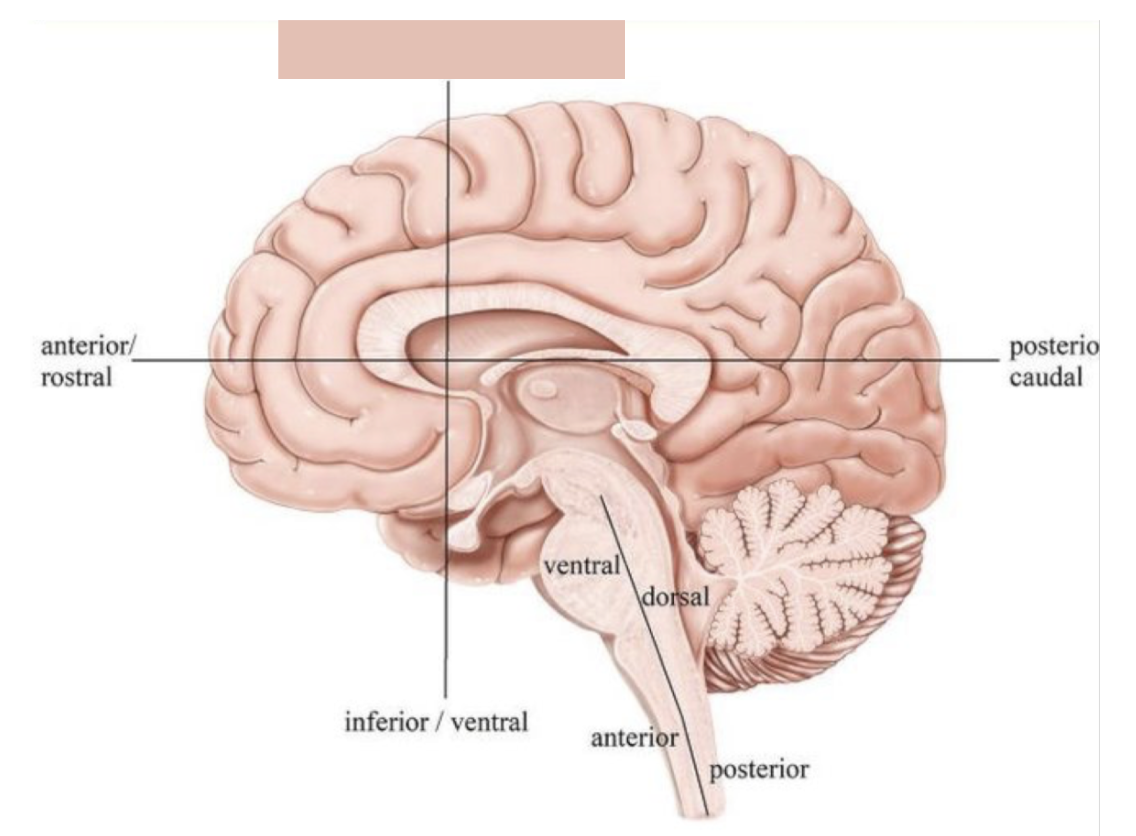
dorsal (brain)
top of the head (superior)
dorsal (body)
toward the backbone from the neck down (posterior)
ventral (brain)
bottom of the head (inferior)
ventral (body)
toward the belly from the neck down (anterior)
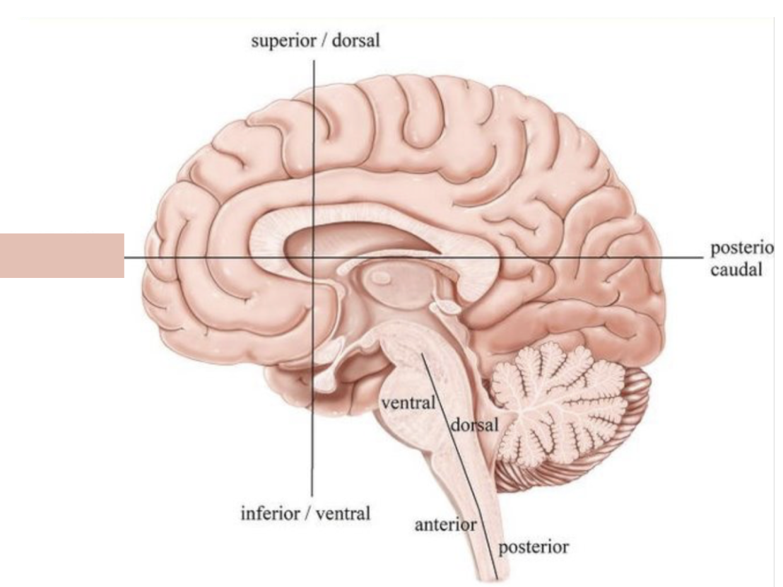
what orientation is this?
superior/dorsal
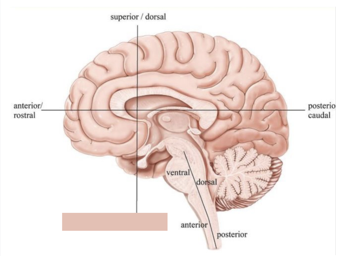
what orientation is this?
inferior/ventral
what orientation is this?
anterior/rostral
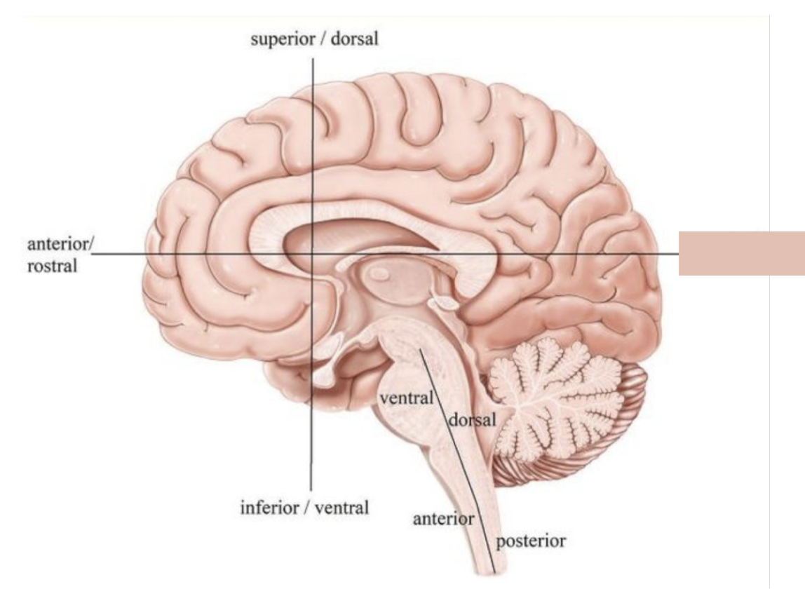
what orientation is this?
posterior/caudal
medial
toward the middle
lateral
toward the edge
frontal or coronal plane
divides into front and back
sagittal plane
divides into right and left
midsagittal plane
evenly divides into right and left
horizontal plane
divides into top and bottom
what view is this section?
coronal
what view is this section?
sagittal (midsagittal)
what view is this section?
horizontal
what are the 3 main components of neurons
dendrites, soma, and axon
dendrites
receive signals
cell body (soma)
contains the nucleus and processes information
axon
transmits signals away to other neurons
what is the direction of information flow?
dendrites > cell body (soma) > axon
describe the location of the myelin sheath
a fatty, insulating layer wrapped around axons in the central (CNS) and peripheral nervous systems (PNS)
describe the function of the myelin sheath
allows action potentials to jump from one node to the next quickly and efficiently along the nerve cells
describe the location of the nodes of Raniver
periodic, 1 μm gaps in the myelin sheath along myelinated axons in both the central and peripheral nervous systems
describe the function of the nodes of Rainier
allow nerve impulses (action potentials) to "jump" from one node to the next
what are glial cells
support cells that provide structure, protection, and waste management
what is the function of glial cells
to maintain homeostasis, protect neurons, and regulate brain development and repair
what does myelin do?
speeds up action potentials. It acts as a protective, insulating sheath that prevents electrical signals from leaking out, ensuring efficient communication across the nervous system.
What happens at the synapse?
Neurotransmitters are released from the presynaptic neuron and bind to receptors on the postsynaptic cell.
What is the difference between afferent and efferent neurons?
Afferent = sensory input to CNS; efferent = motor output from CNS.
What is gray matter?
neuron cell bodies that generate signals (unmyelinated)
What is white matter?
neuron cell axons that carry signals (myelinated)
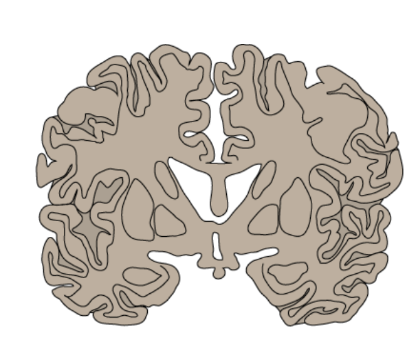
what is the formation of gray and white matter in the brain
gray matter outside, white matter inside
what is the formation of gray and white matter in the spinal cord
white matter outside, gray matter inside
why is the orientation of grey and white matter different in the brain and spinal cord
The brain has gray matter on the outside to maximize information processing and integration, while the spinal cord has white matter on the outside to optimize fast signal transmission between the brain and body.
What is somatotopy?
Organized mapping of body parts onto the cortex with overlap. a "body map,"
Where is the somatotopic map located for motor function?
The primary motor cortex (M1) in the precentral gyrus of the frontal lobe.
Where is the somatotopic map located for somatosensory function?
The primary somatosensory cortex (S1) in the postcentral gyrus of the parietal lobe.
What is the motor homunculus?
A distorted body representation showing how much motor cortex is devoted to controlling each body part.
Why are the hands and face large in the motor homunculus?
They require fine motor control and therefore have greater cortical representation.
What does the size of a body part in the motor homunculus represent?
The amount of motor cortex dedicated to controlling that body part—not its physical size.
What is the somatosensory homunculus?
A cortical map showing how sensory input from different body parts is represented in the somatosensory cortex.
Which sensory modalities are represented in the somatosensory cortex?
Touch, pressure, pain, temperature, and proprioception.
Do somatotopic maps have sharp boundaries?
No. There is overlap between neighboring body part representations.
Why is overlap in somatotopy important?
It allows for flexibility, redundancy, and cortical reorganization (plasticity).
What happens to somatotopic maps after injury or training?
Cortical areas can reorganize, with adjacent regions taking over lost or enhanced functions.
Why does the left brain control the right body (and vice versa)?
Motor and sensory pathways cross in the brainstem/spinal cord.
Which hemisphere controls movement of the right hand?
Left
Which hemisphere controls movement of the left hand?
Right
How do lateralization and somatotopy work together?
Each hemisphere contains a somatotopic map representing the contralateral side of the body.
What are the primary functions of the left hemisphere?
Processing speech sounds
Basic language comprehension
Motor speech
Language dominance
Motor control of the right side of the body
Which hemisphere is typically dominant for language?
Left
What are the primary functions of the right hemisphere?
Visuospatial processing
Emotion
Music
Supralinguistic features (sarcasm, humor, tone)
Attention
Which hemisphere is dominant for emotional processing?
The right hemisphere.
Which hemisphere is dominant for visuospatial skills?
The right hemisphere.
When do hemispheric functional differences typically emerge?
Around 2 years old
What does unilateral functional dominance mean?
Certain cognitive functions become more specialized in one hemisphere than the other.
What does contralateral sensorimotor control mean?
Each hemisphere controls sensory and motor function on the opposite side of the body.
What does ipsilateral mean?
Same side.
What does contralateral mean?
Opposite side.
Damage to the left hemisphere is most likely to affect what functions?
Language, speech production, and right-sided motor control.
Damage to the right hemisphere is most likely to affect what functions?
Emotion, visuospatial awareness, attention, and prosody.
Which hemisphere attends to BOTH sides of space?
Right
Which hemisphere attends to right side of space?
Left
A right hemisphere lesion is most likely to cause what condition?
Left hemispatial neglect.
Why is left neglect more common than right neglect?
Because the right hemisphere processes both left and right space, while the left processes only right space.
True or False: The right hemisphere is dominant for language.
False
True or False: Left-handed people are always right-hemisphere language dominant.
False.
What are Brodmann’s areas?
Functionally distinct regions of cortex defined by differences in cytoarchitecture (cellular organization).
Why are Brodmann’s areas important for understanding brain function?
Areas with different cellular structure serve different functions (motor, sensory, language, etc.).
What is the Brodmann area for the primary motor cortex (M1)?
BA 4
Where is BA 4 located?
The precentral gyrus of the frontal lobe.
What is the main function of BA 4?
Execution of voluntary motor movements.
How is BA 4 organized?
Somatotopically (motor homunculus).
Damage to BA 4 would most likely cause what?
Contralateral weakness or paralysis.
What Brodmann area is associated with premotor function?
BA 6
What is the role of BA 6?
Motor planning and preparation of movements.
Which Brodmann areas make up the primary somatosensory cortex (S1)?
BA 3, 1, and 2
Where is BA 3,1,2 located?
The postcentral gyrus of the parietal lobe.
What sensory information is processed in BA 3,1,2?
Touch
Pressure
Pain
Temperature
Proprioception
How is the primary somatosensory cortex organized?
Somatotopically (sensory homunculus).
What does a larger cortical representation in the sensory homunculus indicate?
Greater sensory receptor density and finer discrimination.
Which Brodmann areas are associated with somatosensory association processing?
BA 5 and BA 7
What is the function of BA 5 and BA 7?
Integration and interpretation of sensory information.
What Brodmann areas correspond to the primary auditory cortex (A1)?
BA 41 and BA 42
Where is BA 41/42 located?
The superior temporal gyrus.
What is the primary function of BA 41?
Initial cortical processing of auditory input.
How is the auditory cortex organized?
Tonotopically (organized by sound frequency).
Which Brodmann area is critical for auditory language comprehension?
BA 22
What is the Brodmann area for Wernicke’s area?
BA 22
Where is BA 22 located?
Posterior portion of the superior temporal gyrus in the left hemisphere.