Cardiology Lab - Exam 1
1/44
There's no tags or description
Looks like no tags are added yet.
Name | Mastery | Learn | Test | Matching | Spaced |
|---|
No study sessions yet.
45 Terms
Inflammatory cells!
What's this?
Lungs trachea esophagus and heart
What makes up the pluck?
The pericardium
What is best to first assess on the heart?
To collect blood/pus
Why might you make a small incision in the pericardium?
True
T/F: Inside the pericardium is sterile
Just a tad
How much fluid is in the pericardium normally?
Contour,
Epicardium,
Great vessels,
Coronary groove, and
Coronary vessels
What can you observe from the outside of the heart, with the pericardium removed?
The left ventricle
In the normal heart, the apex is part of what ventricle?

Epicardium
What layer of the heart is visible?
Saran wrap, light, smooth and glistening
How should the surface of the heart look?

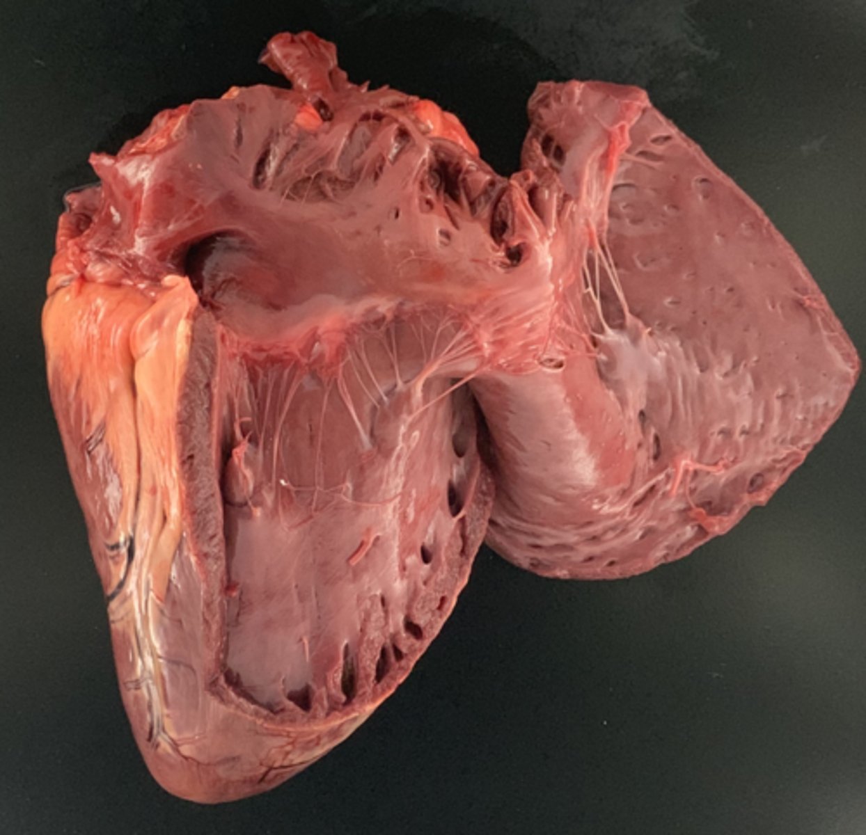
The caudal vena cava into right atrium by incising from auricle to auricle
What place should you open first?
Right ventricle
From the right atrium, where do we look next?
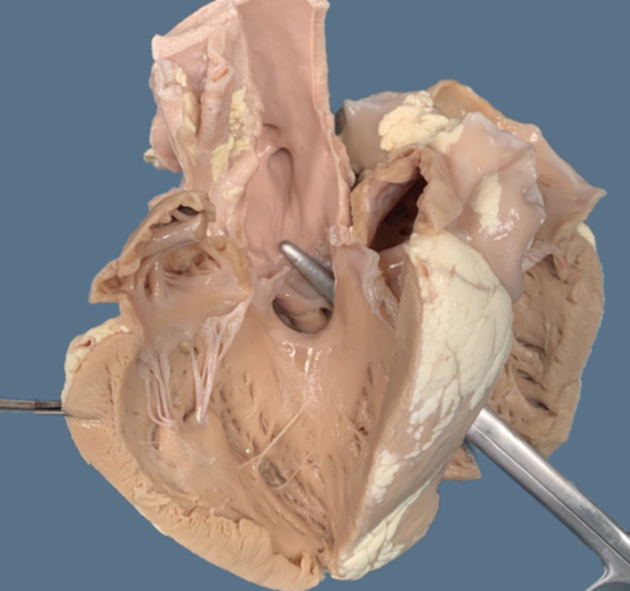
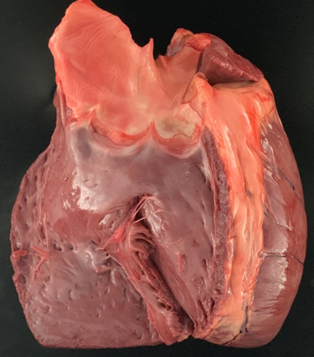
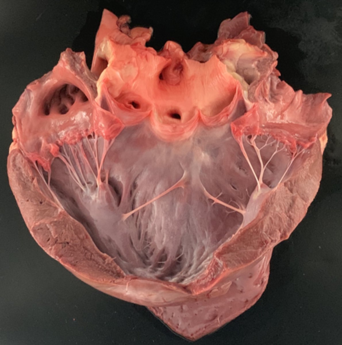
The pulmonary outflow tract, with chordae tendinae and a semilunar pulmonic valve at top (looks like an ear)
What vessel is next to observe?
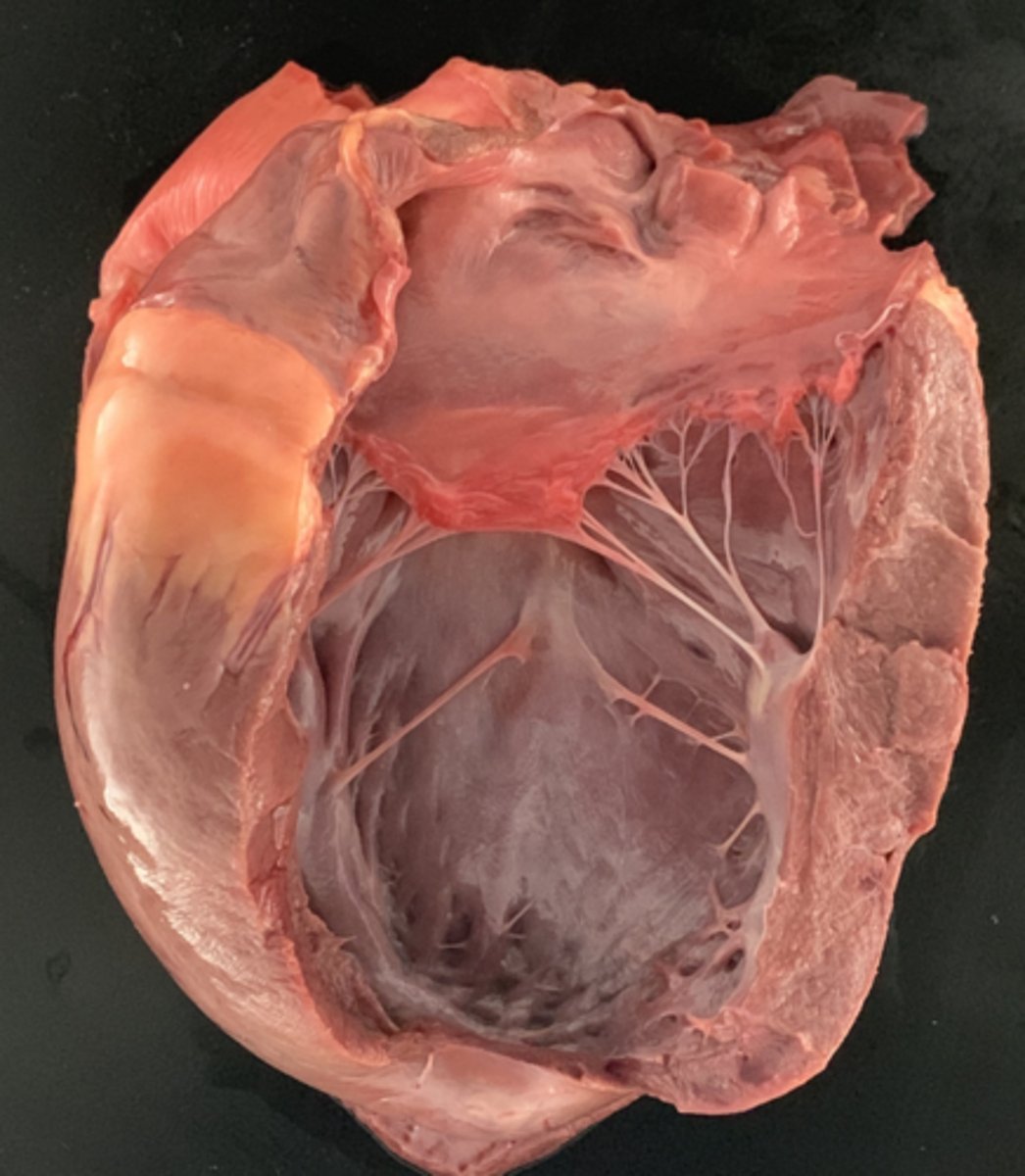
The left atrium and ventricle
What part of the heart is next for examination?
Aorta
Finally, the outflow to the body is the ______
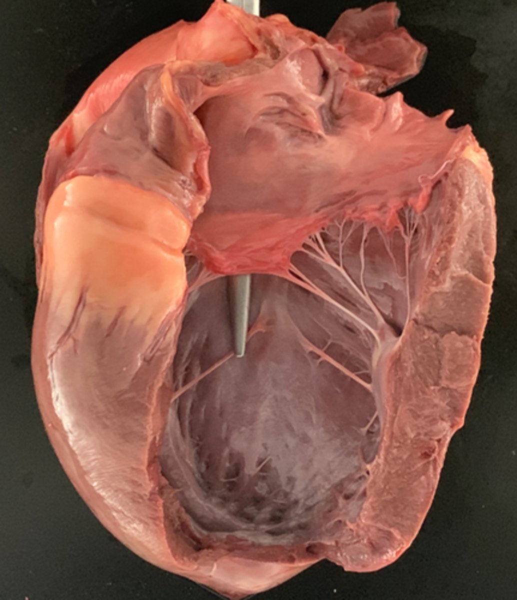
The left wall is three times as thick as the right
How much thicker should the left wall be from the right?
2 - 4% of total body weight
What is the weight of a normal heart?

Ingestion of metal leads to the penetration of the reticulum wall, which infects the heart with bacteria, leading to fibrinous pericarditis and epicarditis, which can become restrictive
What is the pathophysiology of hardware disease?
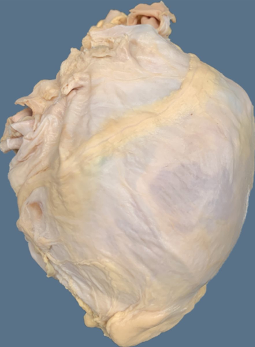
Fibrin
What is on the outside of this heart?
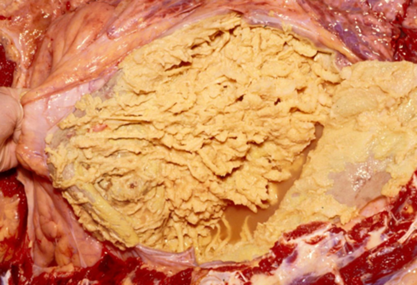
Pericardium with fibrin
What the thick band around the heart?
Fibrinonecrotizing and suppurative epicarditis and pericarditis
Morphologic diagnosis?

Fibrin is an inflammatory response to damage that has changed endothelial dynamic
Fibrous just means dense connective tissue
What is the difference between fibrin and fibrous?
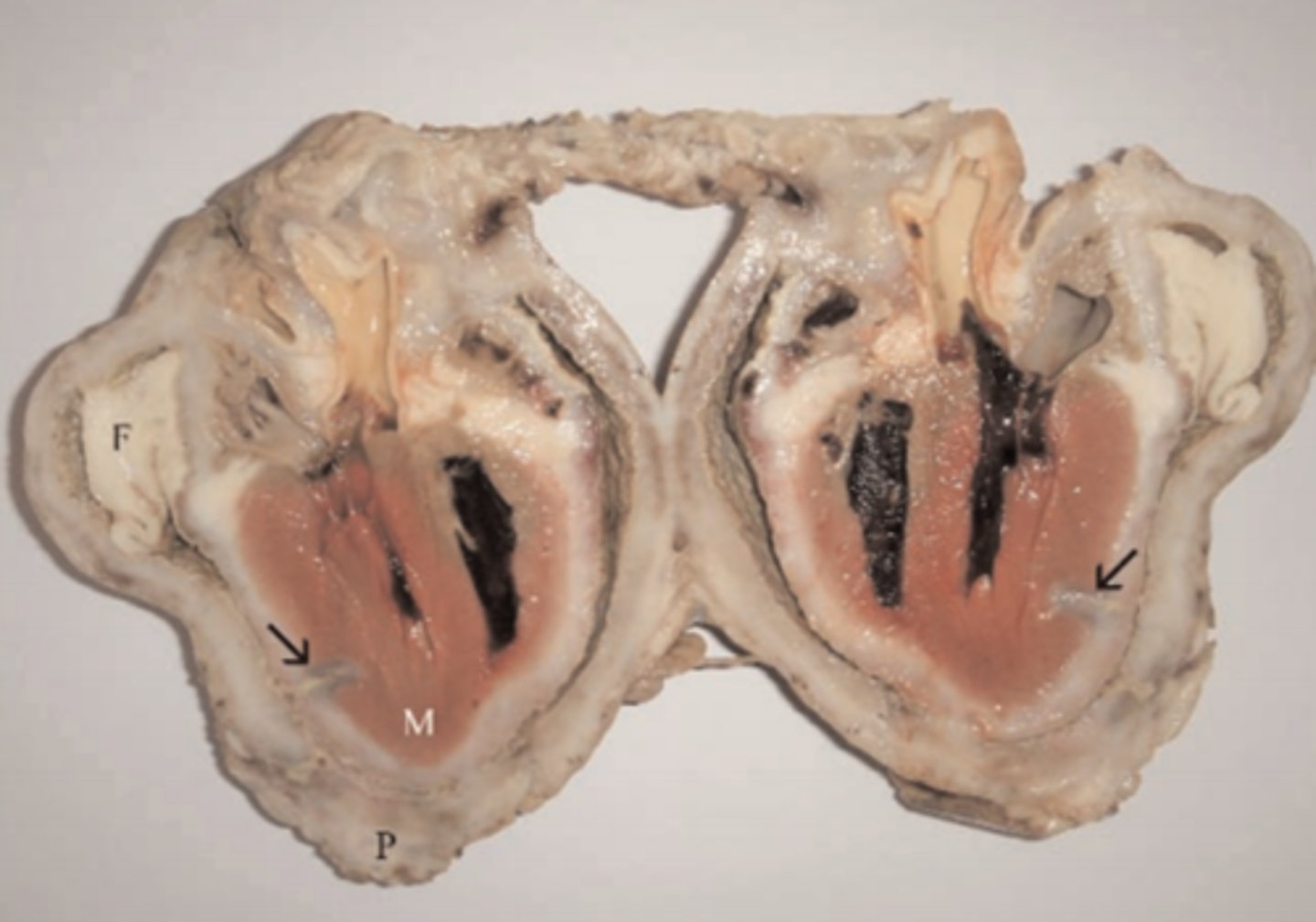
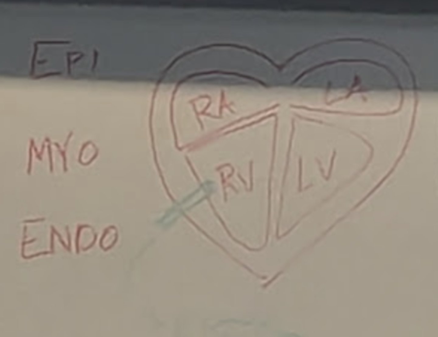
A representative piece of Epicardium, Myocardium, and Endocardium
(See green)
What section should you cut for a sample of this disease?
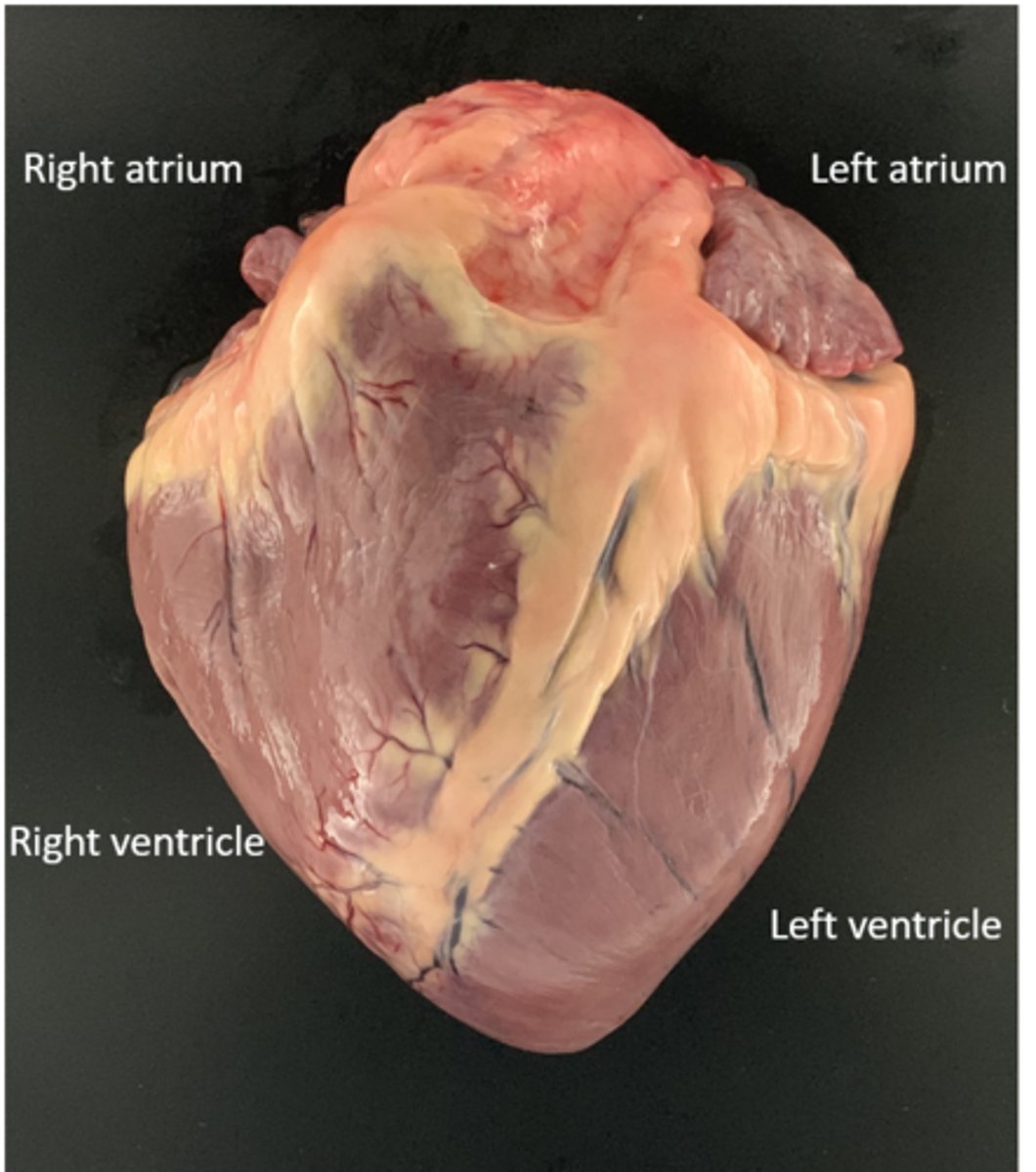
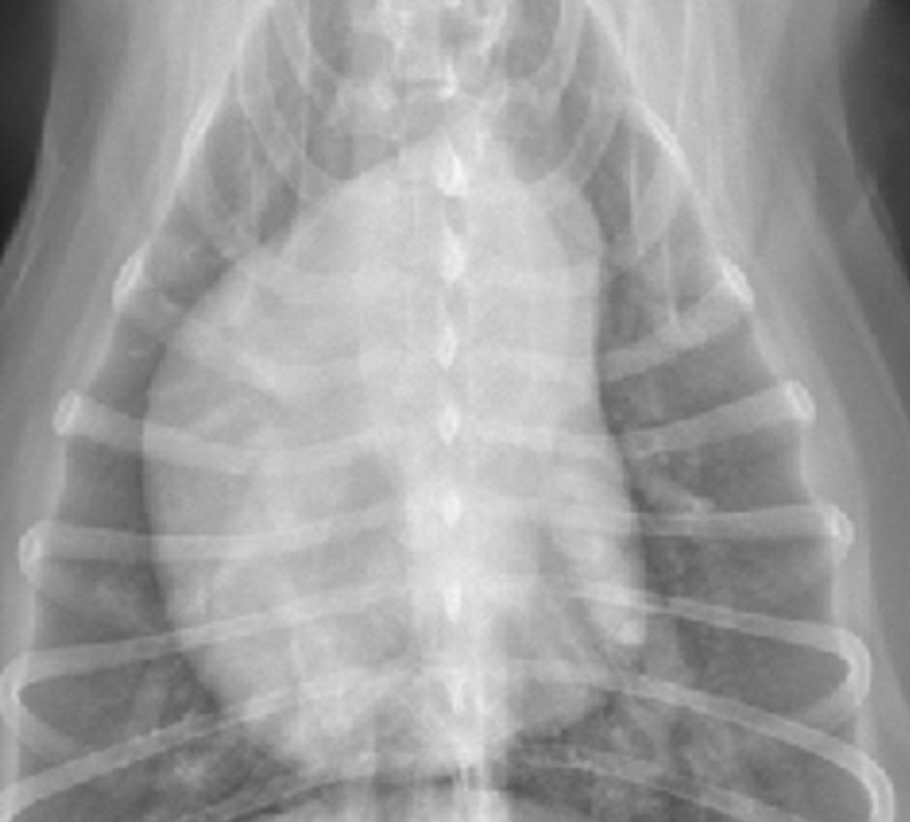
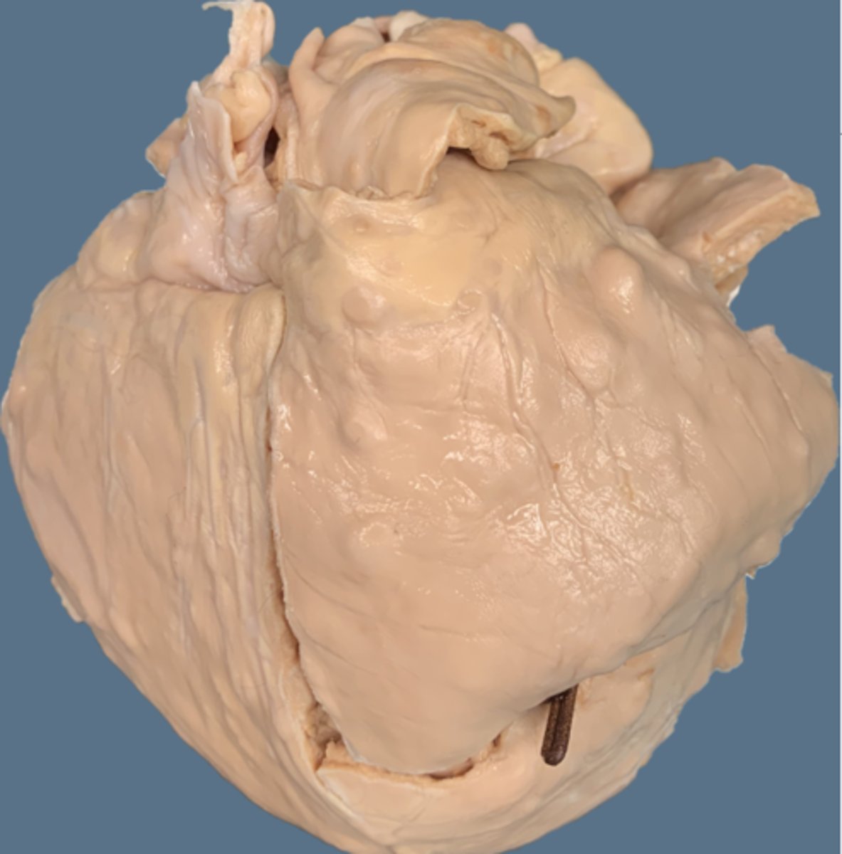
Kinda round, kinda pudgy, one could even call it globoid
What do you see wrong?
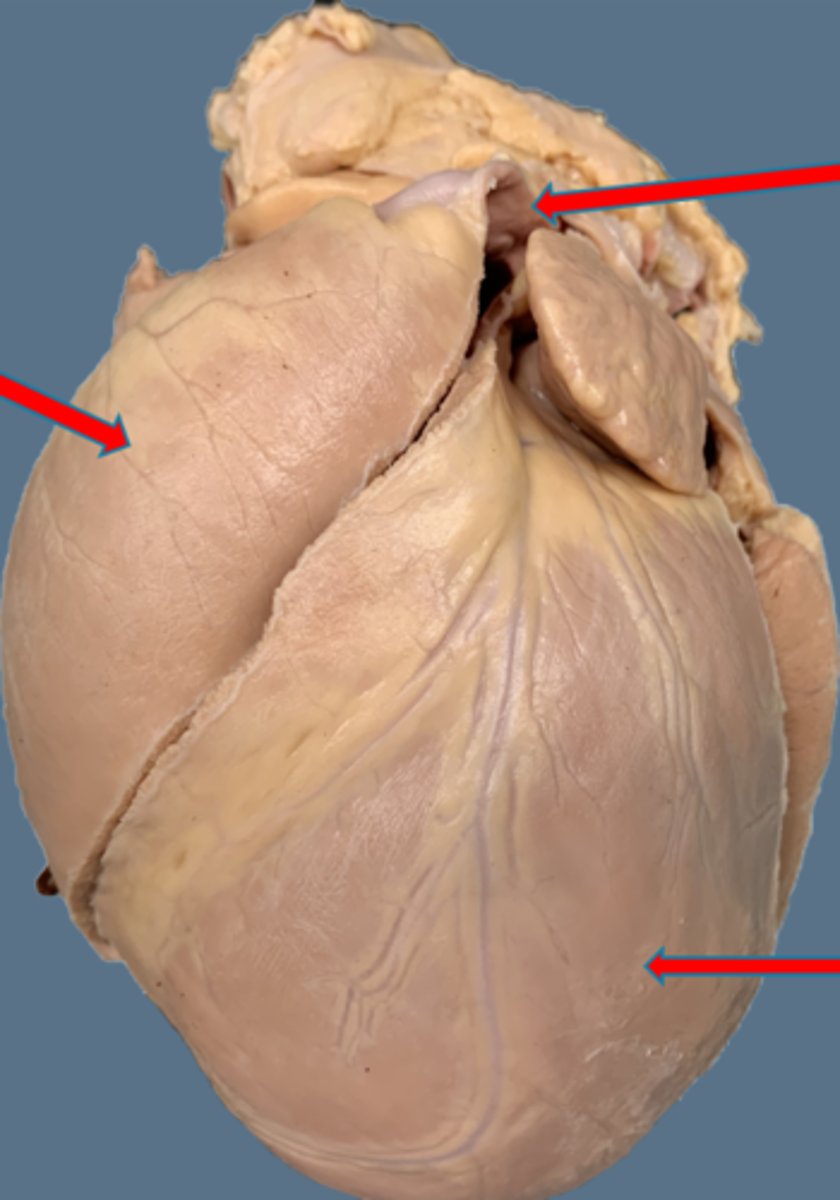
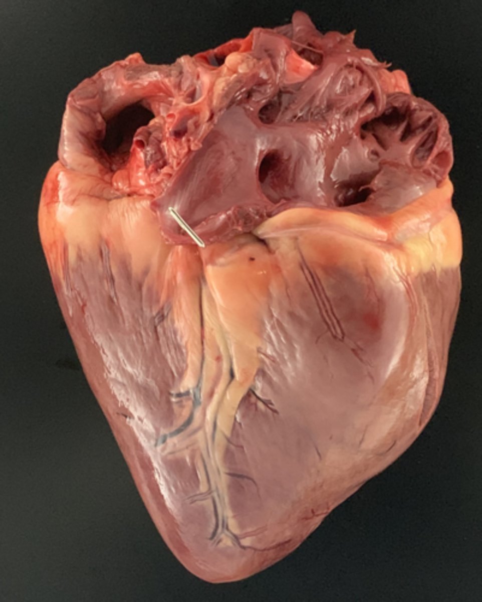
Heartworms
What're those?
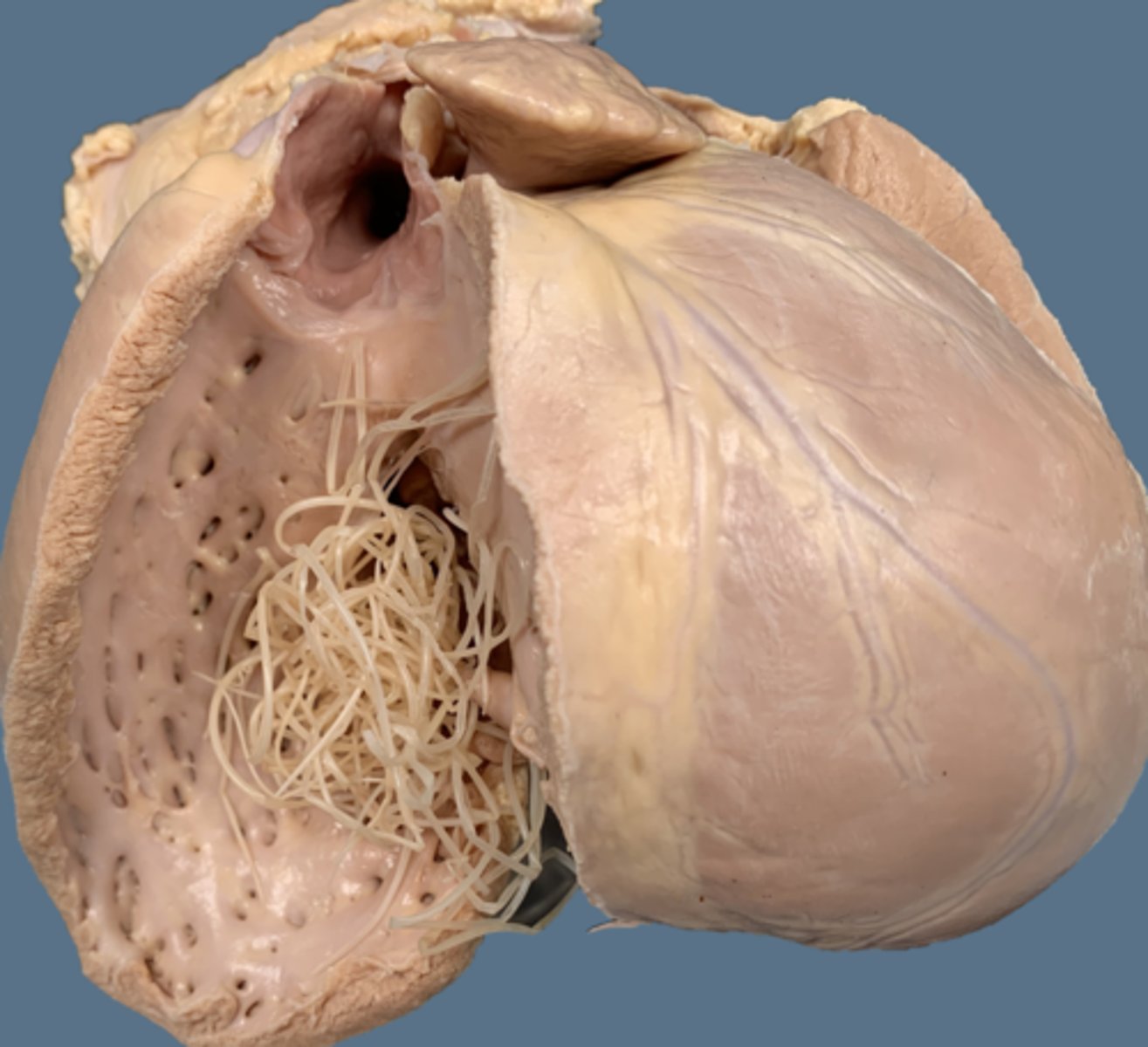
The right atrium and ventricle are typically dilated due to worms, and valvular insufficiency is often present
What is true of the right side of a heart with heartworms?

Hypertrophy
What happens to the left side of a heart with heartworms?

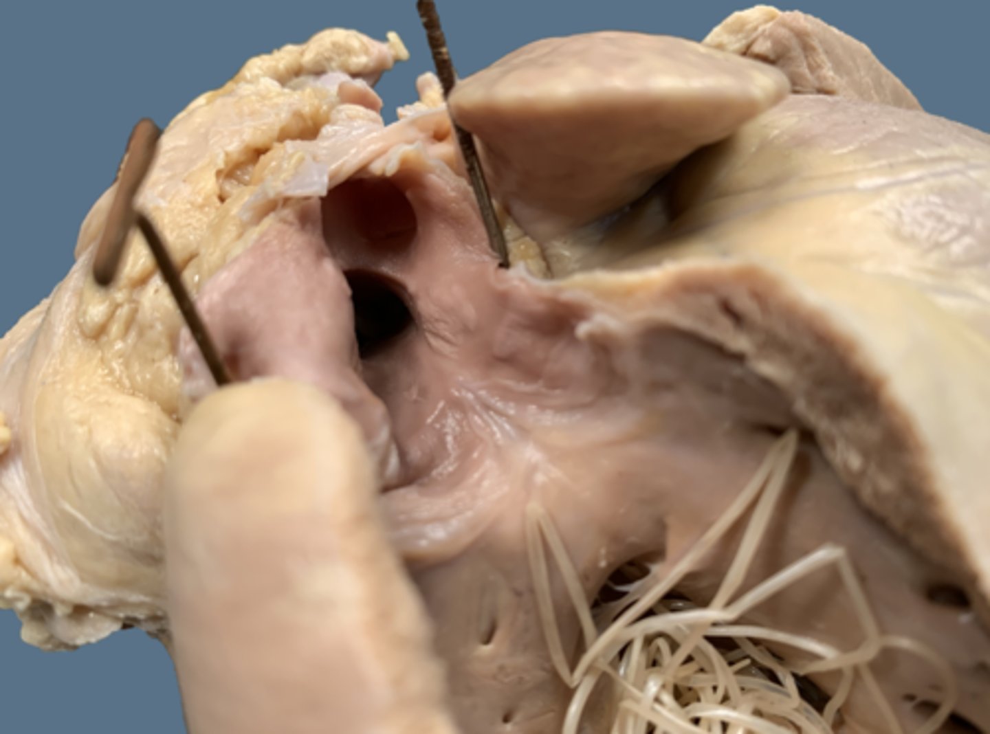
Myointimal proliferation
What is the wrinkle near the opening of the pulmonary tract?

Dirofilariasis (you just said heartworms, didn't you)
What is the morphologic diagnosis?
Reverse D ( ;-D )
What shape do you see in this heart with dirofilariasis?
Right ventricular dilation,
Left ventricular hypertrophy,
Dilated pulmonary artery, and
Myointimal proliferation
What are four morphological changes you see with heartworms?

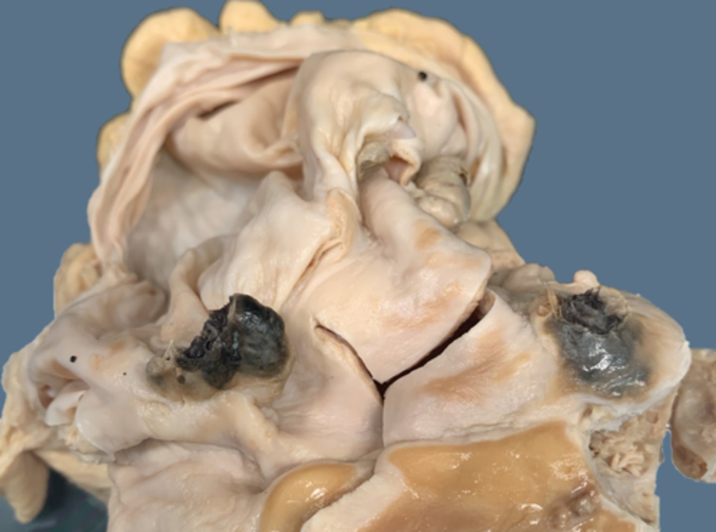
Hemangiosarcoma
Blood filled pericardium
What's Dog 1?
It's black, friable, and bleeding, thus its a hemangiosarcoma!
Find the mass and identify the mass
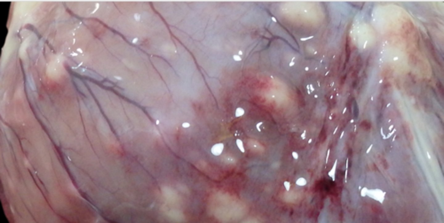
In the endocardium
Where are the nodules embedded?
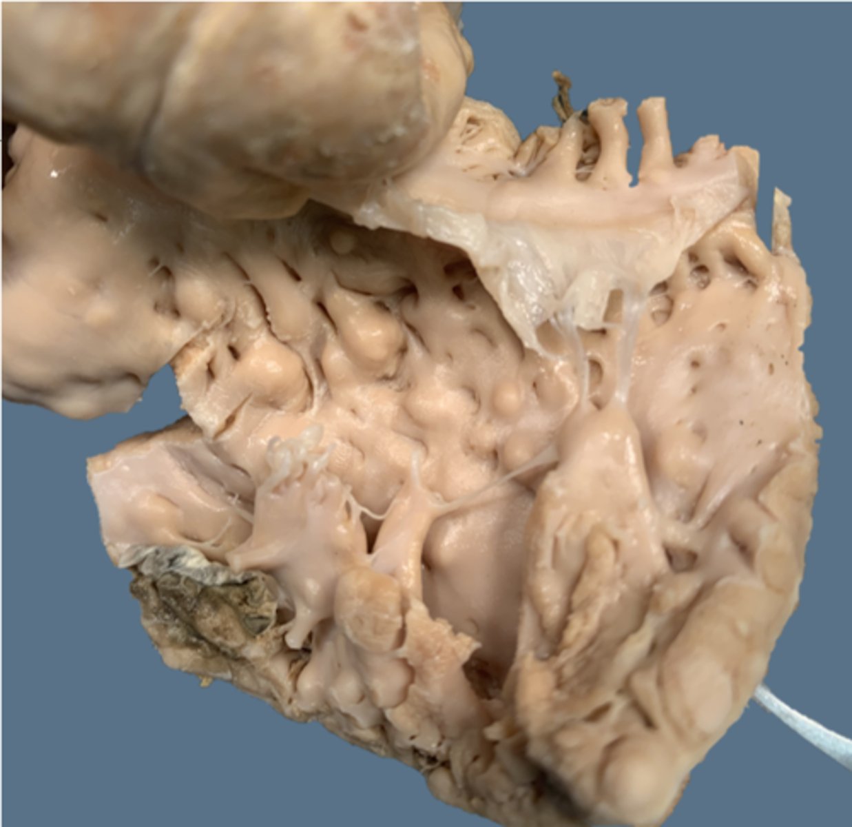
Lymphosarcoma
What are these nodules?
Dark, smooth, round, friable, and blood filled
Describe a gross lesion of a hemangiosarcoma?
White, variably shaped, and firm
Describe a gross lesion of a lymphosarcoma?
Variably sized, highly infiltrative, and vascular channels with plump endothelium
What is the histopathology of hemangiosarcoma?
Densely cellular, poorly demarcated, and neoplastic
What is the histopathology of lymphosarcoma?
Hermy wormies bump against endothelium
What causes a thrombus?

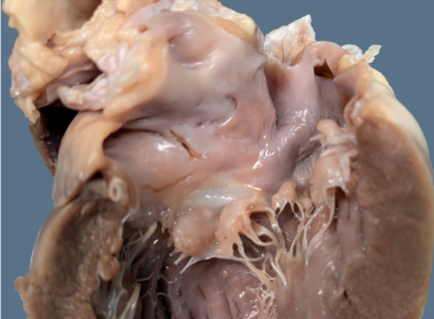
Endocardiosis, rounded leaflets, smooth
What's wrong with this picture?
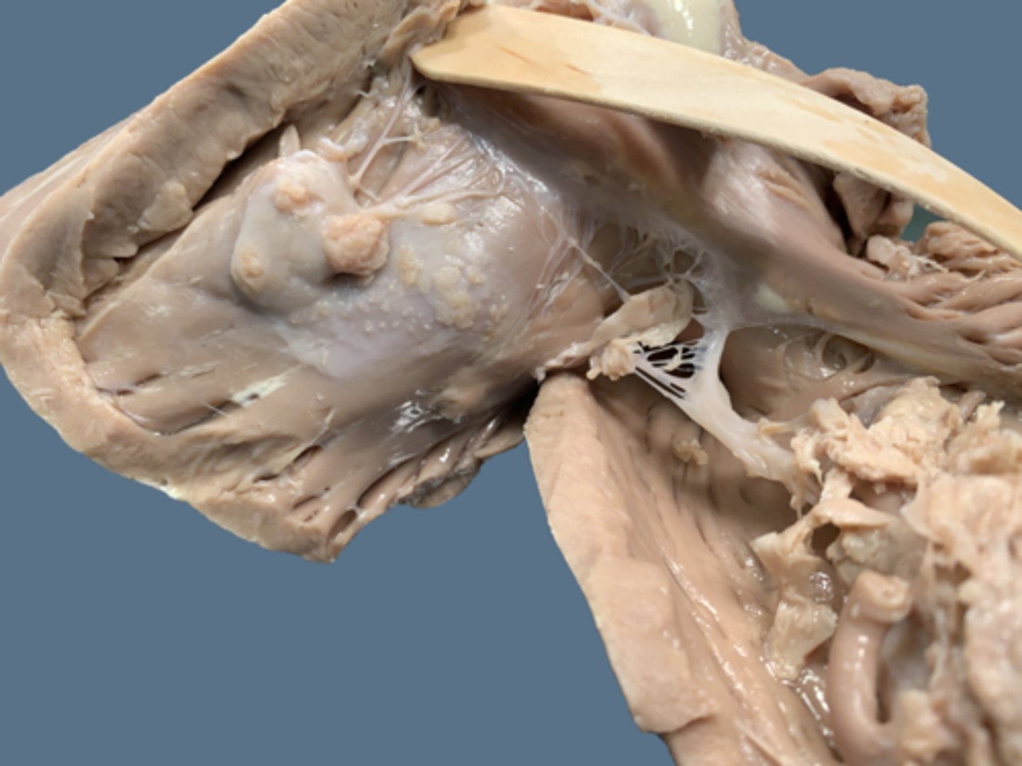
FIlled with fibrin, debris, spiky and spiculous
Endocartitis
What's wrong here?
Endocardiosis on the left, which is just degeneration of the AV valves
vs
Endocarditis on the right, which is inflammation due to infection
Compare
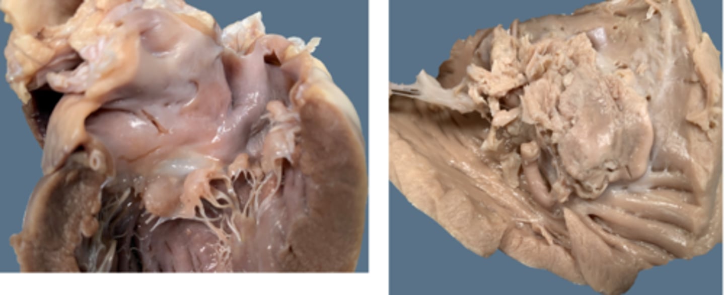
Unalived, due to endocarditis
Diagnose
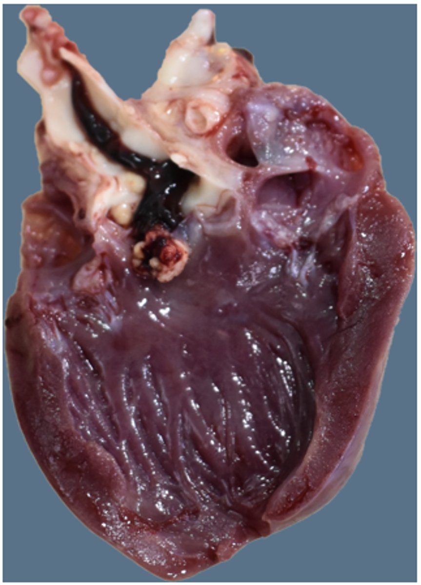
Intraventricular septal defect
(A hole!)
What's wrong