cerebellum
1/24
There's no tags or description
Looks like no tags are added yet.
Name | Mastery | Learn | Test | Matching | Spaced | Call with Kai |
|---|
No analytics yet
Send a link to your students to track their progress
25 Terms
already covered
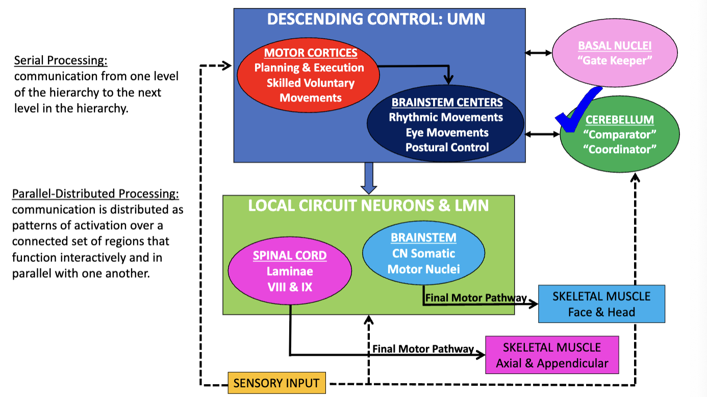
lowest level of the motor hierarchy=
local circuit neurons and LMNs, responsible for sending final motor command to skeletal muscles
highest level of the motor hierarchy=
descending control systems
(1 of the) side loops=
basal nuclei, which acts as a gatekeeper of initiation and termination of movement
dysfunction —> hyperkinetic and hypokinetic movement disorders
remaining information
(2nd of the 2) side loops=
cerebellum, which plays a major role in comparing the intended motor plan developed at the highest level of the motor hierarchy with the actual movement that was produced by the lowest level of the motor hierarchy
in the comparison, it detects errors
correction of these errors produce smooth and coordinated movement
dysfunction —> cerebellar ataxia
modulation of upper motor neurons
the cerebellum, as a general rule, does not project directly to __ or __ of the __ or __ , rather, it modulates the activity of __ and ***plays a major role in the following:
local circuit neurons (lowest level of motor hierarchy); LMNs; spinal cord; brainstem; UMNs (of the cortex and brainstem)
maintenance of posture and balance
coordination of eye movements and gaze fixation
coordination of articulatory movements for speech production
coordination of voluntary limb movements (e.g., reaching, grasping, walking)
motor learning
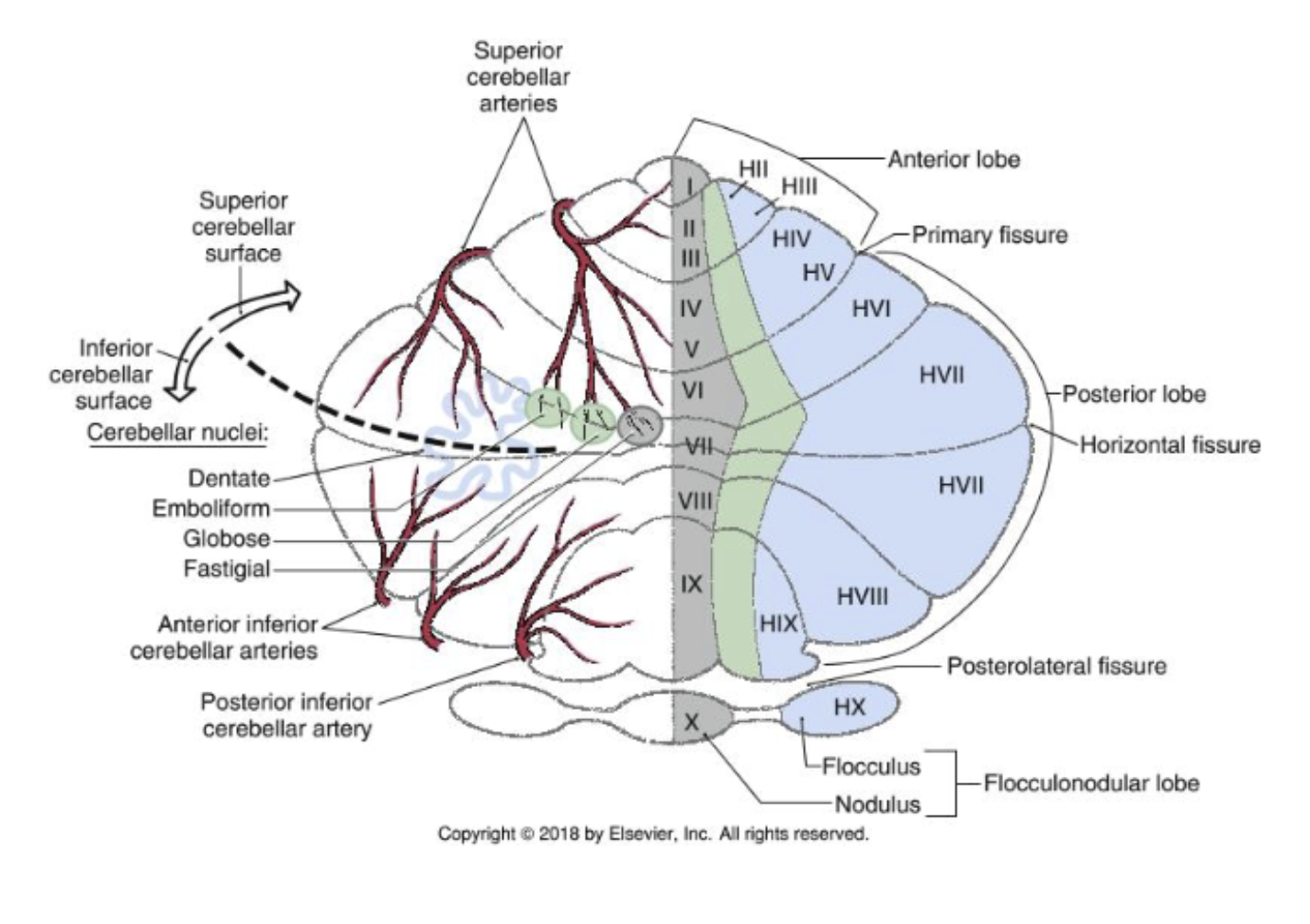
***cerebellum cortical zones, w/ their ***associated deep cerebellar nucleus
***medial (vermal) zone:
***intermediate zone:
***lateral zone:
medial (vermal) zone (grey): narrow strip of cortex adjacent to the midline, extending through the anterior and posterior lobes (also includes nodulus)
***fastigial nucleus
intermediate zone (green): adjacent to the vermal zone, also includes/extends to the anterior and posterior lobes but generally excludes the floccular nodular lobe
***interposed nuclei (globus and emboliform)
lateral zone (blue): the largest zone located just lateral to the intermediate zone, occupying the majority of the cerebellar cortex, will include very large portions of the anterior and posterior lobes, and will include the flocculus
***dentate nucleus
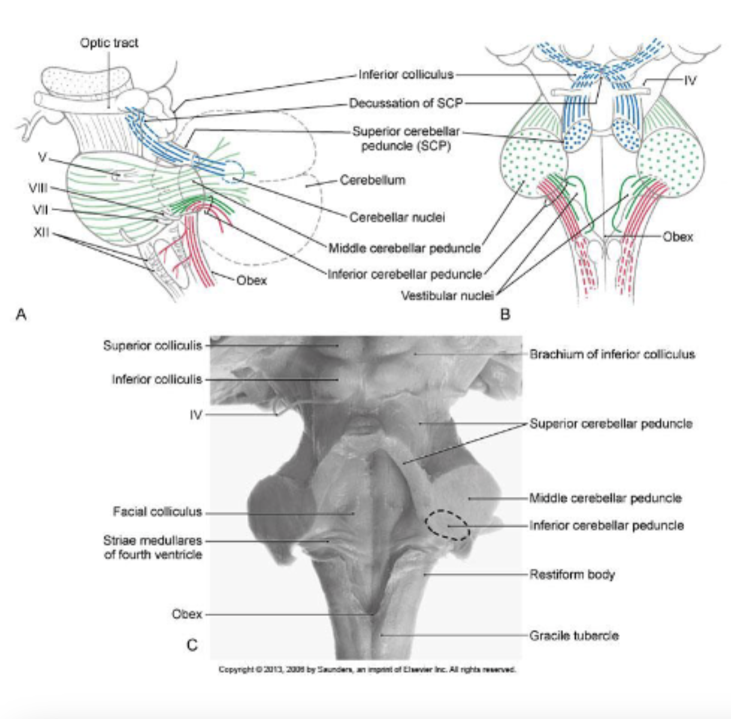
***afferent pathways of the cerebellum
the cerebellum ***will receive __ information from the following sources via both the __ and __ __
***__ peduncle:
***__ peduncle:
***neurons of the deep cerebellar nuclei receive input from 3 main sources- what are they?
***neurons of the deep cerebellar nuclei output to various regions- which nuclei output to which regions and what is the functional significant of this output?
***afferent; ***inferior; ***middle cerebellar peduncles:
***middle peduncle:
largest peduncle
*cortex: conveying afferent information about motor plan, coming from the cortex, via the relay pontine nuclei: mossy fibers
corticopontine tract conveys information about the motor plan from the cortex to the cerebellum
—> projection to pontine nuclei
—> pontine nuclei will send axons that will cross to the opposite side
—> motor plan coming from 1 cortex is conveyed to the contralateral cerebellum (e.g., information from the right motor cortices, conveying motor plans resulting in movement of the left side of the body, ends up in the left cerebellum)
***inferior peduncle:
*vestibular nuclei: conveys sensory information about head and body position to the cerebellum: mossy fibers
*spinal cord: conveys proprioceptive information from the anterior and posterior spinocerebellar + cuneocerebellar tracts: mossy fibers
**inferior olivary complex: information from the inferior olivary complex is used to modulate and coordinate the final output of the cerebellum (error information): climbing fibers
*mossy fibers= axons of the cells carrying information from the….
cortex
vestibular nuclei
spinal cord
**climbing fibers= axons of the cells carrying information from the inferior olivary complex

***efferent pathways of the cerebellum
the cerebellum ***will receive __ information to the following targets via the __ and __ __
***__ peduncle:
***__ peduncle:
***efferent; ***superior; ***inferior; cerebellar peduncles
***superior peduncle:
output from the cerebellum will reach the contralateral cerebral cortex, predominantly targeting the sensorimotor cortices that contain the UMNs that give rise to the…
corticospinal
corticonuclear tracts
***inferior peduncle: conveys output from the cerebellum to the areas of the brainstem that contain UMNs
vestibular nuclei
gives rise to the vestibulospinal tract
reticular formation
gives rise to the reticulospinal tract
red nucleus
gives rise to the rubrospinal tract
—> also projects to the inferior olivary complex, which is important with respect to signaling motor errors
image
blue= projections of the superior cerebellar peduncle
crosses to the contralateral side
green= projections of the middle cerebellar peduncle
containing cross projection coming from the pontine nuclei
red= projections of the inferior cerebellar peduncle
contains mixed afferent and efferent projections
***cerebellar functional modules- function
each module consists of a __, a __ that contains the modules afferent and efferent fibers, and a __ that are functionally related to the cortical area
***vestibulocerebellum:
***spinocerebellum:
***cerebrocerebellum (aka pontocerebellum):
cortical zone; white matter core; nucleus or nuclei
== ***work together in parallel to modulate activity of UMNs (i.e., parallel distributive processing)
***vestibulocerebellum: modulates eye movements (e.g., vestibulo-ocular reflex) and movements that maintain posture and balance
made up of the caudal parts of the cerebellum (flocculus and nodulus)
receive input from the vestibular nuclei
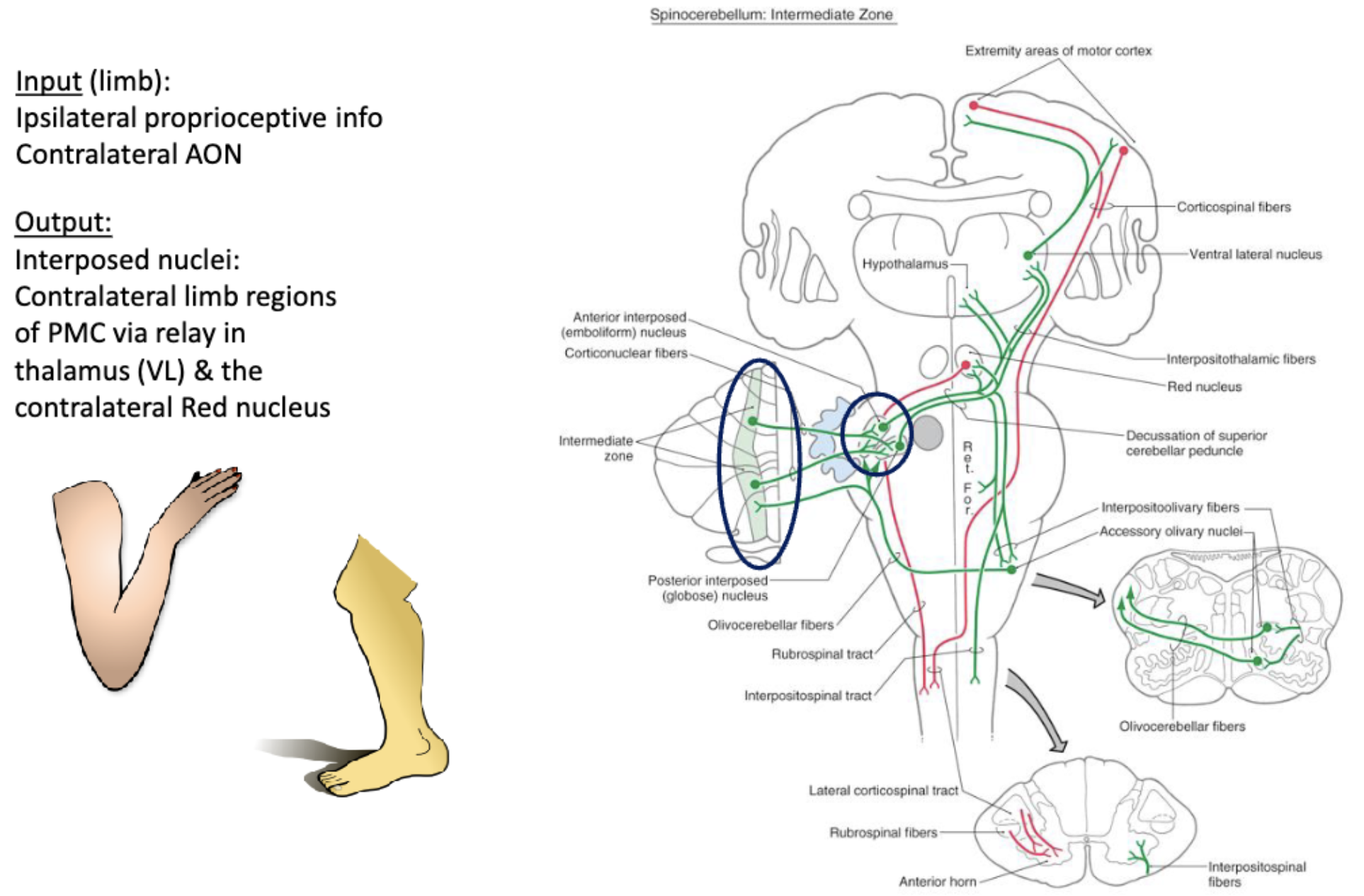
***spinocerebellum: modulates motor commands to adjust and “fine-tune” ongoing limb & truncal movements
located medically to the vestibulocerebellum
only part of the cerebellum that receives direct input from the spinal cord via the spinocerebellar and cuneocerebellar tracts
consists of intermediate (concerned with movements of the limbs) and vermal zones (concerned with movements of the trunk)
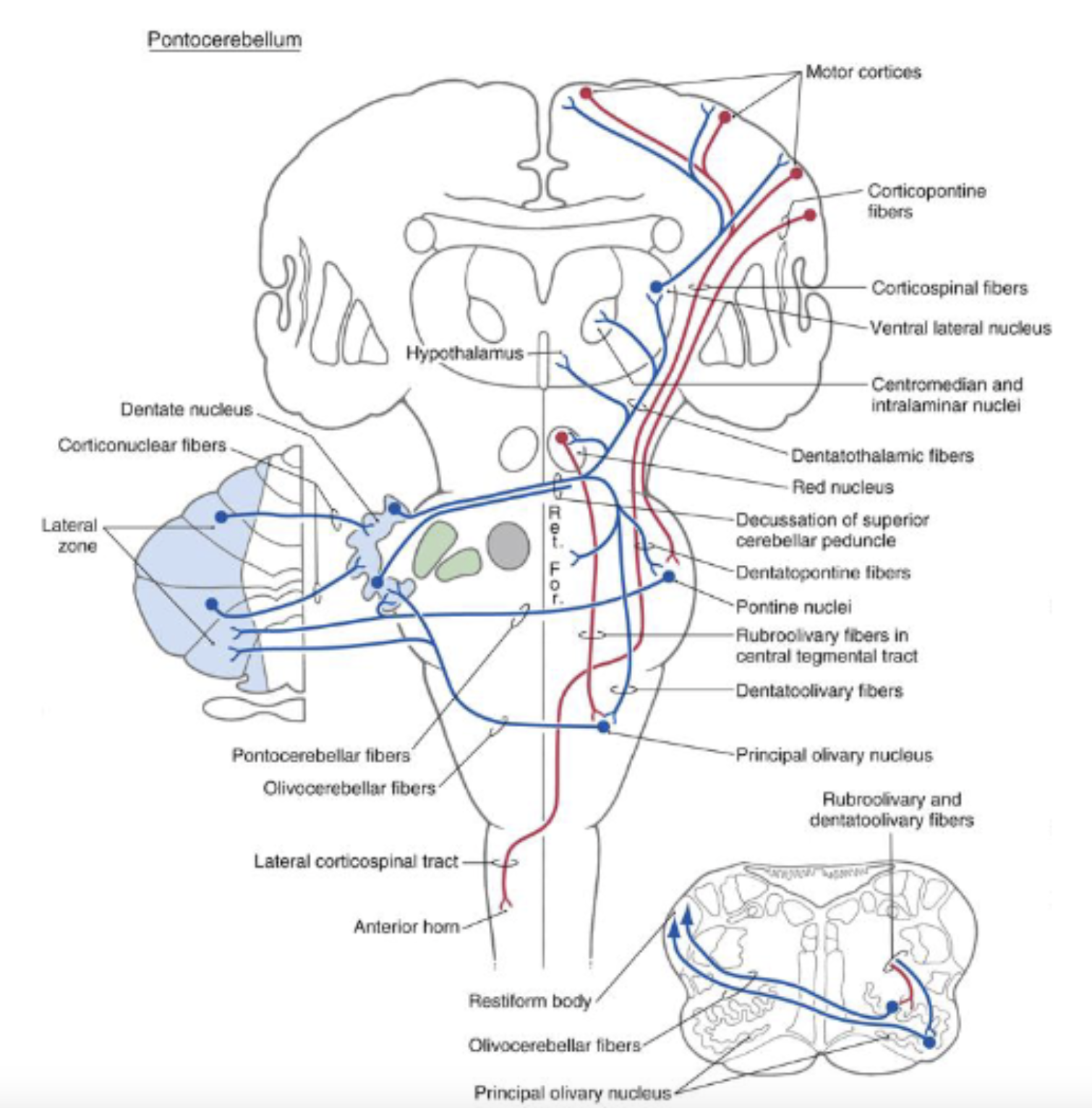
***cerebrocerebellum (aka pontocerebellum): modulates and updates motor plans for highly skilled movements (e.g., tasks involving eye-hand coordination, speech, dexterous movements of the hand) and fine-tune motor patterns so that they become automatic
largest subdivision and occupies the majority of the lateral zone
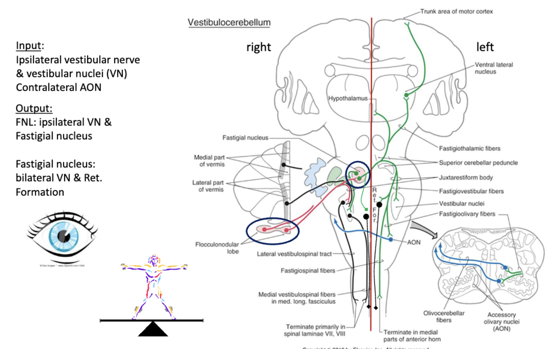
***vestibulocerebellum
components/***structure + function:
***zone
***deep nuclei
***input/output
***which motor systems (e.g., dorsolateral, ventromedial) does each of the cerebellar functional modules modulate?
***which UMNs are involved in these systems?
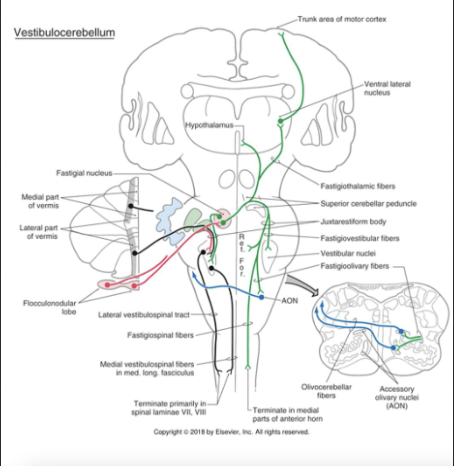
***components: flocculonodular lobe & the fastigial nucleus; vestibular nerve & nuclei
flocculonodular lobe & ***fastigial nucleus receive the majority of their ***INput directly from the…(vestibular system information)
***ipsilateral vestibular nerve & ***vestibular nuclei and
supplementary input from the ***contralateral inferior olivary complex via the ICP
flocculondular lobe ***OUTputs to the ***ipsilateral vestibular nuclei directly (predominantly) & the ***fastigial nucleus
***fastigial nucleus ***OUTputs to the ***vestibular nuclei and ***reticular formation bilaterally
***modulation of motor commands to eye muscles
modulation of ***UMNs of the VST (***vestibulospinal tract) and RST (***reticulospinal tract) for the coordination of axial muscles for postural control
==> ***ventromedial motor system
***structure + function
***zone
***deep nuclei
***input/output
***which motor systems (e.g., dorsolateral, ventromedial) does each of the cerebellar functional modules modulate?
***which UMNs are involved in these systems?
***vestibulocellebellum
= modulate ***UMNs outputting to ***muscles of the eye + ***postural control
***lobe of interest: ***floccular nodular lobe
related ***deep cerebellar nuclei: ***fastigial nucleus
concentrate on ***OUTput:
***floccular nodular lobe OUTputs directly to the ***ipsilateral vestibular nucleus, ***for the regulation of eye movements (e.g., vestibuloocular reflex)
***floccular nodular lobe OUTputs to the ***deep cerebellar nuclei, ***the fastigial nucleus, which ***projects to the vestibularnuclei bilaterally + ***reticular formation for the modulation of the ***UMNs of the ***vestibulospinal tract + ***reticulospinal tracts
== outputs to LMNs ***regulating postural control
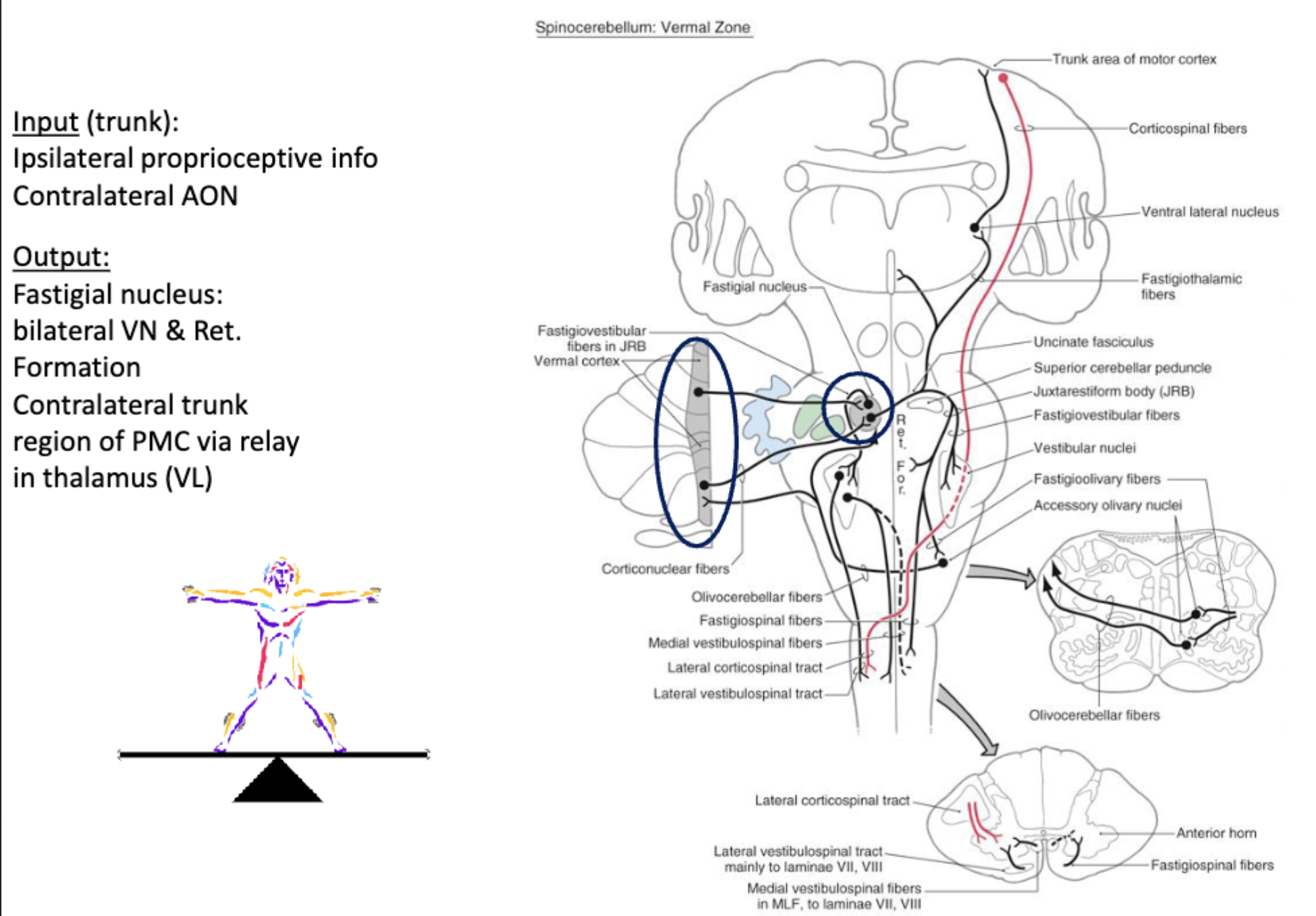
***spinocerebellum
components/***structure + function:
***zone
***deep nuclei
***input/output:
***which motor systems (e.g., dorsolateral, ventromedial) does each of the cerebellar functional modules modulate?
***which UMNs are involved in these systems?
components: *medial (vermal) & **intermediate zones and the ***fastigial (for the medial zone) & ***interposed nuclei (for the intermediate zone)
medial & intermediate zones, & nuclei receive primarily ***ipsilateral
proprioceptive ***INput from the…(***spine- which helps us know where we are in space)***body and head &
supplementary input from the ***contralateral inferior olivary complex
*medial zone will ***OUTput to the *fastigial nucleus & the **intermediate zone will ***OUTput to the **interposed nuclei
*fastigial nucleus projects primarily to RF (reticular formation) & vestibular nuclei & to the trunk region of the PMC (primary motor cortex) via a relay in the VL nucleus of the thalamus
***== for modulation of ***UMNs of the ***ventromedial system for postural control
**interposed nuclei project to the limb regions of PMC (primary motor cortex) via a relay in the VL nucleus of the thalamus and to the red nucleus
***== for modulation of ***UMNs of the ***dorsolateral system for skilled limb movements
***zone
***deep nuclei
***input/output:
***which motor systems (e.g., dorsolateral, ventromedial) does each of the cerebellar functional modules modulate?
***which UMNs are involved in these systems?
spinocerebellum: ***vermal zone —> ***OUTput to (***mediating) postural control
components: vermal zone of the cerebellar cortex
related ***deep cerebellae nuclei: ***fastigial nucleus
main ***INput coming into the cerebellum:
***ipsilateral proprioceptive input
integrated input from the ***contralateral accessory olivary nucleus
concentrate on ***OUTput:
fastigial nucleus
—> outputs to the ***vestibular nuclei bilaterally
—> outputs to the ***reticular formation of the brainstem bilaterally
—> another output conveyed via the relay in VL of the thalamus, ultimately to the ***trunk area of the primary motor cortex, for regulation of ***UMNs ultimately traveling in the anterior part of the spinal tract (anterior corticospinal tract in the ventromedial system)
***zone
***deep nuclei
***input/output:
***which motor systems (e.g., dorsolateral, ventromedial) does each of the cerebellar functional modules modulate?
***which UMNs are involved in these systems?
spinocerebellum: ***intermediate zone —> ***OUTput to dorsolateral system
components: intermediate zone of the cerebellar cortex
related ***deep cerebellar nuclei: ***interposed nuclei
concentrate on ***OUTput:
sent to ***red nucleus of the midbrain
sent to ***ventrolateral nucleus of the thalamus
—> output from the VL will be relayed to the ***extremity areas of the motor cortex
== modulate ***UMNs of the ***dorsolateral system, impacting the **corticospinal fibers which become the ***lateral cortical tract + modulating ***UMNs giving rise to the ***rubrospinal tract
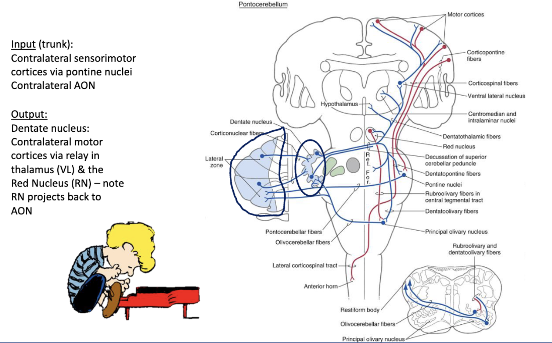
***cerebrocerebellum
components***structure + function:
***zone
***deep nuclei
***input/output:
***which motor systems (e.g., dorsolateral, ventromedial) does each of the cerebellar functional modules modulate?
***which UMNs are involved in these systems?
components: ***lateral zone & the ***dentate nucleus; sensorimotor cortices; red nucleus; pontine nuclei
***INputs from…
***sensorimotor cortices that reach contralateral cerebellum via a relay in the pons
also receive input from the ***contralateral inferior olivary complex
lateral zone ***OUTputs to the ***dentate nucleus
—> ***OUTput from the dentate nucleus: axons cross the midline & ascends to the contralateral motor cortices via a relay in the VL nucleus of the thalamus; also sends ***OUTput collaterals to contralateral red nucleus, which projects back to the inferior olivary complex
modulate ***UMNs of the motor cortices impacting
***corticospinal and ***rubrospinal systems***== appears to be important for…
modulating the duration of muscle contraction and the timing of muscle activation
inactivation important for smooth, coordinated movements
***also very important for updating motor plans related to motor learning
***zone
***deep nuclei
***input/output:
***which motor systems (e.g., dorsolateral, ventromedial) does each of the cerebellar functional modules modulate?
***which UMNs are involved in these systems?
cerebrocerebellum/pontocerebellum (relay that occurs w/ respect to input coming into the cerebellum in the pons): ***lateral zone —> ***OUTput to dentate nucleus, which modulates activity of UMNs
components: lateral zone of the cerebellar cortex
related ***deep cerebellar nuclei: ***denate nucleus
concentrate on ***OUTput:
output sent to ***VL in the thalamus
—> VL will project to ***motor cortices
—> how cerebellum modulates the activity of ***UMNs giving rise to the ***corticospinal tract, which will ultimately become the ***lateral corticospinal tract
== modulates ***UMNs related to highly skilled movements
also ***OUTput sent to ***red nucleus in the midbrain
—> some modulation of the ***rubrospinal tract here
—> more importantly, red nucleus will ***project to the contralateral inferior olivary complex
—> provides ***input into the cerebellum (cerebellum cortex & deep cerebellar nuclei)
***== loop between the cerebellum, the red nucleus, and the inferior olivary complex is an important loop with respect to motor learning + learning from error information
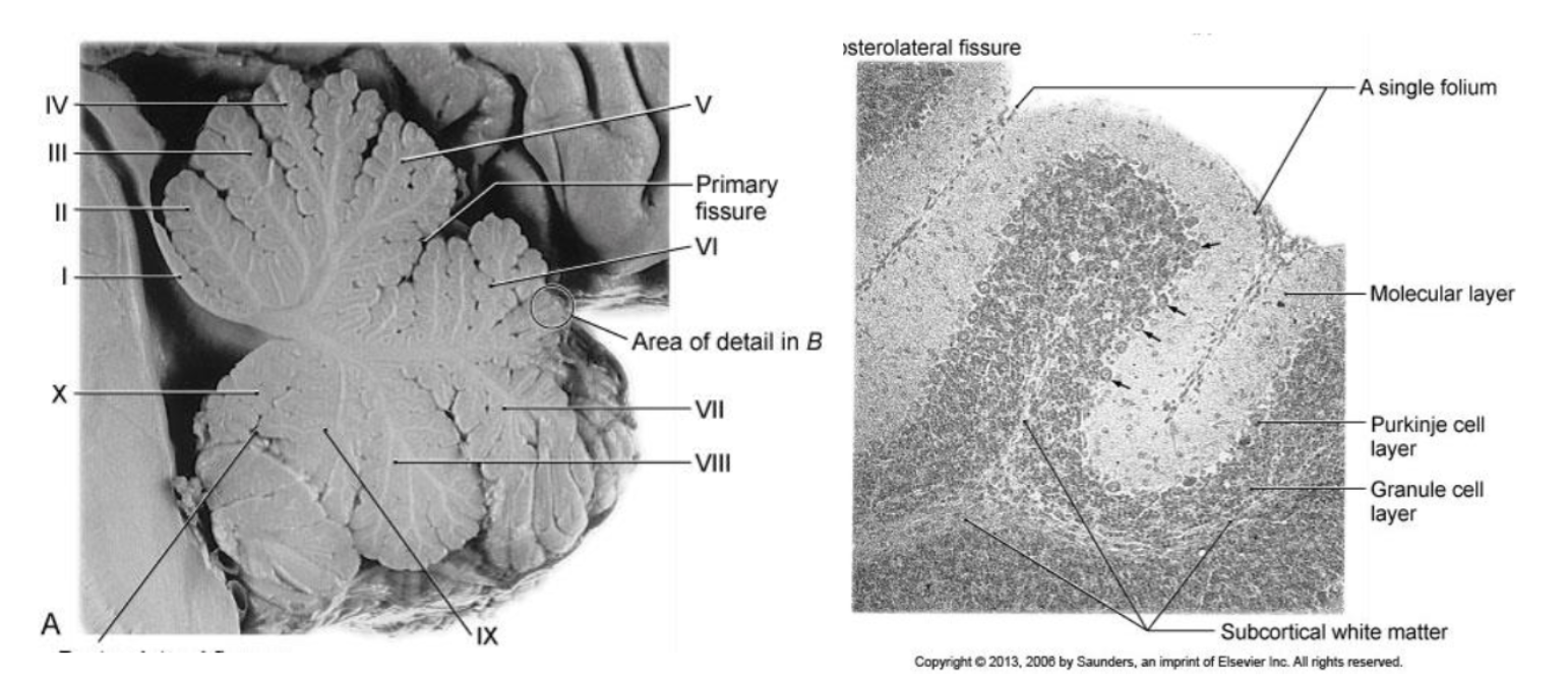
cerebellar cortex: cells of importance
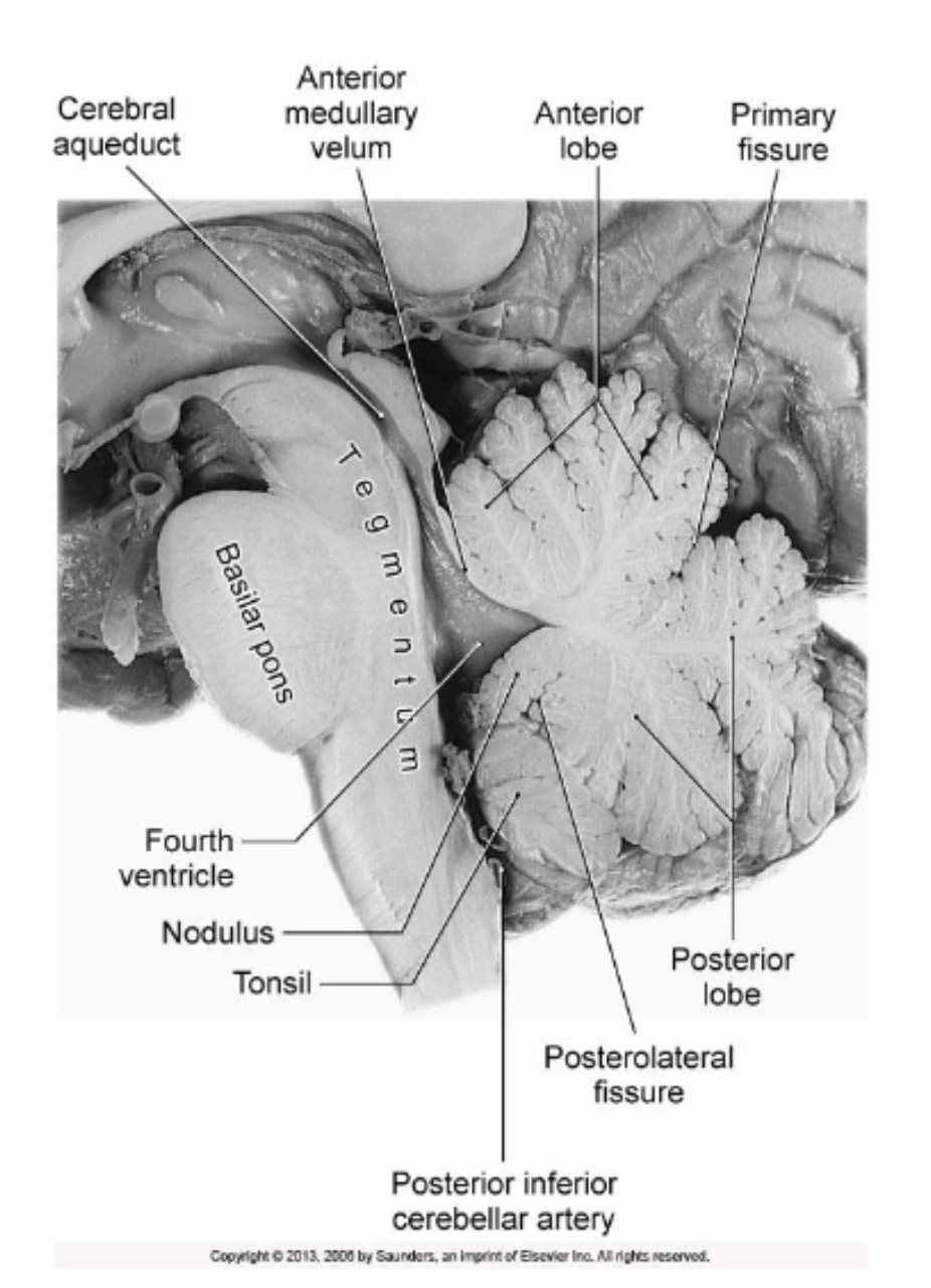
hemispheres —> broken down into anterior and posterior lobes, and a floccular lobe —> broken down into lobules —> broken down into folia
image on the right: blown up picture of a single folium, for viewing of the 3 layers of the cerebellar cortex
3 layers
molecular layer
outermost layer
purkinje cell layer (arrows pointing to the large cell bodies of the purkinje cells)
outputs to the deep cerebellar nuclei
granule cell layer
inner most layer
subcortical white matter
where deep cerebellar nuclei are buried
deep cerebellar nuclei output to the structures that will allow the cerebellum to modulate UMN activity
cerebellar cortex
consists of three layers from superficial to deep: __ layer, __ layer, and __ layer
cerebellar cortex contains 6 cell types:
__ layer:
__ cells:
__ layer:
(image= showing single folium)
molecuar; purkinje; granular
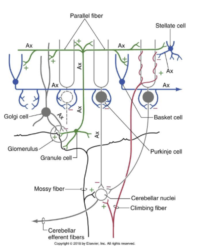
molecular layer: contains **basket and **stellate cells that are inhibitory
neurons that synapse with Purkinje cells
these cells release the neurotransmitter GABA
**purkinje cells: largest cell in the cerebellar cortex and the ultimate target
of all input to the cerebellum; they are the efferent cell of the
cerebellar cortex, sending inhibitory projections to the neurons in the
deep cerebellar nuclei
also releasing GABA
granular layer: deepest layer with 3 different cell types
*granule cells: receive afferent input from mossy fibers; their axons project to the molecular layer of the cortex, sending excitatory neurons/input that synapse with all other neurons of the cortex (e.g., predominantly the purkinje cells but also the stellate and basket cells)
release neurotransmitter glutamate
**golgi cells: inhibitory interneurons that synapse with all other
cells in the cortexrelease GABA
*unipolar brush cells: found predominantly in the flocculodular lobe and are an excitatory neurons
release neurotransmitter glutamate
*all excitatory cells in the cerebellar cortex (i.e., granule cells and brush cells) release neurotransmitter glutamate
**all inhibitory cells in the cerebellar cortex release neurotransmitter GABA
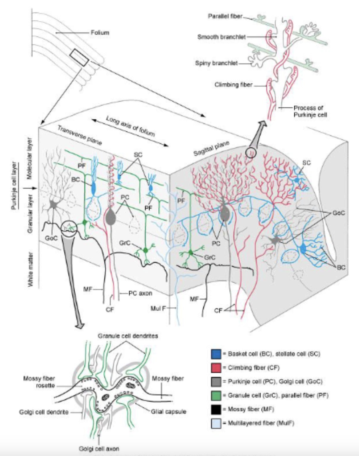
neural circuitry
2 main types of cerebellar input:
__ fibers:
__ fibers:
2 main types of cerebellar input:
mossy fibers: convey information originating in the pontine nuclei (motor planning information), the spinal cord (proprioceptive information) & the brainstem (proprioceptive information)– they make excitatory projections onto neurons in the cerebellar nuclei and granule cells of the cerebellar cortex (i.e., excitatory input from mossy fibers is conveyed to both the deep cerebellar nuclei, as well as the cortex)
granule cells, in turn, send projections called parallel fibers that run perpendicular to the Purkinje cell dendrites in the molecular layer – they make excitatory projections onto purkinje cells, as well as basket and stellate cells
excitatory input from granule cells can be modified by basket and stellate cells
climbing fibers: originate in the inferior olivary complex – they make excitatory projections onto neurons in the cerebellar nuclei and onto Purkinje cells in the cortex; research suggests they provide an important “training” signal that modulates the effectiveness of the parallel fiber connections – thought to provide feedback about the sensory consequences of movement and motor error
difference between mossy and climbing fibers when connecting to purkinje cells
mossy fibers
granule cells will make contact with multiple purkinje cells
each parallel fiber can make contact with tens of thousands of purkinje cells
huge degree of divergence of inputs form the mossy fibers that impact the firing rate of purkinje cells
climbing fibers
wrap around the dendrites of the purkinje cells, like a climbing vine
each purkinje cell is going to receive multiple synapses, but from only 1 climbing fiber
this climbing fiber makes for a great resource to modulate excitatory input to the purkinje cell (i.e. intimate relationship between climbing fiber and purkinje cells)

neural circuitry
activity of the cells in the __ modulate the activity of the cells in the __
excitatory outflow of the deep cerebellar nuclei varies in
response to:
cerebellar cortex; deep cerebellar nuclei
excitatory outflow of the deep cerebellar nuclei varies in
response to:excitatory input coming in from afferent collaterals of the mossy
and climbing fibersinhibitory input coming in from the cerebellar cortex mediated by the Purkinje cells (PCs);
== thus, increases or decreases in inhibitory input from the cortex modulates the activity of the deep nuclei, ultimately modifying the output of UMNs
cerebellum and motor learning
cerebellum is thought to play a key role in the learning of…
relatively simple reflexive motor behaviors (e.g., VOR (vestibular ocular reflex), reflexes to aversive stimuli)
and complex voluntary movements (e.g. riding a bike)
using error information, it is thought that the cerebellum recalibrates movements for successful performance;
experimental examples –
adjustment of the VOR (vestibular ocular reflex) to magnifying glasses;
when we move our head, the VOR will compensate for the head movement by rotating the eyes in the equal and opposite direction to the head movement, keeping the image stable on the retina
adjustment of reaching trajectories to novel force fields;
adjustment of pointing movements to a visual target during prism adaptation (Tedxtalk video)
allowing compensation for shift in visual image during pointing task
proposed that input from the climbing fibers to purkinje cells can induce plastic changes that modify the responsiveness of these cells to specific inputs from the parallel fibers of granule cells
—> improved accuracy, pruning away defective responses
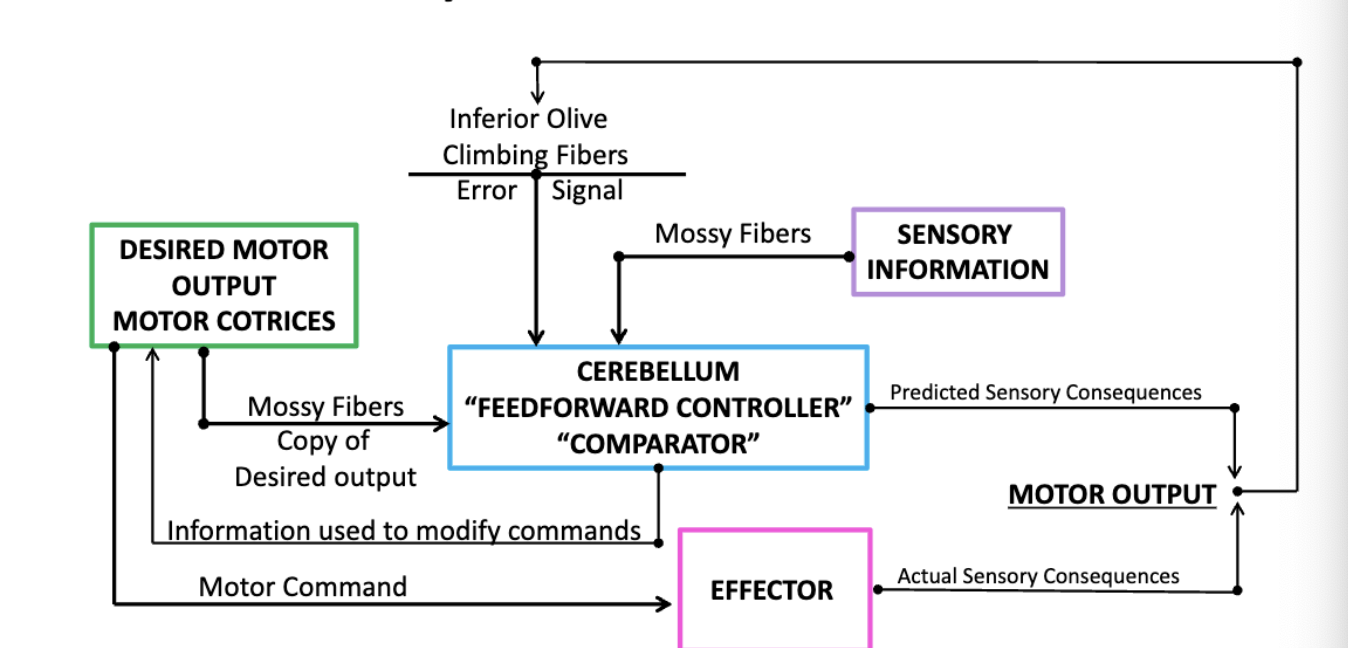
neural circuitry: role of cerebellum as a “feedforward controller/comparator”
we have a desired motor output that comes from motor cortices
through mossy fibers, relay in the pons, a copy of the desired input will be fed into the cerebellum
—> cerebellum will take information about the motor plan + information about the state of the system/body to develop predictive sensory consequences (i.e. cerebellum can predict what the sensory consequences of that motor plan would be)
—> as commands are sent down to LMNs, ultimately to affect the effectors (e.g., limb and body movement), we will get actual motor output
—> anticipated and predicted motor sensosry consequences are compared:
if there’s an error between what was predicted and what actually occurred, the error information will be fed into the inferior olivary complex via climbing fibers
error information will be used by the cerebellum in 2 ways:
1. modify motor commands online
2. update motor plans for future use
== we learn over time!
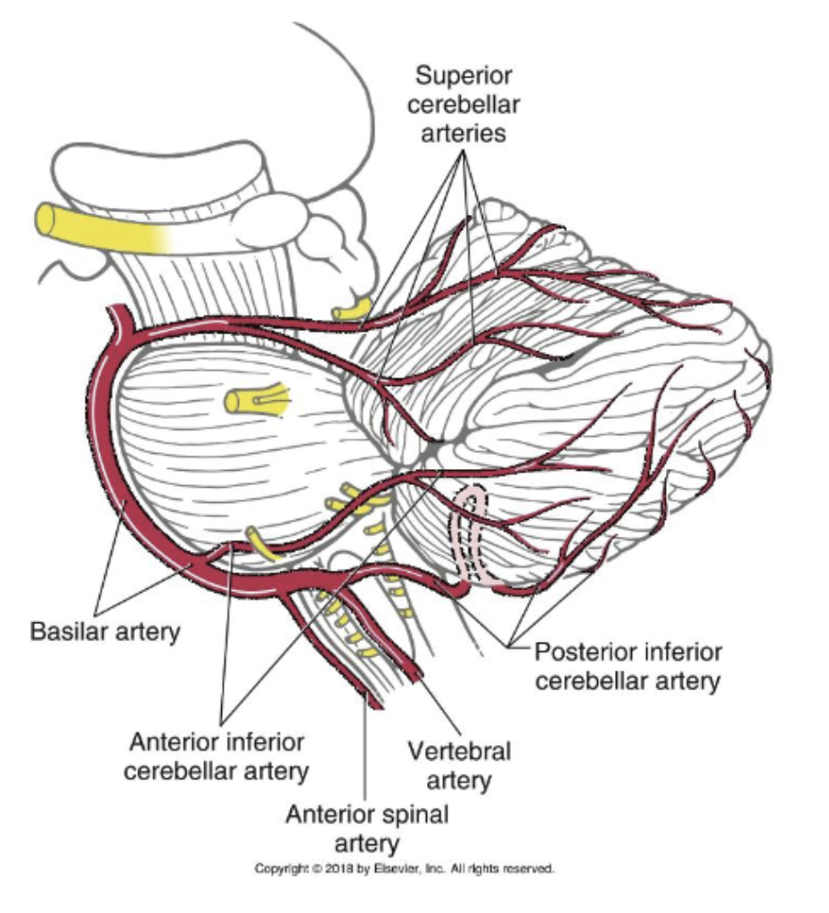
blood supply
superior cerebellar artery:
anterior inferior cerebellar artery
posterior inferior cerebellar artery
superior cerebellar artery: supplies the…
entire superior surface of the cerebellum
majority of the deep cerebellar nuclei
superior cerebellar penducle
most rostral part of the middle cerebellar peduncle
(arising from the *basilar artery)
anterior inferior cerebellar artery: supplies the…
caudolateral parts of the inferior cerebellar surface
caudal part of the middle cerebellar peduncle
(arising from the *basilar artery)
posterior inferior cerebellar artery: supplies the…
caudomedial parts of the inferior cerebellar surface (including vermis)
inferior cerebellar peduncle
(arising from the *vertebral artery)
*stroke involving any one of these arteries can produce cerebellar dysfunction, most often impacting the intermediate and lateral zones of the cerebellum
deficits = cerebellar ataxia, will be seen ipsilateral to the stroke
1 exception in strokes impacting the midline (vermal region), in which we will see bilateral deficits with trunkal ataxia being the predominant clinical sign
clinical notes- ***what are common cerebellar signs
ataxia:
disturbance of posture/gait:
dysmetria:
dysdiadochokinesia:
dysarthria:
intention tremor:
titubation:
***ataxia: impairment in coordination and accuracy of movements that accompany cerebellar damage; deficits are predominantly seen ipsilateral to the lesion
***disturbance of posture/gait: characterized by a wide base stance; staggering gait pattern with frequent loss of balance toward the side of the lesion
***dysmetria: (aka past-pointing) inappropriate force and distance of targeted movements; characterized by hypometria (undershooting of target) or hypermetria (overshooting of target).
***dysdiadochokinesia: inability to perform rapid alternating movements (e.g., rapid supination or pronation, opening/closing hand, finger tapping)
***dysarthria: slurred/garbled speech or scanning speech (staccato nature of speech - slow/disjointed)
***intention tremor: oscillatory trajectory during target-directed movements – pronounced at the end point of the movement; tremor is absent at rest
***titubation: type of essential tremor that causes uncontrollable, rhythmic shaking
clinical notes- ***what are common cerebellar signs
hypotonia
decreased deep tendon reflexes
are also possible with cerebellar damage
(hypotonia= ***abnormally low muscle tone)
clinical notes- ***what are common cerebellar signs
dysfunction in eye movements:
nystagmus, disturbed pursuit movements or difficulty maintaining visual fixation on a target are possible with cerebellar damage
***a stroke or lesion of the cerebellum on the right side of the brain would lead to motor deficits on which side of the body?
stroke or lesion on the right side of the cerebellum would primarily cause motor deficits on the right side of the body; this is because the cerebellum controls movement on the same side of the body (ipsilateral)
***what would you expect to see with damage to the midline structures of the cerebellum (vermal zone/fastigial nucleus)?
damage to the midline structures of the cerebellum, including the vermal zone and fastigial nucleus, primarily leads to truncal (trunk) ataxia, which is characterized by uncoordinated movements and a wide-based, unsteady gait; additionally, patients may exhibit truncal tremors, difficulty maintaining balance, and impaired postural control