Looks like no one added any tags here yet for you.
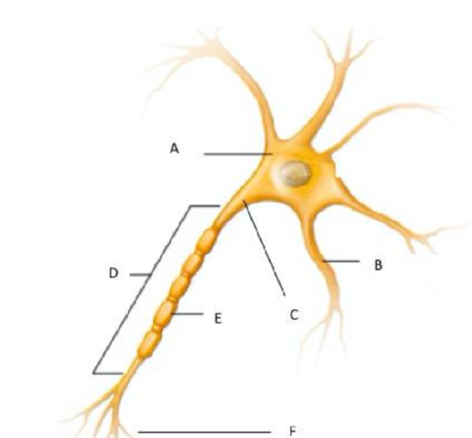
Identify the structures of the neuron
A. Soma/Cell body
B. Dendrite
C. Axon hillock
D. Axon
E. Myelin sheath
F. Axon terminal
Where are voltage gated Na+ Channels
axon hillock and axon
Where are voltage gated K channels
axon hillock and axon
where are voltage gated Ca2+ channels
axon terminal
where are ligand gated gated Na channels
dendrite and soma
where are ligand gated K channels
dendrite and soma
where are ligand gated Ca channels
there are none!
What are EPSPs and where do they happen?
Excitatory postsynaptic potentials that basically means that the neuron gets excited and depolarizes. They happen in the dendrite. This is a graded potential
Neurotransmitter released from the presynaptic neurons bind to the postsynaptic neuron receptors which are chemically gated cation channels, causing them to open. Then Na flows into the neuron faster than K can flow out and the inside of the neuron becomes more positive and the EPSP propagates toward the initial segment.
What are IPSPs and where do they happen
inhibitory postsynaptic potentials that basically means that the neuron hyperpolarizes and doesn’t lead to an action potential. This also happens in the dendrite and is a type of graded potential.
action potential
depolarizing of neuron leading to this signal. This is all or nothing and occurs in the axon hillock and axon. This all happens within 4 seconds due to brain using a lot of ATP.
very brief, large depolarizations that travel for long distances through a neuron without losing strength.
saltatory conduction
jumping of a signal from nodes of ranvier to other nodes due to insulation of axon. This happens in the axon as well.
continuous conduction
conduction that occurs when neurons have no myelin sheaths. This is slower and leads to signals being lost as the channels rush in negative ions leading to hyperpolarization in the axon.
exocytosis
occurs in the axon terminal with K leaving the cell and hyperpolarizing it.
Nernst equation
61/z (charge of the ion) x log (ion out)/log (ion in)
only calculates permeability for 1 ion inside and outside the cell.
GHK equation
calculates the permeability of the cell membrane for each ion. (Permeability of ion times the voltage of the ion) + that for each one.
cerebellum
part of brain that influences muscle activity; inhibits unnecessary motor movements; coordinates slow sustained contractions. This is involved in physical balancing of your body and is easily influenced by alcohol. However, this is not where motor signal starts.
Basal nuclei/ganglia
found throughout brain stem; reticular activating system; also involved in motor activities and visceral activities. This releases dopamine for motor control. Also involved in balancing. Less release of dopamine from here leads to Parkinson’s disease, affecting movement.
cerebral cortex
memory, integration, interpretation, discrimination, localization, language.
hypothalamus
controls several major endocrine functions → temperature, thirst, hunger, sexual desire, osmolarity.
limbic system
functional system responsible for emotional behavior. This includes the amygdala (panic button for emotion and memories), hippocampus (involved in learning and memory, for new memories, not storing previous memories), and thalamus (sending signals to other areas).
Information is input into short term memory and processing (requiring the hippocampus) allows for recall and storage into long term memory.
medulla oblangata
vital reflex centers: cardiac, vasomotory, respiratory
midbrain
controlled in eye reflexes, such as pupillary reflex, consensual response, blinking (pupil reflex, also called cat brain???)
pons
controls our biological clock which regulates our daily patterns.
spinal cord
contains many major reflex centers: withdrawal and stretch
thalamus
edits sensory information which is passed on to cerebral cortex; relays motor signals coming out of the cerebral cortex.
Which membrane protein is the main contributor to the resting potential
K leak channel (prevents cell from becoming too negative)
Graded potential
Potentials that occur with molecules flowing into or out of the neuron and these can be of varying strengths and can add up together. The cell becomes more positive inside due to this and Na channels opening. They work to trigger action potentials and can decay.
subthreshold potentials start as strong but decay and never trigger action potentials, dying at the axon hillock.
suprathreshold ones are opposite and are very very strong, they decay but end up triggering an action potential ultimately.
variable strength signals that travel over short distances and lose strength as they travel through the cell.
Depolarizations or hyperpolarizations
Size or amplitude is directly proportional to the strength of the triggering event.
Lose strength as they move through the cytoplasm due to current leaks (open leak channels that allow positive charge to leak out to the ECF) and cytoplasmic resistance
Reach a region of Trigger zone and the first part of the axon called the initial segment.
Subthreshold graded potentials are triggered by a single stimulus
Suprathreshold graded potentials cause an action potential.
Membrane potential
the difference in electrical charge across a plasma membrane. This does not equal the concentration of ions across membrane. This is measured in milivolts. Resting membrane potential is higher Na outside and higher K inside.
Sodium Potassium Pump
ATPase that pumps 3 Na outside and 2 K inside to keep cell membrane relatively negative.
depolarization
inside of membrane becomes less negative and graph deflects upward
repolarization
inside of membrane becomes more negative and the graph returns to resting potential
hyperpolarization
membrane becomes more negative than resting.
refractory period
ion channels remain closed and no AP can be generated.
synapse
a small gap between neurons where electrical signals are transferred to chemical signals. VG Ca channels open to trigger NT release.
Neurotransmitters
chemical messenger molecules that follow principles of protein based lipophobic ligand action. These can trigger chemically gated ion channels to open and a secondary messenger cascade.
They can be excitatory (make inside of cell positive) or inhibitory (make inside of cell more negative) depending on the receptor. These are influenced by drugs and diseases.
ACh - Acetylcholine
excitatory or inhibitory neurotransmitter that works by unblocking or blocking ligand gated sodium channels. This works at neuromuscular junctions, in the ANS, CNS, and is degraded by AChE since we already have so much, we don’t need reuptake of it.
dopamine
neurotransmitter part of our reward and happiness circuitry
serotonin
neurotransmitter part of our emotions
Glutamate
Major excitatory neurotransmitter in the CNS that also works by opening ligand gated sodium channels. This is linked to memories and learning.
when we forget, a synapse disappears and when we remember, the synapse appears and forms again
synapse grows and stays with repetition.
Norepinephrine
excitatory (mainly) or inhibitory neurotransmitter that works in the sympathetic part of the ANS for the fight or flight response. In the CNS, it works for mood, motivation, alertness. This is reuptaken and recycled.
GABA
primary inhibitory neurotransmitter in the brain that works to trigger open the ligand gated Cl- channel and it basically makes it so that the neurons fire less. This is used in sedative medicine and triggered with alcohol intake. There is reuptake into axon terminal and glial cells by the GABA transporter.
Temporal Summation
When one neuron leaks more and more neurotransmitters over time and leads to an action potential eventually.
Spatial summation
when many synapses provide a smaller amount of neurotransmitters, leading to a sum of a large amount that leads to an action potential.
Divergence
1 neuron is activated and sends a signal to multiple neurons. This is very sensitive.
Convergence
When a signal from multiple neurons is converged into 1 in order to save neurons and resources. Brain loses the power to know exactly where the stimulus is coming from (not as accurate).
Neurons
nerve cells that carry electrical signals rapidly and sometimes over long distances.
They are uniquely shaped and have thin extensions called processes that can extend up to a meter in length.
Release neurotransmitters that are chemical signals released into the ECF to communicate with neighboring cells.
Nerves
the long axons of both afferent and efferent peripheral neurons bundled together with connective tissue into cordlike fibers, extend from the CNS to targets of the component neurons.
Sensory nerves and motor nerves have specific directions (listed above) but mixed nerves can carry signals in both directions.
Axonal transport
when proteins are moved in vesicles down the axon.
Forward/anterograde transport moves vesicles and mitochondria from the cell body to the axon terminal.
Retrograde/Backwards transport returns old cellular components from the axon terminal to the cell body for recycling.
Uses stationary microtubules as tracks along which transported vesicles and mitochondria "walk" with aid of attached motor proteins.
Fast axonal transport goes in both directions and can move material at rates of up to 400 mm per day.
Slow axonal transport moves soluble proteins and cytoskeleton proteins from the cell body to the axon terminal at a rate of 0.2-0.8 mm/day.
CNS
brain and spinal cord. This is well protected and doesn’t engage at all with the outside or physical stimuli directly.
PNS
cranial and spinal nerves in the body, has a lot of divisions.
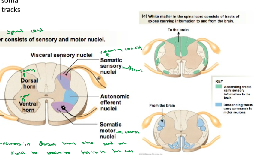
All of the spinal cord weird root and horn stuff
When reflexes or quick responses are happening, this is what takes over and sends it.
The Dorsal root ganglion is a cluster of cell bodies (sensory part) that receives stimuli and sends this information to the brain (late) to fill it in but it sends a direct signal to the dorsal horn in teh spinal cord which goes to the… (see below)
The ventral horn which is a motor neuron that sends signals out through the ventral root.
the lateral horn is in the middle and is a part of the fight or flight response. This is close to either root or horn depending on what you want to do
The horns are the somas and cell bodies and the roots and everything else are tracks.
Vision and Occipital Lobe
Vision goes to other side of brain than eyes. The occipital lobe is the visual association area and the visual cortex.
Sensory part of Parietal Lobe
This is in the front of the parietal lobe. More neurons are dedicated to the face and hands as this leads to more accurity. The back has too small number of neurons to cover such a large area so this leads to less resolution and accurity. There is a homunculus (uneven distribution) for this.
Frontal Lobe Motor cortex
the back part of the frontal lobe that works to control movement, where signal starts. We have more neurons in our faces and hands and thus a homunculus for this as well.
Prefrontal cortex: reasoning and decision making
Area involved in decision making that takes about 16 years to mature, sometimes even longer. We are the only animal with the ability to delay gratification and make decisions for the long run.
Frontal Lobe lobotomy
Procedure that would cut the prefrontal cortex and change it permanently, leading to no emotions and numbness and a different person.
Reflexive implicit memory
Recall is automatic and does not require conscious attention. This is acquired slowly through repetition and is hard to forget. This includes motor skills and rules and procedures.
Declarative Explicit Memory
Recall requires conscious attention and depends on higher level thinking skills such as inference, comparison, and evaluation. Memories can be reported verbally.
Sleep stages and descriptions
We repeat the sleep cycle 5-7 times in sleep.
awake and eyes closed
Stage 1: REM, rapid eye movement, dreams, body is active but paralyzed essentially
sleep
sleep
deep sleep with slow waves and delta waves. Neurons fire synchronously and forms synapses, releasing glutamate and forming memories during sleep
The deepest sleep occurs in the first 3 hours. Depression causes sleep abnormalities and an onset of REM sleep and shorter sleep cycles overall.
Lateral inhibition
a process that stops other neurons from sending signals, blocking them. Brain knows location only from the neurons sending the signal and not blocked neurons. This helps the brain pinpoint where signals are coming from very specifically. (We don’t increase signal of one neuron, just block others to make the first one stand out).
Rods
Sensory cell of the eye that has a large receptive field, high convergence, has dark current, and uses less cellular resources.
120 million per retina, more numerous in periphery and less accurate, high sensitivity to light waves and activated by 1 photon even. Vision is in shades of gray
cones
sensory cell of the eye that is less sensitive to light, is involved in color vision and concenrated in fovea of retina, has a small receptive field with low convergence and uses more cellular resources. There are 6 million per retina, used for bright light and day vision.
3 kinds of cones for humans that make up all the light we see → blue green and red. If we miss one then that color and the combinations we can make with it are completely gone. This is usually the red cone being gone.
Dark current
in the dark rhodopsin is inactive and that leads to an increase in cGMP and activates CNG channels to increase the membrane potential and send out inhibitory neurotransmitters so that the signal doesn’t go to the brain and we see less in the dark
in the light, rhodopsin is active, decrease in cGMP, deactivates CNG channels, cells doesn’t depolarize and no inhibitory neurotransmitters are sent out and signals are sent to the brain allowing us to see in light.
Simple receptors
neurons with free nerve endings. They have myelinated or unmyelinated axons. Used for touch.
Complex receptors
Complex neural receptors have nerve endings enclosed in connective tissue capsules. Used for touch.
Special Senses REceptors
Cells that release neurotransmitters onto sensory neurons initiating an action potential. Used for all complex 4 senses.
Nature/modality
what type of stimulus it is (light, smell, pain) and this is distinguised through the brain distinguishing signals arriving to it from different areas and going to different parts of the brain to be processed.
Location
Location of a stimulus is determined and controlled by lateral inhibition and divergence/convergence.
stimulus intensity and duration
receptor potential strength and duration vary with the stimulus
receptor potential is integrated at the trigger zone
frequency of action potentials is proportional to stimulus intensity. Duration of a series of action potentials is proportional to stimulus duration.
Neurotransmitter release varies with the pattern of action potentials arriving at the axon terminal.
Receptors also adapt to a sustained stimulus with tonic receptors slowly adapting and phasic receptors rapidly adapting and completely turning off.
Somatosensory pathway
3 neurons in this pathway (1 from spinal cord to brain stem, brain stem to thalamus, and thalamus to parietal or whatever cortex).
The primary somatosensory cortex called S1 is located on the postcentral gyrus in the parietal lobe.
Pain receptors and Substance P pain Pathway
mechanical receptors that are involved in sensing cutting, crushing, pinching and this involves thermal nocireceptors that sense temperature extremes as well. Polymodial nocireceptors sense all damaging stimuli.
A stimuli is sensed at a nocereceptor and substance P is secreted to the dorsal horn.
Reticular formation is a group of neurons in the brain responsible for pain modulation and this received the signal as well as the thalamus next which perceives the pain.
The thalamus sends the signal to the somatosensory cortex to localize it and make sense of it, where it came from.
the reticular formation sends the signal to the hypothalamus and limbic system to form emotions and memories with this pain and avoid it next time.
Phantom limb is caused by other neurons firing and the brain firing the neurons of the limb cut off so the person feels phantom pain.
Changes in lens shapes are controlled by the…
ciliary muscle. THe lens curvature also decreases as we get older which causes our vision to decrease in quality and efficiency.
Fovea
the center of the retina with the most cones and the region of the greatest visual acuity.
3d vision and predator and prey
Predators need eyes in front of them, seeing only 180 degrees, compared to prey that have eyes on 2 sides seeing almost 360 degrees as we need depth perception. 3d vision adn depth perception are necessary and only occur with 2 eyes at the front of the head.
adrenal medulla
neuroendocrine tissue in the adrenal glands that produces and releases epinephrine and norepinephrine.
Sound waves
pressure waves with regions of compression of air particles and regions of rarefaction.
frequency/pitch is how many waves there are per second and amplitude is the loudness correlated to the length of the peaks of the sound waves.
Structure and functions of ear
pinna and ear canal collect sound waves only
ear drum is the starting point of the middle ear and it moves due to the sound waves, also moving the ossicles (all mechanical).
cochlea transfers sound to neuronal signals
We need 2 ears to compare the loudness of sounds and see where they are coming from (comparing a milisecond time difference in when sound reaches one ear vs another)
eustachian tube
tube that connects the ear to the throat from the middle ear and it allows for fluid exchange?? Easier for babies to get ear infections because this tube is smaller for them and bacteria can easily travel from the throat to the ear because of this.
Cochlea
Bony labyrinth in inner ear that is connected to the vestibule and semicircular canals. The cochlea is filled with fluid (endolymph which is high in K+).
sound waves are sent and collected by ear
tympanic membrane vibrates in response to sound wave
vibrations are amplified across the ossicles and they push liquid into the cochlea through this
vibrations against the oval window set up standing wave in the fluid of vestibuli
pressure bends the membrane of the cochlear duct (basilar membrane) at a point of max vibration for any given frequency, causing hair cells and the organ of corti to vibrate. Organ of corti is activated by fluid flowing through the cochlear duct and moving its hair cells.
tectorial membrane and basilar mebrane both move on the cochlear duct adn the hair cells touch the tectorial membrane on top, causing mechanically gated K channels to open and depolarize in the endolymph.
The vestibular sense
all about balance. The vestibule (made up of the utricle and saccule) is a structure in the inner ears and the semicircular canals oriented in 3 different directions. These hair cells in the vestibule don’t use liquid, the otoliths move based on head rotation causing hair cells hair to move and depolarize essentially.
Otoliths were little things in gelatinous layer that vestibule contains, no actual fluid.
hair cells from the equilibrium receptors
vestibular branch of CN 7 vestibulocochlear
eye movements needed to balance, vestibular nucleus in the medulla oblongata, and vestibulospinal tract (skeletal muscles needed for balance)
thalamus and cerebellum
cerebral cortex to be aware of body position.
Auditory nerve (cranial nerve 8)
transmits impulses from the cochlea and the vestibule to the A1.
5 tastes and their receptors
sweet, umami, bitter, sour (H in the sour taste blocks the K leak channel and makes the cell positive which opens up the VG Ca+2 channels leading to depolarization), salty (Na itself depolarizes the cell through an Na channel). The other 3 use GPCRs and secondary messengers.
Olfactory receptor cells and how they work
odorant binds with receptor → activates GPCR → cAMP mechanism → nonspecific cation channels open → Na and Ca influx → receptor potential → AP
Bypasses the thalamus and the bulb is directly connected to the limbic system. Pheromones are sexual chemicals used for that kind of communication between animals.
Parasympathetic
rest and digest, functions from from the cervical or sacral part of spine.
Long preganglial neuron and short postganglial neuron (close to target cells)
pupils constrict
salivation
hr slows
bronchioles constrict
increased digestive motility and secretion
increased enzyme secretion from pancreas
stimulates insulin secretion
release of urine
erection
Sympathetic
Fight or flight, functions come from the thoracic or lumbar part of spine, for long term survival
short preganglial neuron and long postganglial neuron (close to CNS)
pupils dilate
less salivation
arterioles constrict
HR increases
bronchioles dilate
decreases motility and secretion of digestive tract as well as enzyme secretion
inhibits insulin secretion
secretes catecholamines from adrenal medulla (epi)
urinary retention
fat breakdown
no sexual desire or erection
Cranial nerve 3
Lacrimal gland for crying and for the eyes
cranial nerve 7
for lacrimal gland and crying and for salivation
cranial nerve 9
the parotid salivary gland
cranial nerve 10
vagus nerve, for the cardiac plexus, heart, lungs, and GI.
nicotinic receptors
receptors not on the target tissue for both sympathetic and parasympathetic pathways. ACh is used for this receptor in both pathways. This receptor is an ion channel at autonomic ganglion.
Target tissues and their receptors
in sympathetic pathways, the postganglionic neuron releases norepinephrine which binds to adrenergic receptors on target tissues
in parasympathetic pathways, the postganglionic neuron releases ACh which binds to muscarinic receptors on the target tissues.
Both Adrenergic and Muscarinic receptors are GPCRs
Somatic motor pathways:
postganglionic neuron releases ACh that binds to nicotinic receptor on target skeletal muscle.
Parasympathetic pathway - ANS:
preganglionic neuron releases ACh and binds to nicotinic receptor on ganglion and postganglionic neuron releases ACh to muscarinic receptor on target tissue.
Sympathetic Pathway - ANS:
preganglionic neuron releases ACh and bidns to nicotonic receptor on ganglion and postganglionic neuron releases Norepinephrine to bind to alpha and beta 1 receptors on target tissues (adrenergic receptors)
preganglionic neuron releases ACh and binds to nicotinic receptor on ganglion and postganglionic neuron releases ACh to muscarinic receptor on target tissue (FOR SWEAT GLANDS ONLY)
preganglionic neuron releases ACh and binds to nicotinic receptor on ganglion and postganglionic neuron releases ACh to nicotinic receptor on adrenal medulla cells.
alpha 1 adrenergic receptor
GPCR that vasoconstricts blood vessels to the GI, contracts walls of the reproductive organs. overall vasoconstriction.
beta 1 adrenergic receptor
GPCR that increases CO and increases contractility of heart muscle. Vasodilates BV to the heart
Beta 2 adrenergic receptor
GPCR that bronchodilates the lungs and vasodilates BV to the lungs, decreases GI motility, vasodilates BV to skeletal muscle.
beta blockers
medication that counters epi and NE as it is an antagonist for B1 receptors.
Atropine
antagonist for muscarinic receptors as it inhibits ACh and increases heart rate. dual