Muscle Physiology Test
1/38
There's no tags or description
Looks like no tags are added yet.
Name | Mastery | Learn | Test | Matching | Spaced | Call with Kai |
|---|
No analytics yet
Send a link to your students to track their progress
39 Terms
Name the functions of the muscle
Movement of the body and its parts
Maintains posture
Generates heat
Stabilizing joints
Name the characteristics of the skeletal muscle
Location: Most are attached by tendons to bones
Shape: Long, cylindrical, multinucleate, visible striations
Regulation contraction: Voluntary, subject to conscious control
Speed contraction: Slow to fast
Rhythmic: no rhythmic contraction
Name the characteristics of the smooth muscle
Location: Found mainly in the walls of hollow organs (ex.stomach, bladder, respiratory passageway)
Shape: No striations, spindle-shaped cell, single nucleus
Regulation contraction: involuntary
Speed contraction: Very slow
Rhythmic: Yes, in some
Name the characteristics of the cardiac muscle
Location: Only in the heart
Shape: striations, single nucleus, joined to another muscle cell at an intercalated disc
Regulation contraction: involuntary
Speed contraction: slow
Rhythmic: Yes
Name the muscle
Smooth
Name the muscle
Skeletal
Name the muscle
cardiac
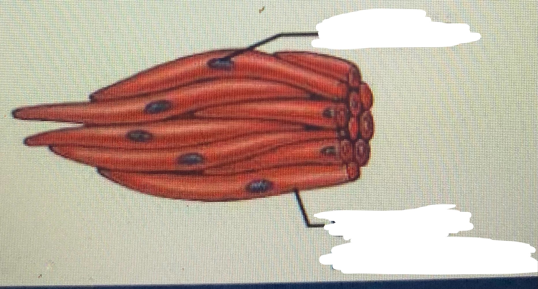
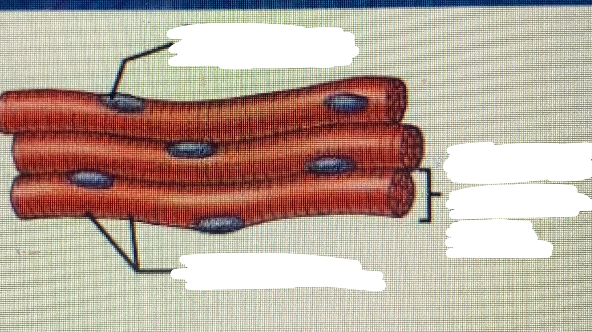
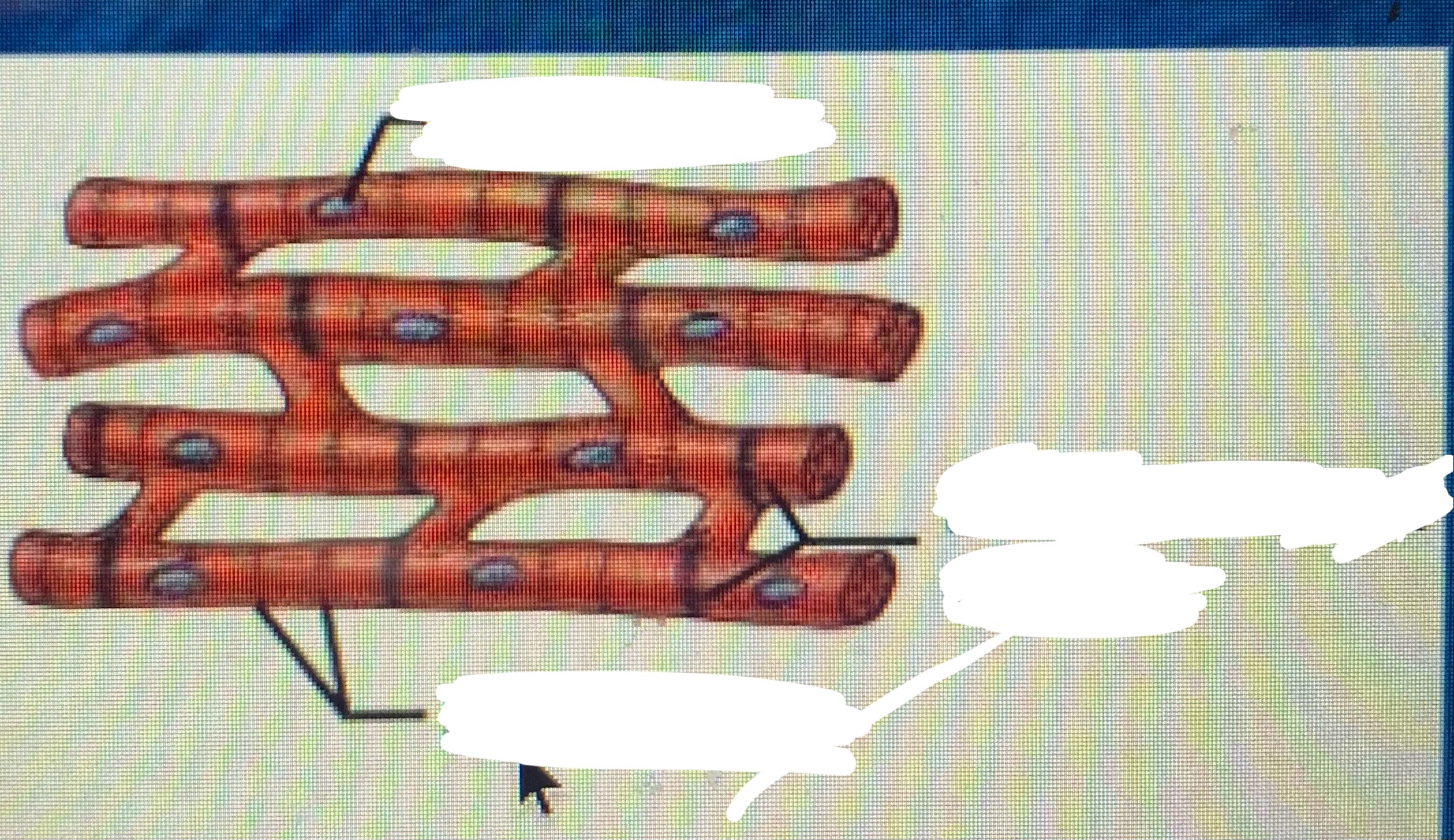
Name the wrappings of the skeletal muscle
Muscle fiber
Endomysium
Fascicle
Perimysium
Epimysium
Tendons
What does the endomysium wrap?
Surrounds and supports each individual muscle fiber (muscle cell) within the skeletal muscle (inside)
What does the perimysium wrap?
Bundles of muscle fibers, that are the fascicles (around)
What does the epimysium wrap?
The entire skeletal muscle
Microscopic anatomy of skeletal muscle
Cells are multinucleated
The nuclei are beneath the sarcolemma
What is the sarcoplasmic reticulum?
It is found beneath the sarcolemma
For storage of calcium
*Calcium is key for muscle contraction*
What are myofibrils?
Bundles of myofilaments
Myofibrils are aligned to give distinct bands I band: light band
A band: dark band
What are sarcomeres?
A contractile unit of a muscle fiber
Explain the organization of the sarcomeres
Thick filaments= myosin filaments, composed of the protein myosin, has ATPase
Thin filaments: actin filaments
Composed of the protein actin
Continuation of the sarcomere
Myosin filaments have heads, extension or cross bridges
Myosin and actin overlap somewhat
At rest there is a bare zone that lacks actin filaments
Name the properties of the skeletal muscle
Irritability: ability to receive and respond to a stimulus
Contractility: ability to shorten when an adequate stimulus is received
Extensibility: stretch
Elasticity: Recoil
Why do skeletal muscles have to stimulated?
Skeletal muscles must be stimulated by a nerve to contract
A motor unit
One neuron and the muscle cells stimulated by that neuron
What is the neuromuscular junction and what does it consist of?
An association site of nerve and muscle
Synaptic cleft: gal between nerve and muscle
*The nerve and muscle do not make contact, area between nerve and muscle is filled with interstitial fluid
Name the transmission of nerve impulse to muscle
Neurotransmitter, chemical released by nerve upon arrival of nerve impulse
The neurotransmitter for skeletal muscle is acetylcholine (aCh)
The neurotransmitter attaches to receptors on the sarcolemma (plasma membrane)
Sarcolemma becomes permeable to sodium
Continuation of transmission
Sodium rushing into the cell generated an action potential
Once started, muscle contraction cannot be stopped
What is the sliding filament theory
An activation by nerves causes myosin heads (cross bridges) to attach to binding sites on the thin filament
Myosin heads then bind to the next site of the thin filament
This action causes a sliding of the myosin along the actin
The result is that the muscle is contracted
Basic steps of the neuromuscular junction
Message sent
Neurotransmitter
Depolarization
Calcium + Troponin= Actin exposed
Actin + Myosin= contraction
Relaxation
Nervous sys. Sends message to the effector organ
Neurotransmitter released ACh
ACh binds to sarcolemma of muscle fiber
ACh initiates opening of sodium-potassium
Depolarization
Cause: the binding of ACh to sodium-potassium channels, opening of channels + movements of sodium and potassium across sarcolemma
Involves the movement of charges
More sodium moves in and potassium moves out
Imbalance of charges =electrical current (action potential)
SR and Calcium
Cause: depolarization
Action potential across the SR causes the release of calcium
Once calcium is released from membrane of SR
The calcium binds with troponin
Troponin- tropomyosin conformation
Troponin and tropomyosin no longer cover actin binding sites thus exposing the sites
Myosin interacts with actin
Cause: Troponin and tropomyosin unveils actin (binding sites exposed)
Actin exposed
Myosin releases inorganic phosphate and ADP
Inorganic phosphate + ADP= ATP
Myosin changes conformation
Myosin binds actin
Myosin and actin slide towards each other and a contraction occurs
Relaxation
ATP binds back with myosin
Myosin detaches and repositions
Troponin and tropomyosin cover up actin
Calcium moves back into SR
Repolarization= sarcolemma stable again
Contraction of skeletal muscle
Muscle fiber contraction is “all or none”
Within a skeletal muscle, not all fibers may be stimulated during the same interval
Different combinations of muscle fiber contractions may give differing responses
Muscle response to strong stimuli
Muscle force depends upon the number of fiber stimulated
Muscles can continue to contract unless they run out of energy
Energy for muscle contraction
muscles used stored ATP for energy
Bonds of ATP are broken to release energy
Only 4-6 seconds worth of ATP is stored by muscles
After this time, other pathways must be utilized to produce ATP
Direct phosphorylation
Muscles cells contain creatine phosphate (CP)
CP is a high energy molecule
After ATP is depleted, ADP is left
CP transfers energy to ADP, to regenerate ATP
CP supplies are exhausted in about 20 seconds
Anaerobic glycolysis
Reaction that breaks down glucose without oxygen
Glucose is broken down to pyruvic acid to produce some ATP
Pyruvic acid is converted to lactic acid
Anaerobic glycolysis continued
This reaction is not as efficient but is fast
Huge amounts of glucose are needed
Lactic acid produces muscle fatigue
Muscle fatigue and oxygen debt
When a muscle is fatigued, it is unable to contract
The common reason for muscle fatigue is oxygen debt
Oxygen must be repaid to tissue to remove oxygen debt
Oxygen is required to get rid of accumulated lactic acid
Increasing acidity (from lactic acid) and lack of ATP causes the muscle to contract less
What are two types of muscle contractions?
Isotonic contractions and Isometric
What are the differences between the two types of muscle contraction?
Isotonic contractions: myofilaments are able to slide past each other during contractions, the muscle shortens
Isometric contractions: tension in the muscles increases
The muscle is unable to shorten
Muscle tone
Some fibers are contracted even in a relaxed muscle
Different fibers contract at different times to provide muscle tone
The process of stimulating various fibers is under involuntary control