8 Transport in mammals
1/23
There's no tags or description
Looks like no tags are added yet.
Name | Mastery | Learn | Test | Matching | Spaced | Call with Kai |
|---|
No study sessions yet.
24 Terms
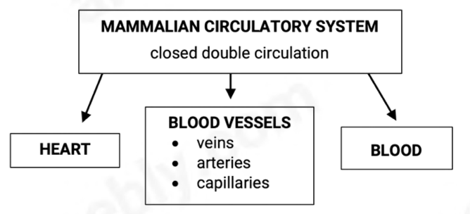
The circulatory system
It’s called closed circulation as the blood remains within blood vessels.
Double Circulation
DOUBLE CIRCULATION | |
SYSTEMIC CIRCULATION | PULMONARY CIRCULATION |
left ventricle → AORTA → body (except lungs) → VENACAVA | right ventricle → PULMONARY ARTERIES → lungs → PULMONARY VEIN→ left atrium |
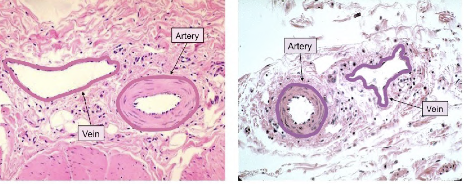
Detailed Diagram of veins and arteries
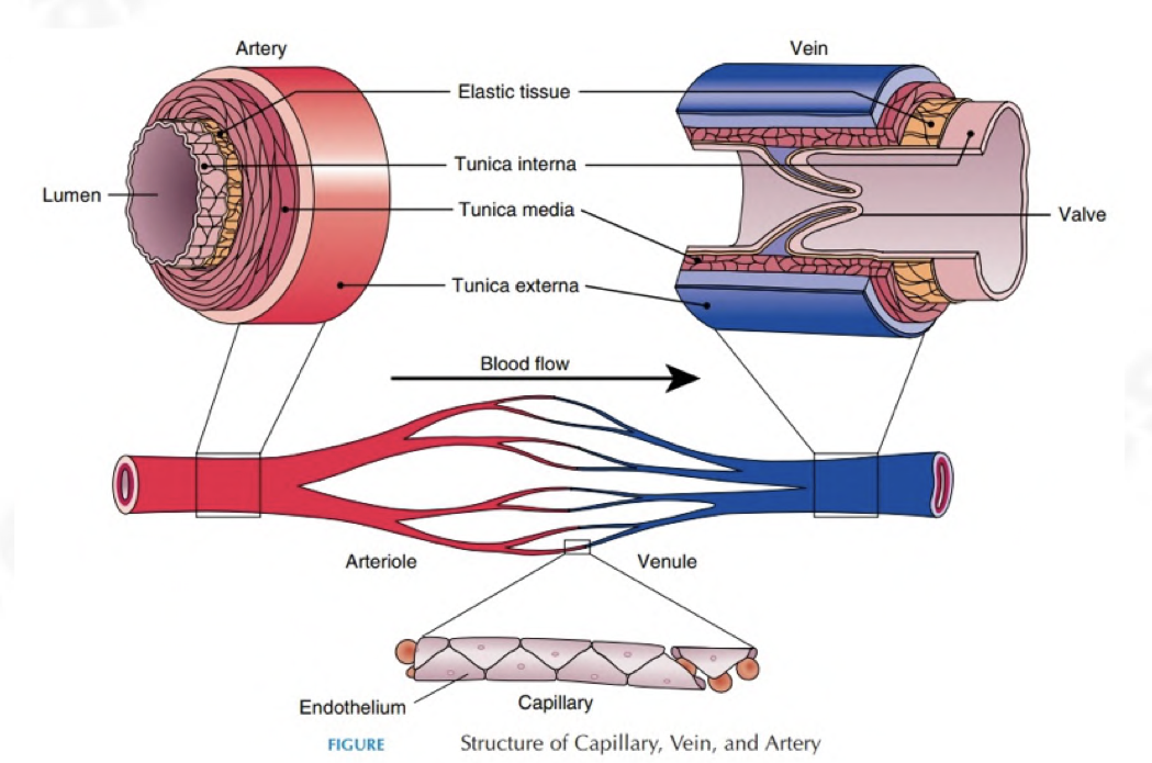
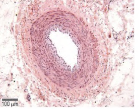
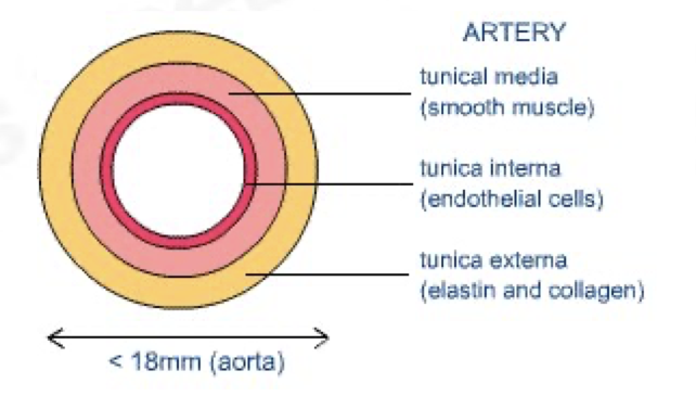
Outline the structre of the artery
transports oxygenated blood at high pressures to tissues
pulmonary artery and aorta have semilunar valves in the beginning
tunica intima/interna – very smooth, single layer of flat cells
tunica media – smooth muscle, collagen fibres, elastic fibres
tunica media contains large amounts of elastic fibres to allow the artery wall to stretch as blood surges through at high pressure
tunica media is the thickest in arteries
tunica externa – collagen fibres, elastic fibres
depending on the pressure, thickness of arteries’ walls
differs
artery wall can recoil inwards if the pressure drops
as blood at high pressure enters, it can widen,
reducing pressure slightly and vice versa
arteries branch out into arterioles
arteriole walls have more smooth muscle which can contract, narrowing the diameter and reducing blood flow
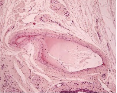
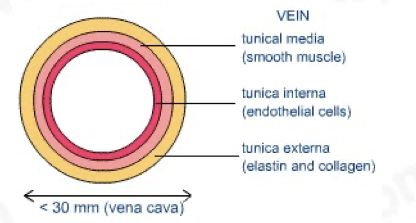
Outline the structre of the vein
tunica intima – flat cells, smooth and not crinkly
tunica media – smooth muscle, collagen, and elastic fibres, tunica media in veins is thin
tunica externa – elastic and collagen fibres, the thickest in veins
blood is transported at low pressures, no need for thick walls
contain semi-lunar valves (formed from their endothelium)
large lumen
irregular shape
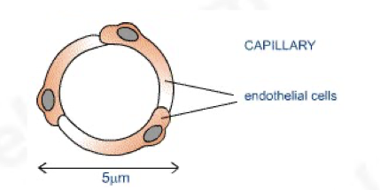
Outline the structure of capillaries
takes blood really close to cells allowing exchange of materials
network of capillaries is called the capillary bed
wall made of endothelial cells and is one cell thick
gaps are present between individual cells that form the endothelium
gaps allow some components of blood to seep through into spaces between cells (tissue fluid)
What are blood plasma & tissue fluid
as blood flows through capillaries within tissues, some plasma leaks out due to the pressure and seeps out into places between the cells of the tissues
this plasma that leaks out is called tissue fluid
if blood pressure is too high, too much fluid may be forced out of capillaries and the fluid may accumulate, this results in oedema
it’s through tissue fluid that the exchange between cells and blood occurs
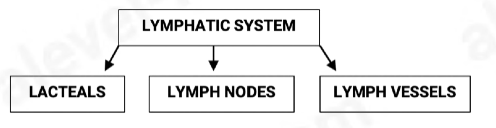
Outline the lymphatic system
it is the drainage system
it is digestive (assimilation of fatty acids)
immunity – produces lymphocytes
lymphatics are tiny, blind-ended vessels
they contain valves which allow tissue fluid to flow in but not out
walls are wide enough to allow larger protein molecules to pass through
fluid inside lymphatics is called lymph
lymph is transported to subclavian vein
lymph vessels have smooth muscle in their walls which contract to push lymph along
Outline the red blood cells (erythrocytes)
contain haemoglobin which gives red colour and transports oxygen
produced in the bone marrow
have a biconcave, disc shape – dent increases surface area in relation to volume
spongy and flexible – have specialised cytoskeleton made of protein filaments that allow them to be squashed
have no nucleus, endoplasmic reticulum, mitochondria – more space for haemoglobin, maximising amount of oxygen which can be carried
broken down in spleen
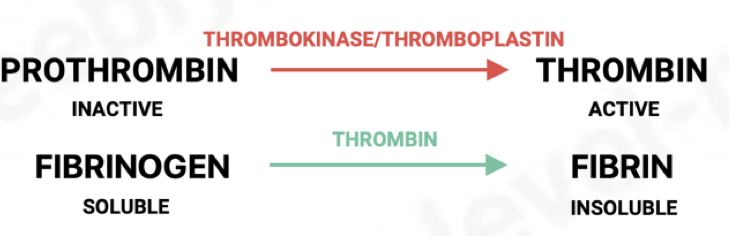
Outline the platelets (thrombocytes)
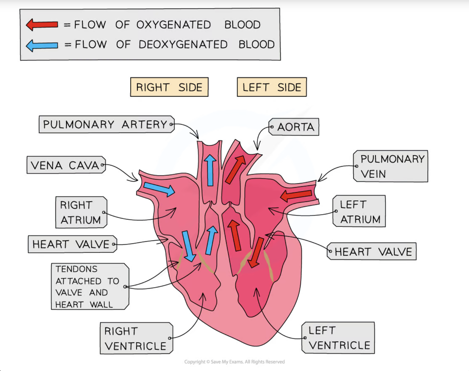
Outline the structre of the heart
consists of 2 atria and 2 ventricles
right and left side separated by septum
made of cardiac muscle
papillary muscles contract to pull on valve tendons to prevent inversion of valves during systole
atria and ventricles have valves between them called atrioventricular valves: RIGHT SIDE – TRICUSPID, LEFT SIDE – BICUSPID / MITRAL
2 types of valves: ATRIOVENTRICULAR – TENDONS, SEMI-LUNAR – POCKETS
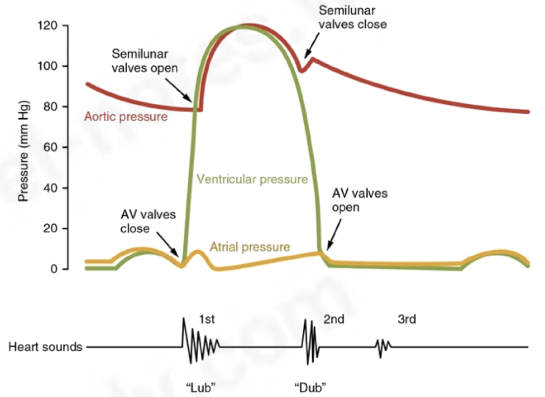
Outline the cardiac cycle
The cardiac cycle is the series of events that take place in one heart beat, including muscle contraction and relaxation
The contraction of the heart is called systole, while the relaxation of the heart is called diastole
One cardiac cycle is followed by another in a continuous process
There is no gap between cycles where blood stops flowing
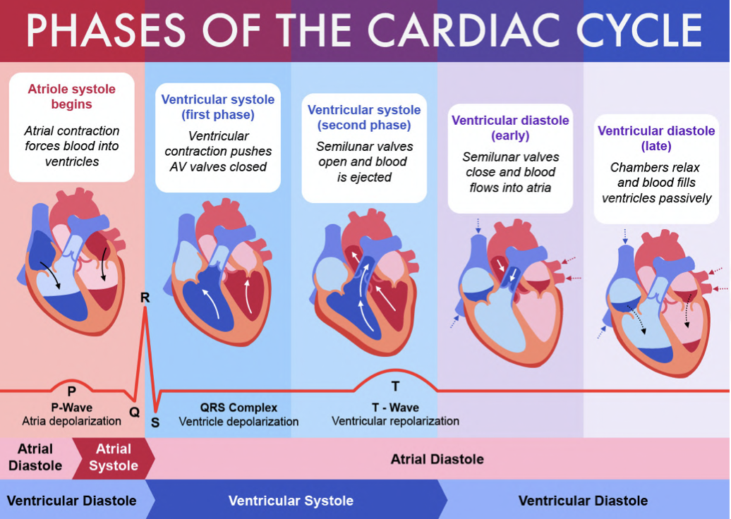
Outline the process of Atrial systole
The walls of the atria contract
Atrial volume decreases
Atrial pressure increases
The pressure in the atria rises above that in the ventricles, forcing the atrioventricular (AV) valves open
Blood is forced into the ventricles
There is a slight increase in ventricular pressure and chamber volume as the ventricles receive the blood from the atria
The ventricles are relaxed at this point; ventricular diastole coincides with atrial systole
Outline the process of Ventricular systole
occurs about 0.1s after atria contract
The walls of the ventricles contract
Ventricular volume decreases
Ventricular pressure increases
The pressure in the ventricles rises above that in the atria
This forces the AV valves to close, preventing back flow of blood
The pressure in the ventricles rises above that in the aorta and pulmonary artery
This forces the semilunar (SL) valves open so blood is forced into the arteries and out of the heart
During this period, the atria are relaxing; atrial diastole coincides with ventricular systole
The blood flow to the heart continues, so the relaxed atria begin to fill with blood again
Outline the process of Ventricular diastole
The ventricles and atria are both relaxed
The pressure in the ventricles drops below that in the aorta and pulmonary artery, forcing the SL valves to close
The atria continue to fill with blood
Blood returns to the heart via the vena cava and pulmonary vein
Pressure in the atria rises above that in the ventricles, forcing the AV valves open
Blood flows passively into the ventricles without need of atrial systole
The cycle then begins again with atrial systole
force produced in the right ventricle must be relatively small as –
blood goes only to the lungs which are at a shorter distance + less resistance to overcome
if a too-high pressure was developed, tissue fluid would accumulate in lungs hampering gas exchange
Outline the cardiac cycle
Cardiac muscles are myogenic which means it naturally contracts and relaxes without receiving impulses from a nerve.
SAN (sinoatrial node)/pacemaker sends out waves of excitation which stimulates atria to contract
non-conducting tissue between atria and ventricles prevents atria and ventricles from contracting at the same time
AVN (atrioventricular node) delaying the impulse allows it to flow from atria into ventricles
AVN sends an impulse down to the bundle of His and along purkine fibres
this causes ventricles to contract from the base upwards
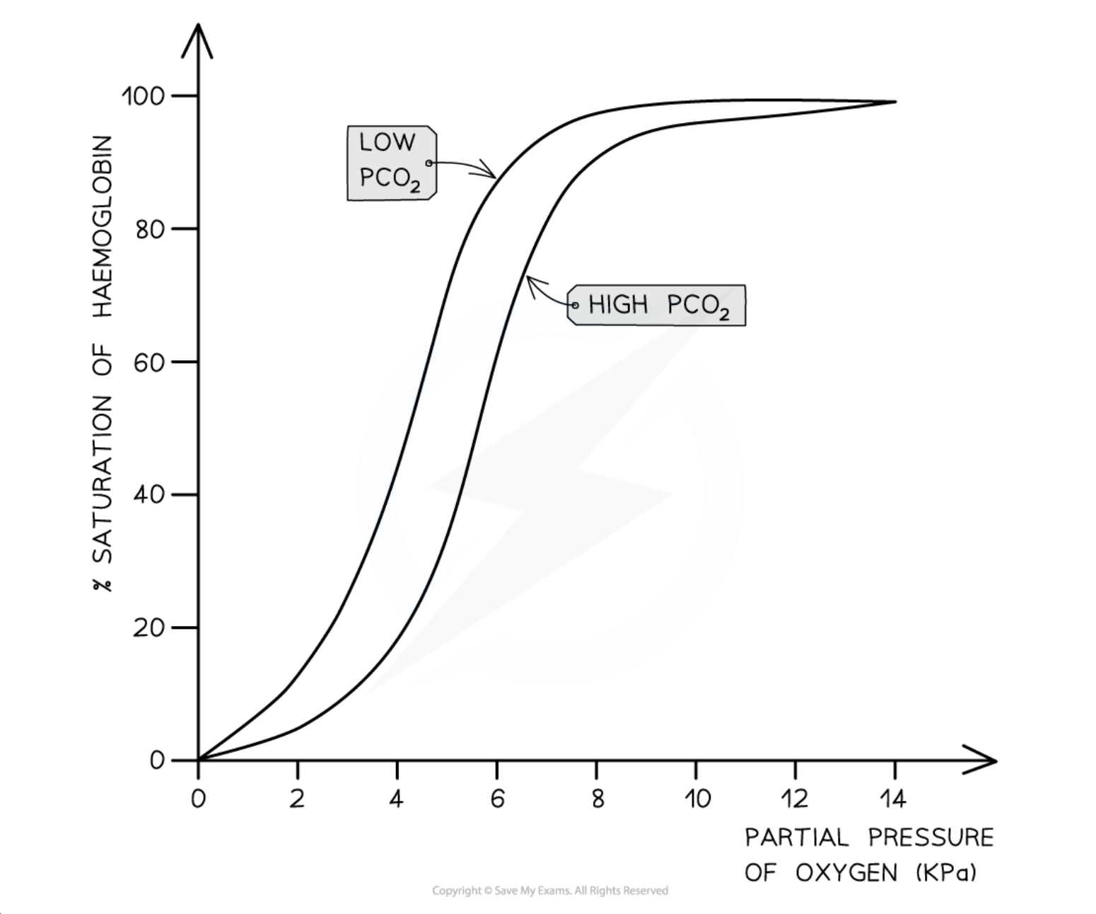
Outline the Oxygen dissociation curve
once an O2 molecule combines with haemoglobin, it becomes easier for more molecules to combine therefore, the curve rises very steeply
a small change in the partial pressure O2 causes a very large change in amount of O2 carried by haemoglobin
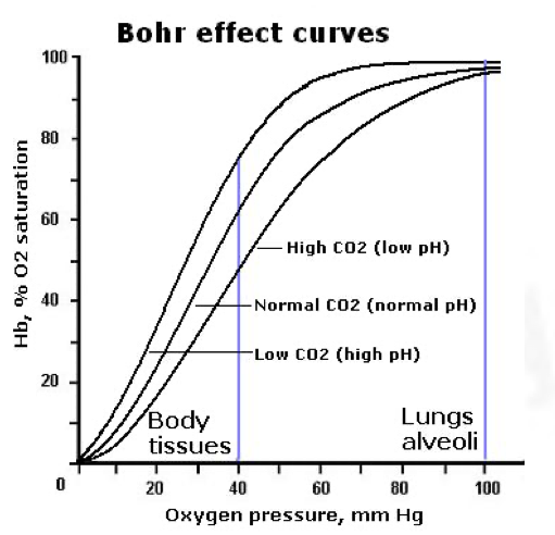
Outline the process of the Bohr shift
The shift in the curve of oxyhaemoglobin due to concentration of CO2 at a given partial pressure of O2 is the Bohr effect
the amount of O2 haemoglobin carries is affected by the partial pressures of both O2 and CO2
the presence of high partial pressure of CO2 causing Hb to release O2 is the Bohr’s effect
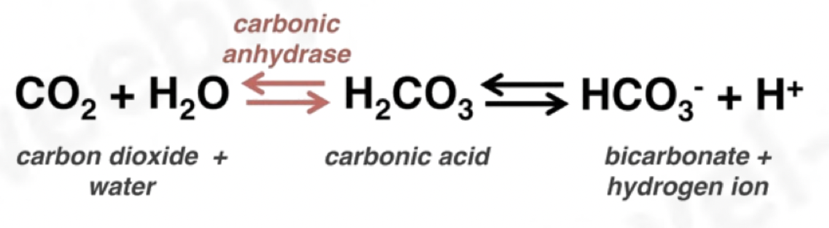
In the cytoplasm of red blood cells, CO2 is catalysed by carbonic anhydrase enzyme when it reacts with water to form carbonic acid (REFER TO DIAGRAM)
When the carbonic acid dissociates; haemoglobin combines with H+ ions forming haemoglobunic acid (HHb) and releases the O2 it’s carrying
Haemoglobin combining with H2 ions maintains blood pH as if the ions were left in solution, pH of the blood would’ve been less and turns acidic
presence of high partial pressures of CO2 causes haemoglobin to release O2
high concentration of O2 is found in respiring tissues which need O2
high concentration of CO2 causes Hb to release O2, curve lies below and to the right
85% of CO2 – diffuses out of RBC into blood plasma and are carried in solution
5% of CO2 – CO2 that hasn’t dissociated and remains as CO2 dissolves in blood plasma
10% of CO2 – CO2 diffuses to RBC and combines directly with amine groups (–NH2) of some haemoglobin molecules forming carbaminohaemoglobin
What is formed when oxygen binds with haemoglobin?
Oxygen + Haemoglobin ⇌ Oxyhaemoglobin
4O2 + Hb ⇌ Hb4O 2
The binding of the first oxygen molecule results in a conformational change in the structure of the haemoglobin molecule, making it easier for each successive oxygen molecule to bind; this is cooperative binding
The reverse of this process happens when oxygen dissociates in the tissues
Outline the Chloride shift
The movement of chloride ions into red blood cells that occurs when hydrogen carbonate ions are formed
How are hydrogen carbonate ions formed?
Carbon dioxide diffuses into red blood cells
The enzyme carbonic anhydrase catalyses the combining of carbon dioxide and water to form carbonic acid (H2CO3)
CO2 + H2O ⇌ H2CO3
Carbonic acid dissociates to form hydrogen carbonate ions and hydrogen ions
H2CO3 ⇌ HCO3- + H+
What happens to negatively charged hydrogencarbonate ions
Negatively charged hydrogencarbonate ions formed from the dissociation of carbonic acid are transported out of red blood cells via a transport protein in the membrane
To prevent an electrical imbalance, negatively charged chloride ions are transported into the red blood cells via the same transport protein
If this did not occur then red blood cells would become positively charged as a result of a build up of hydrogen ions formed from the dissociation of carbonic acid
How and where is waste carbon dioxide excreted?
Waste carbon dioxide produced during respiration diffuses from the tissues into the blood
This waste carbon dioxide is transported around the body in different ways
In the blood plasma in the form of hydrogen carbonate ions (HCO3-); around 85 % of carbon dioxide is transported in this way
Around 5 % of carbon dioxide dissolves directly in the blood plasma
Bound to haemoglobin as carbaminohaemoglobin; this accounts for around 10 % of carbon dioxide transport in the blood
Outline Carbon Dioxide in the plasma
Carbon dioxide released as a waste product from respiring cells diffuses into the cytoplasm of red blood cells
Inside red blood cells, carbon dioxide combines with water to form H2CO3
CO2 + H2O ⇌ H2CO3
Red blood cells contain the enzyme carbonic anhydrase which catalyses the reaction between carbon dioxide and water
Without carbonic anhydrase this reaction proceeds very slowly
The plasma contains very little carbonic anhydrase hence H2CO3 forms more slowly in plasma than in the cytoplasm of red blood cells
Carbonic acid dissociates readily into hydrogen ions (H+) and hydrogen carbonate ions (HCO3-)
H2CO3 ⇌ HCO3– + H+
Hydrogen ions can combine with haemoglobin, forming haemoglobinic acid and preventing the H+ ions from lowering the pH of the red blood cell
Haemoglobin is said to act as a buffer in this situation
The hydrogen carbonate ions diffuse out of the red blood cells into the plasma to be transported in solution