1019DOH - Applied Oral Biology (HEAD AND NECK ANATOMY)
1/313
Earn XP
Description and Tags
part 2 of the oral biology flashcard series! please study these unshuffled:) happy studying
Name | Mastery | Learn | Test | Matching | Spaced | Call with Kai |
|---|
No analytics yet
Send a link to your students to track their progress
314 Terms
the CNS (central nervous system) begins developing when the embryo is __1__ WIU and at the ___2__ end of the embryo
note: central nervous system will be referred to as CNS from this point onwards
4
head
the CNS development in the embryo begins as a thickening in the ectoderm in the…
neural plate
flip card over for visualiser of CNS embryonic development
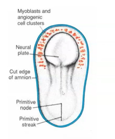
at 4 WIU, how long is the embryo?
3-4mm long
describe the embryonic development of the CNS neural tube
the edges of the neural plate begin to thicken, producing raised margins to the structure. between these margins lies the neural groove
neural folds continue to develop until they meet on top of the neural fold to produce a neural tube.
this will become the ventricles and central canal of the nervous system
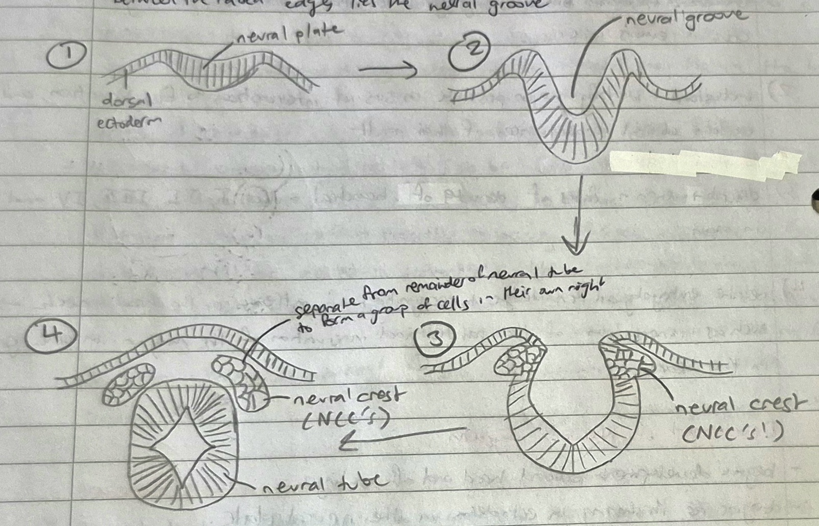
what does the neural tube eventually become?
the spinal cord
during development of the CNS, when do neural crest cells (NCC’s) form?
form after the fusion of the 2 edges of the neural folds

after formation of the neural tube, the NCC’s give rise to…
embryonic connective tissue of the facial region and to branchial arch cartilage
what is ectomesenchyme?
the name for the embryonic connective tissue given rise to by the NCC’s.
called ectomesenchyme to differentiate it from the mesenchyme produced at the primitive streak (think back to the visualiser card)
called ‘ecto’ to account for ectodermal origin of NCC’s - not pure connective tissue anymore
why is ectomesenchyme regarded as the 4th germ layer by some authors?
it influences the structures of the oral cavity and human body
has properties slightly different to mesenchymal structures elsewhere in the body
eg) bones of face and cranium (except base of skull) form by intravenous ossification rather than the endochondral method common in other bones
of 6 branchial arches, how many survive in man?
5
as the first branchial arch is lined by ectoderm on the inside and outside, at which branchial arch does the inner lining of endoderm start?
arch #2
each branchial arch contains:
portion of primitive striated muscle tissue
some nervous tissue from the neural crest
some vascular tissue
bar of cartilage in mesodermal core
∴ each arch contains an artery and a nerve
branchial arches support the…
lateral wall of the primitive pharynx
the first branchial arch (mandibular arch) forms the mandibular division of the ________ nerve
trigeminal
3 divisions of the trigeminal nerve
opthalmic
maxillary
mandibular
how do bones of the maxilla and mandible form (as they don’t form from remnants of Meckel’s cartilage?
from intramembranous ossification following degeneration of the cartilage
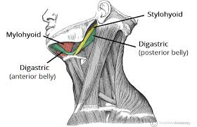
muscle tissue from the first branchial arch forms what 4 structures?
where do these structures get their motor nerve supply from?
masticatory muscles
tensor tympani + tensor veli tympani
mylohyoid muscle
anterior belly of the digastric
motor nerve supply of these four structures are from branches of the mandibular division/nerve of the trigeminal nerve
does the artery of the first branchial arch survive?
no
the maxillary and mandibular nerve are both supplied by which artery for blood?
the maxillary artery
the 2nd branchial arch (the hyoid arch) develops around the future _____ bone
hyoid
what is the cartilage in the second (hyoid) arch called?
Reichert’s cartilage (after the anatomist who first described it)
the muscles of facial expression all share a common…
nerve supply - the facial nerve
muscle tissue of the second branchial arch forms…
stapedius muscle
stylohyoid muscle
posterior belly of the digastric
the face develops from _ embryonic tissue masses, also known as _____
5
processes
name the 5 processes for facial development
frontonasal process (1 part)
maxillary process (2 parts)
mandibular process (2 parts)
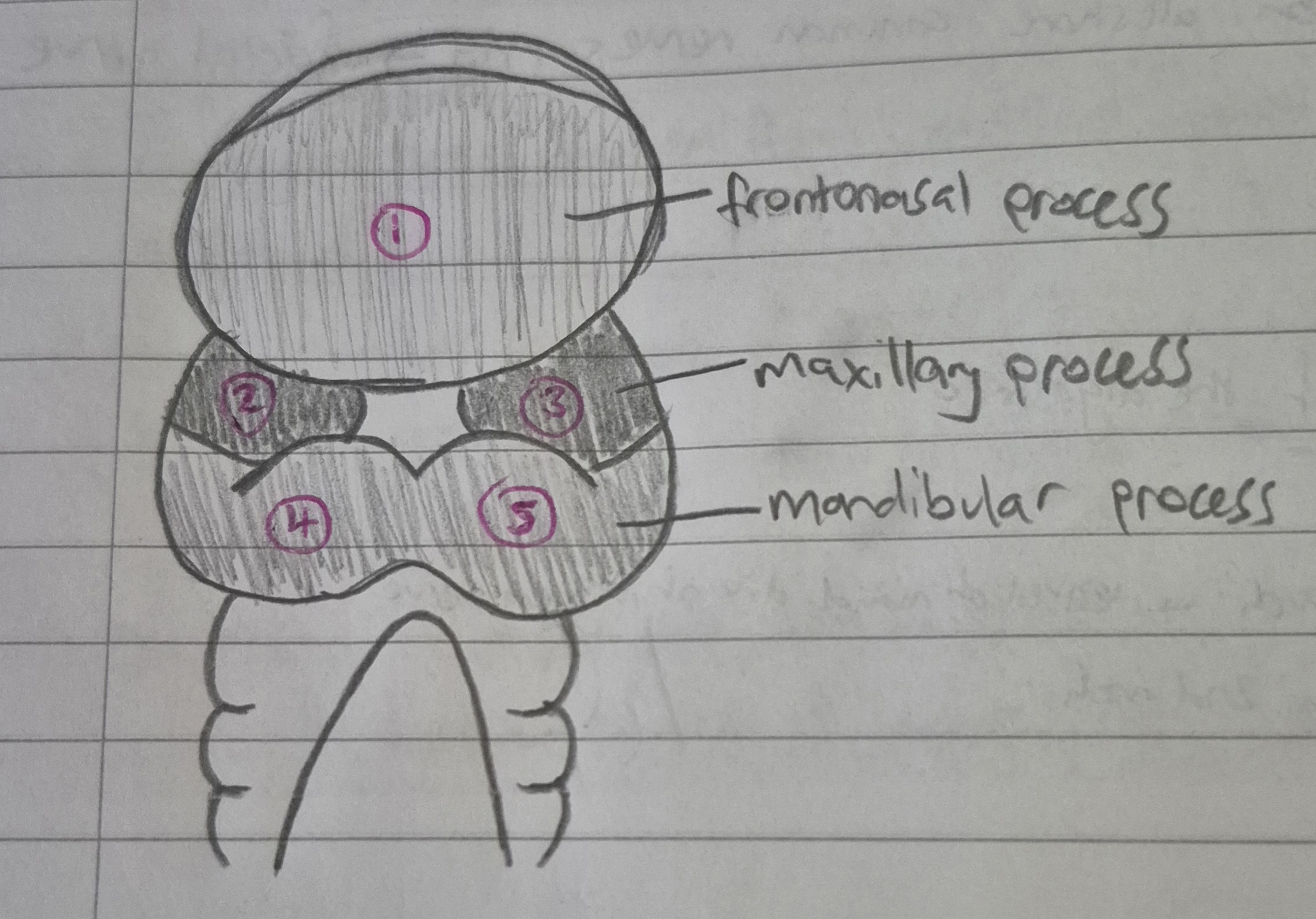
all processes (embryonic tissue masses) all arise by what?
rapid multiplication of NCC’s (neural crest cells) which originate from ectoderm
the 5 processes split to form how many processes that contribute to facial development?
7
3 processes (4 parts) that make up the lips
MIDDLE OF UPPER LIP: frontonasal process
LATERAL PARTS OF UPPER LIP: maxillary processes
LOWER LIP (all): mandibular process
the stomodeum/stomatodeum appears as a ___1___ on the embryonic surface at around _2_ weeks of development. the stomodeum is formed around the same time as the _3_
depression
4
CNS
the floor of the stomodeum (as it appears as a depression) pushes against what? what is the wall that separates these two structures called? what does it represent?
the floor of the stomodeum pushes against the developing gut
the wall that separates these two is called the buccopharangeal membrane
represents meeting of ectoderm and endoderm
why does the buccopharangeal membrane need to be broken?
so that direct connection/access between mouth and guts can be established
the stomodeum can then open directly into the primitive pharynx of the foregut
the face develops between the ___th and the ____th day of gestation
24th, 38th
in the early stages, face development is dominated by changes that create the….
from which processes do these changes occur from?
primitive nasal cavities
nasal pits (primitive nostrils), medial and lateral nasal processes from the frontonasal processes
eventually, the maxillary processes will push the __1___ and __2___ processes together, so there will be _3__ medial nasal process/es and __4_ lateral nasal process/es
medial
lateral
1 process
2 processes
think back to the card that said the 5 processes split into 7…
which process splits so this can happen?
the frontonasal process
(frontonasal → 2 lateral and 1 medial process)
maxillary (2 parts)
mandibular (2 parts)
altogether, that’s 7 parts
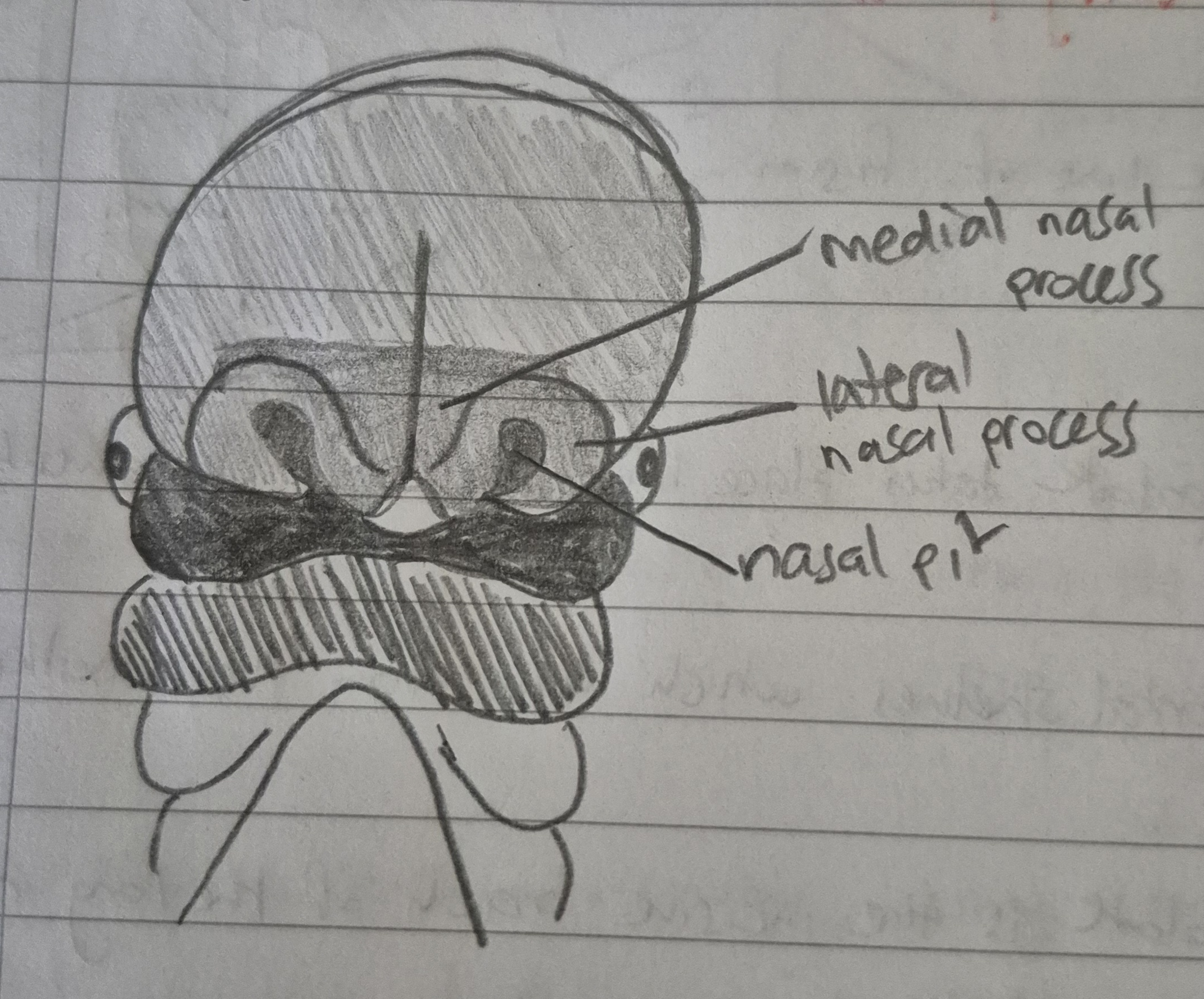
the frontonasal process develops 2 ___ (1)____. tissue builds up around them in a horseshoe shape to form the ___(2)___ and ____(3)_____ nasal processes.
nasal pits
lateral
medial
the maxillary process grows medially and approaches the lateral and medial nasal processes. at this stage, it is separated from them by a _____
groove
the continued central growth of the _____ process pushes the ______ nasal process towards the midline
maxillary
medial
from the process of pushing the medial nasal processes together, they fuse to create….
the middle part of the nose
the middle part of the upper lip
the anterior part of the maxilla
the primary palate
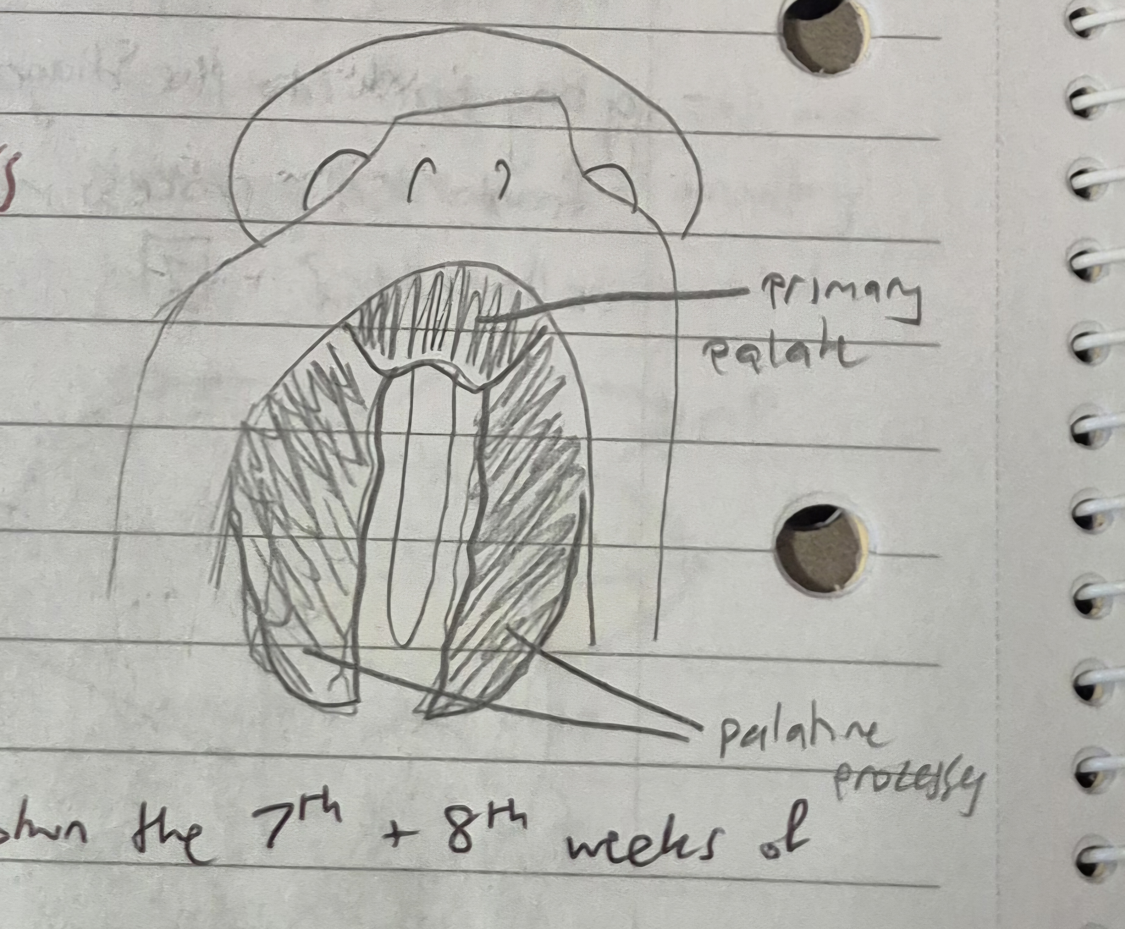
the primary palate is formed by what? what does the primary palate carry? what else can you refer this structure to as?
formed by horizontal shelves which fuse together
carries odontogenic epithelium for maxillary incisors
can refer to as ‘pre-maxilla’
what does the secondary palate form as a result of?
the fusion of palatine process (horizontal shelves from the maxillary process)
end up with Y-shape fusion
the primary palate is formed as a bonus/extra together during the facial development and fusion of the 2 ____ ____ processes into 1
medial nasal
the secondary palate will form as a result of the fusion of the _______ _______. these processes are horizontal shelves extending from the _____ ______. the end result will be a _-shape line of fusion.
palatine process
maxillary processes
Y

only after the formation of WHAT can the distinction between the oral and nasal cavities be seen?
the secondary palate
formation of the secondary palate takes place between the ___th and ___th weeks of development
7th
8th
the nerve of the primary palate is the….
(i broke the answer into bits because reading it all in one sentence makes my head spin ahah)
incisive branch of the
long nasopalatine branch of the
maxillary division of the
trigeminal nerve
the nerve of the secondary palate is the…
greater palatine branch of the
maxillary division of V
what is the tuberculum impar?
a mesenchymal swelling in the midline of the mandibular process of the first branchial arch
how many other swellings appear either side of the tuberculum impar, and what do these swellings do?
2 other swellings appear
these enlarge rapidly and merge with each other and with the tuberculum impar to cover it and form a large mass
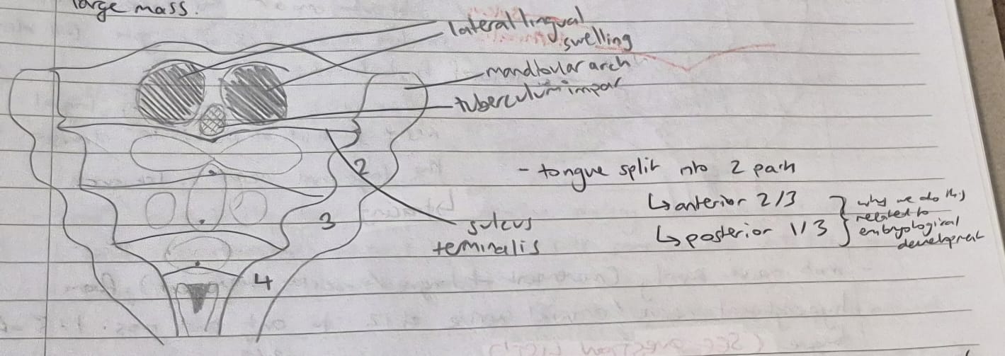
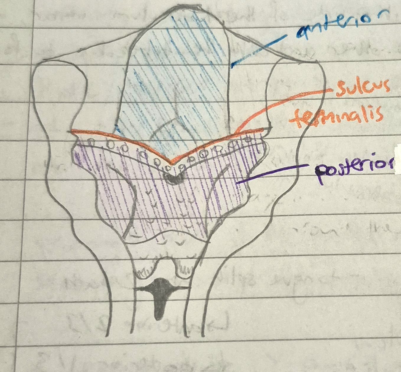
the tongue, during its development, has elements from which arches? what structures do these arches contribute to?
elements from first, second and third arches
FIRST ARCH
formation of anterior 2/3rds of the tongue
SECOND ARCH
eventually disappear and get overpowered by the 1st and 3rd branchial arches
mainly contribute to taste sensation
smaller elements compared to 1st and 3rd branchial arches
THIRD ARCH
posterior 1/3rd of the tongue
all the tongue’s cell divisions, multiplications and swellings are all due to the…
NCC’s
what is median rhomboid glossitis?
where tuberculum impar isn’t fully covered (incomplete fusion of 2 lateral swellings)
results in rhomboidal shape on dorsal surface of the tongue at the junction of the anterior 2/3rds and the posterior 1/3rd
congenital condition
once lateral swellings fully cover the tuberculum impar, what is formed? what branch is this formed from? what is the nerve supply for the structure formed?
the mucous membrane structures of the anterior 2/3rds of the tongue is formed
all elements of the first branchial arch
all supplied by lingual nerve (branch of mandibular division of trigeminal nerve → 1st arch)

the anterior 2/3rds and the posterior 1/3rd of the tongue gets its motor nerve supply from…
what muscles does this motor nerve supply for?
the hyperglossal nerve (cranial nerve #12)
supply for all intrinsic and extrinsic muscles of the tongue except for the palatoglossus muscle
sensory sensation, special sensation and motor sensation of the tongue definitions
SENSORY
temperature
pain
pressure
touch
SPECIAL
taste sensation
MOTOR
contraction of muscles
tongue movement
the nerve of the second arch contributes _____ _______ to the anterior 2/3rds of the tongue via the _____ _______
taste fibres (for taste sensation)
chorda tympani
what is the chorda tympani?
branch of the facial nerve
nerve supply for the second branchial arch
the posterior part of the tongue arises from a __________(1)_ __________ (a large midline swelling in the 3rd branchial arch).
the nerve of which becomes the ______(2)_______ nerve (cranial nerve #_(3)__). this is the main nerve supply for the __(4)_rd branchial arch.
the posterior part of the tongue comes to be supplied by _(5)___, and develops from __(6)__ piece, unlike the anterior 2/3rds which develop from __(7)__ pieces.
hypobrachial eminence
glossopharangeal
9
3 (3rd)
IX
1 (1 piece)
3 (3 pieces)
what is the sulcus terminalis?
fusion of the anterior and posterior parts of the tongue
V-shape line
what is the last tissue to develop during embryological life?
bone
list the order that tissues form during embryological life?
nerves
blood vessels
some muscles (fill in spaces between first two structures)
bone (will form tubes, lamina, canals etc around vessels)
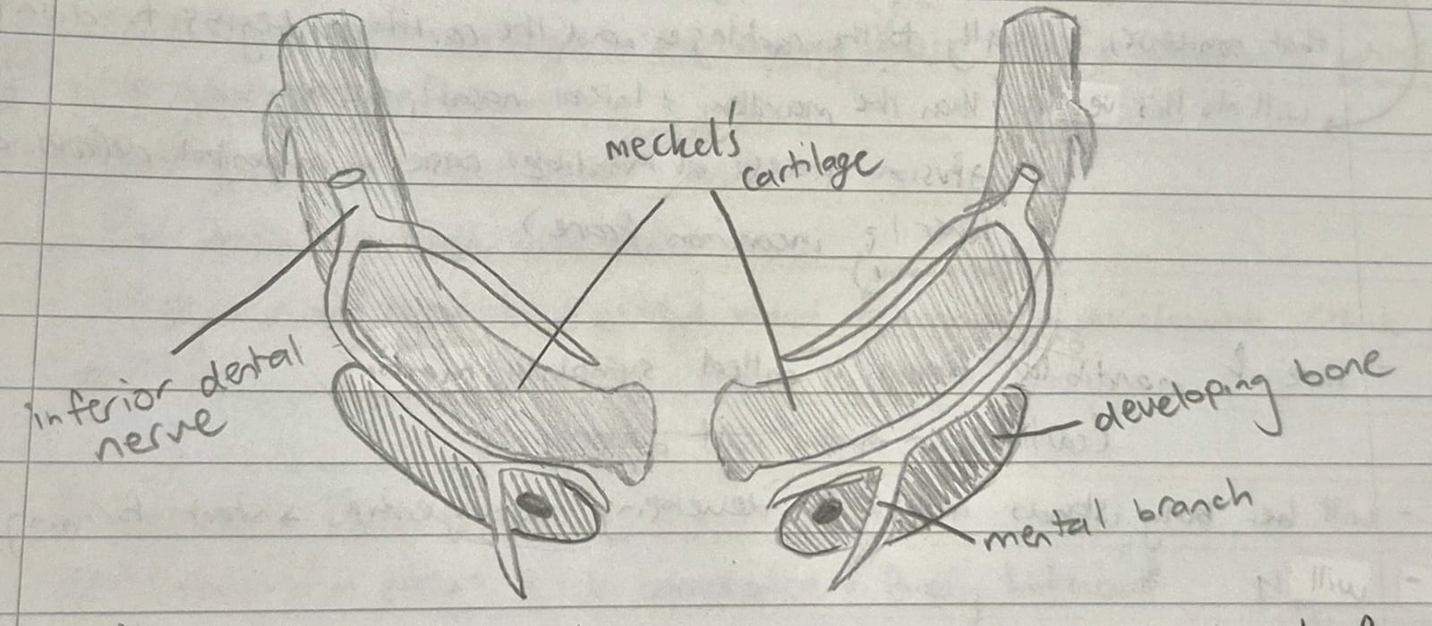
how does the mandible form?
under influence of NCC’s, the 2 mandibular processes fuse in the midline to form a mandibular arch
line of fusion is called symphisis menti
the bone of the mandible begins to form in the mesenchymal tissue that condenses laterally to the cartilage
the cartilage (Meckel’s) begins to disappear
there will be bony islands around the developing tooth germs (start forming the crypts)
~ link between oral histology and head and neck anatomy ~
link the formation of the mandibular crypts to the tooth germ
in the tooth germ: enamel organ, dental papilla and dental sac
from dental sac, there will be mesenchymal condensations from developing tooth germs (bits of bone fuse and form the crypt where developing tooth germs sit)
once the tooth erupts, the crypts turn into sockets (accommodating for roots)
by __ weeks of age, the rudimentary mandible is formed almost entirely from ____________ __________ with little direct involvement from _______ _______.
10
intramembranous ossification
Meckel’s cartilage
although Meckel’s cartilage does not contribute much to mandible formation, what structures’ development does it contribute to?
malleus of the ear and its ligament
sphenomandibular ligament
the maxilla also develops from a ________ of ______ from the ____ branchial arch
condensation
mesenchyme
first
is there cartilage present in the maxillary process?
what is bone formation made entirely by?
no cartilage
made entirely by intramembranous ossification
when does the maxillary sinus form?
in the 16th week
do the mandible and the maxilla develop at the same time? if not, which develops earlier?
not at the same time
mandible always ahead of maxilla in development
think about eruption dates of teeth!
what are congenital defects?
malfunctions in the development of a person
defects can be genetically or environmentally caused
list the 5 groups of environmental factors that affect the embryo
infectious agents
ionising radiation
drugs
hormones
nutritional deficiences
they could exaggerate an existing genetic factor, or trigger failure in a step of embryological development
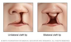
________ clefts are among the most common congential defects
orofacial
what is the difference between a complete and an incomplete cleft lip?
complete extends into the nasal cavity
incomplete does not
what is a unilateral cleft upper lip?
the failure/incomplete fusion of the maxillary process and the lateral nasal processes
can be uni or bilateral
can never have a midline cleft, however, because 1 solid piece of medial nasal process in the middle
are…
unilateral or bilateral cleft lips more common?
left or right unilateral cleft lips more common? why?
unilateral
left - defects tend to occur in the nondominant side of the individual, as there are more right-handed people globally, unilateral left is more common
can cleft palates and cleft lips occur together?
yes
what is a cleft palate?
the 2 palatine processes fail to fuse together
if a lower lip cleft lip occurs (as its very rare), where would it be present on on the lip?
in the midline
describe the TMJ’s structure
bilateral synovial joint
site of articulation between the mandible and the cranium
serves to open and close the jaws and approximate teeth of each jaw during mastication
what does TMJ stand for?
temporomandibular joint
what is the articular mandibular/glenoid fossa?
the fossa at the base of the temporal bone
the condyle of the mandible sits here
why can the TMJ be correlated with the knee joint?
bilateral
synovial
ball and socket joint
what does elevating the mandible do?
close the jaws
between the two bones of the TMJ is the ________ _____ ____, breaking the joint into 2 separate synovial-lined compartments.
fibrous articular disc
what are the muscles of mastication and the accessory muscles of mastication?
the several pairs of muscles attached to the mandible that produce movements required to suckle, ingest and masticate food, swallow, yawn and produce speech
one has direct attachment to the TMJ components, but the other muscles are just close enough to the TMJ and are responsible for movements in different directions
the mandible possesses 2 articular surfaces, what are they and where are they located? what do they articulate with?
the condyles
located on the upper end of each of the bilateral condylar processes
articulate with a meniscus (articular disc) which lies between it and the temporal bone
how many compartments does the TMJ have? what are they called?
2 compartments
upper and lower compartment
what happens to the condyle and articular disc during jaw movement, and what are the consequences if the movement is not properly limited?
between condyle and articular disc
condyle will slightly rotate against the articular disc and push it towards the upper compartment
when they reach the upper compartment, bodily movement occurs
however, movement must be limited - otherwise if the condyle goes past the eminence, you get a dislocation
the rotated condule and disc will move downwards and forwards (or anteriorly)
what shape are the condyles? what does their angle ensure?
football shaped
their oblique angle ensures that if the planes of the long axes are combined they would meet at the foramen magnum
describe the articular disc
compact, dense fibrous connective tissue (fibrocartilage) plate
roughly oval in shape
lies between mandibular condyle and articular eminence of the temporal bone
uneven thickness because it follows the outline of the fossa/eminence
inferior surface concave to fit convex surface of the condyle
superiorly, the surface is concavo-convex
thickest at periphery, thinnest in stress-bearing part of the joint
what are the coverings of the articular surfaces of the condyle and articular eminence composed of?
fibrous connective tissue
how do the fibrocartilaginous structures of the TMJ get nutrients if the structures are all essentially avascular?
they are bathed in synovial fluid which provides nourishment as well as lubrication
what are the two types of ligaments around the TMJ?
true ligaments
accessory ligaments
ligaments that are very close to or directly attached to the TMJ are ______ ligaments, whereas ligaments further away from the TMJ are _______ ligaments
true
accessory
which ligaments of the TMJ are stronger, true or accessory?
true
by which 2 ways could you identify if a ligament is true or false?
can do by location (closer = true, further = accessory)
can do by strength (embracing joint + strong = true, thickening of sheets/rely on stretch receptors = accessory)
the ligaments of the TMJ joint comprises of the _____ ______ and the ________ and _______ _________
joint capsule
medial and lateral ligaments
describe the joint capsule
dense fibrous tissue on the outside of the joint
dense, strong and very close to the TMJ = true ligament
describe the TMJ ligament
runs a figure 8 around the joint in all directions
adds a layer of protection