BIOLOGY- AS Level- 3- exchange and transport
1/51
There's no tags or description
Looks like no tags are added yet.
Name | Mastery | Learn | Test | Matching | Spaced |
|---|
No study sessions yet.
52 Terms
Why do organisms need exchange?
Organisms must take in food, oxygen and water, and other essential substances, from the environment as well as the need to remove waste substances.
What is the difference between exchange in multi and single cellular organisms?
In single-celled organisms, substances diffuse directly across the cell membrane while in multi-celled organisms they need exchange surfaces to get the essential nutrients they need
Why do multi-celled organisms need exchange surfaces?
Cells are not in direct contact with the external environment.
Diffusion distances between cells and their environment are large.
Larger organisms have higher metabolic rates, so they need more oxygen and glucose.
What are features of specialised exchange surfaces?
A large surface area - This provides a larger area across which substances can be exchanged
Thin walls - These minimise the diffusion distance
An extensive blood supply and/or ventilation - This maintains steep concentration gradients
Being surrounded by selectively permeable plasma membranes - This controls what substances are exchanged
What are 3 examples of specialised cells?
Root hair cells- large surface area
Alveoli- thin layer
Gills- good blood supply/ ventalation to maintain a good gradient
What is the order in which air passes through the human respiratory system?
Nasal passage/ Mouth cavity, Trachea, Bronchi, Bronchioles, Alveoli
What is the ciliated epithelium?
A layer of cells which have cilia
What is the function of cilia on the ciliated epithelium?
These waft the mucus, containing dust and micro organisms upward to the mouth so it can be swallowed and destroyed by the stomach acid
Where are goblet cells found?
In the ciliated epithelium
What are the function of goblet cells?
Cells that secrete mucus
What is the function of mucus?
To trap dust and microorganisms
What is the trachea?
A large tube that carries air from the throat down to the lungs
What are the adaptations of the trachea?
Rings of cartilage- to keep the airway open
Smooth muscle- which can contract or relax to constrict or dilate the airway and change airflow
Elastic tissue- which contains elastic fibres with elastin that allows stretching and recoiling
Lined with ciliated epithelial cells and goblet cells
What is the bronchi?
Two main branches extending from the trachea that carry air into each lung
What are the adaptations of the bronchi?
Reinforced with cartilage- which keeps the airways open
Smooth muscle- which can contract or relax to constrict or dilate the airway and change airflow
Elastic tissue- which contains elastic fibres with elastin that allows stretching and recoiling
Lined with ciliated epithelial cells and goblet cells
What are the bronchioles?
Smaller airways branching from the bronchi that carry air to the alveoli
What are the adaptations of the bronchioles?
No cartilage- so they can change shape
Smooth muscle- which can contract or relax to constrict or dilate the airway and change airflow
Elastic tissue- which contains elastic fibres with elastin that allows stretching and recoiling
Simple squamous epithelium (only larger bronchioles have a ciliated epithelium)
What are alveoli?
Tiny air sacs clustered at the ends of the bronchioles, they are folded to increase surface area
What is the structure of alveoli?
Alveoli are tiny air sacs which are clustered at the end of bronchioles, their walls are made of squamous epithelial cells and are surrounded by a network of capillaries so gases can be exchanged efficiently
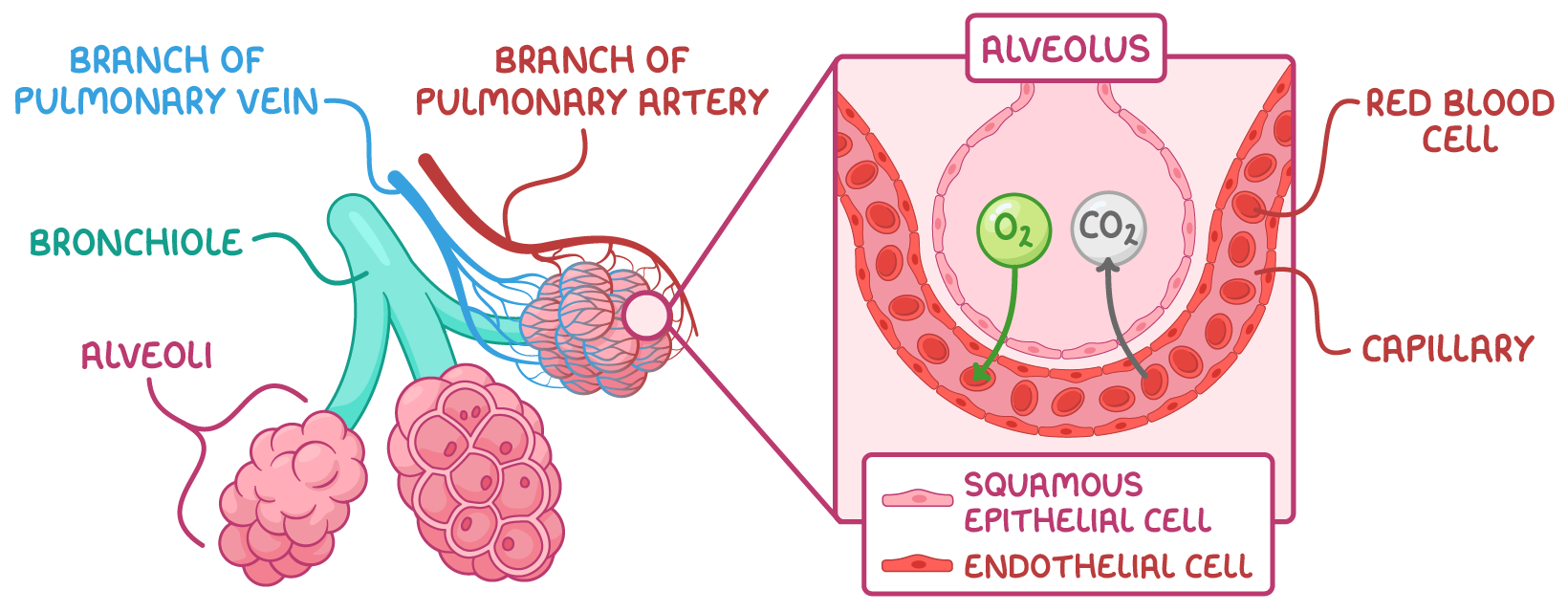
How do alveoli carry out gas exchange?
Oxygen diffuses from the alveoli into the pulmonary capillaries where it binds to haemoglobin in red blood cells
Carbon dioxide dissociates from haemoglobin and diffuses from the blood into the alveoli
What are the the three pulmonary blood vessels involved in the circulation of the lungs and hat are their functions?
The pulmonary artery - This delivers deoxygenated blood from heart to pulmonary capillaries.
The pulmonary vein - This delivers oxygenated blood from capillaries to heart.
The pulmonary capillaries - These are the site of gas exchange between blood and alveoli.
What are the adaptations of the pulmonary capillaries for gas exchange?
Thin walls (one endothelial cell thick) - This maintains a short diffusion distance
Red blood cells pressed against capillary walls - This reduces diffusion distance
Large surface area - This increases diffusion speed
Movement of blood - This maintains steep diffusion gradient
Slow blood movement - This allows more time for diffusion
What is ventilation?
The constant movement of air into and out of the lungs. It consists of inspiration (breathing in) and expiration (breathing out).
What is the structure of the ribcage?
The ribcage is made up of bones called ribs which enclose the thorax and the thoracic cavity, where the lungs are located. There are three different muscles attached to the ribcage which control ventilation- the diaphragm and the internal and external intercostal muscles.
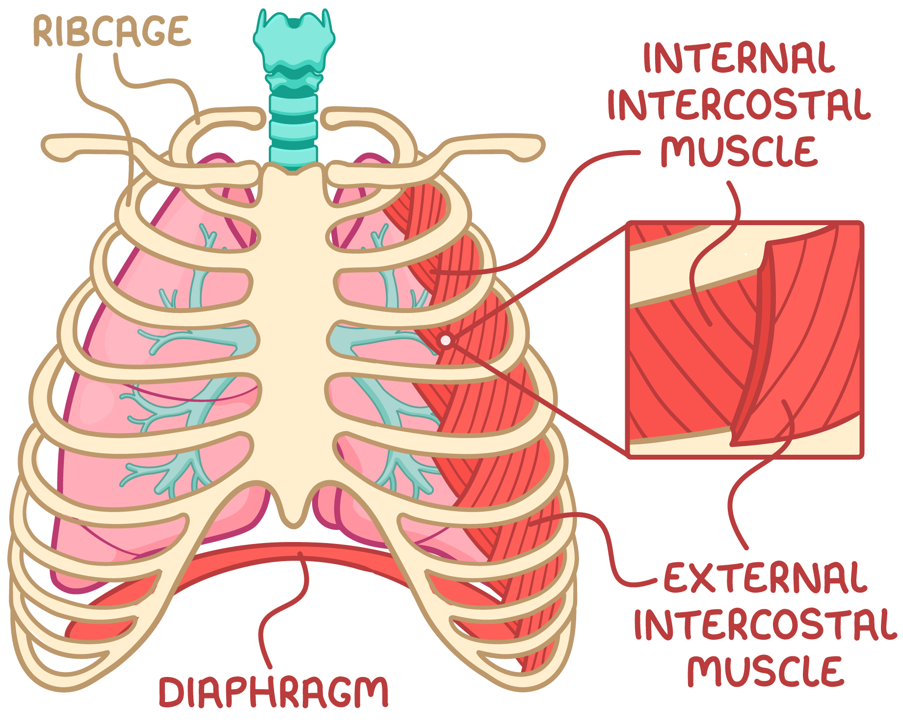
what are the three muscles which act on the ribcage?
The diaphragm - This is a sheet of muscle that moves the ribcage up and out when it contracts
The external intercostal muscles - These are found between the ribs and pull the ribcage up and out when they contract
The internal intercostal muscles - These are found between the ribs but pull the ribcage down and in when they contract
How do the internal and external intercostal muscles work during respiration?
They have opposite effects on the ribcage.
The external muscles expand the ribcage during inspiration, while the internal muscles shrink it during expiration.
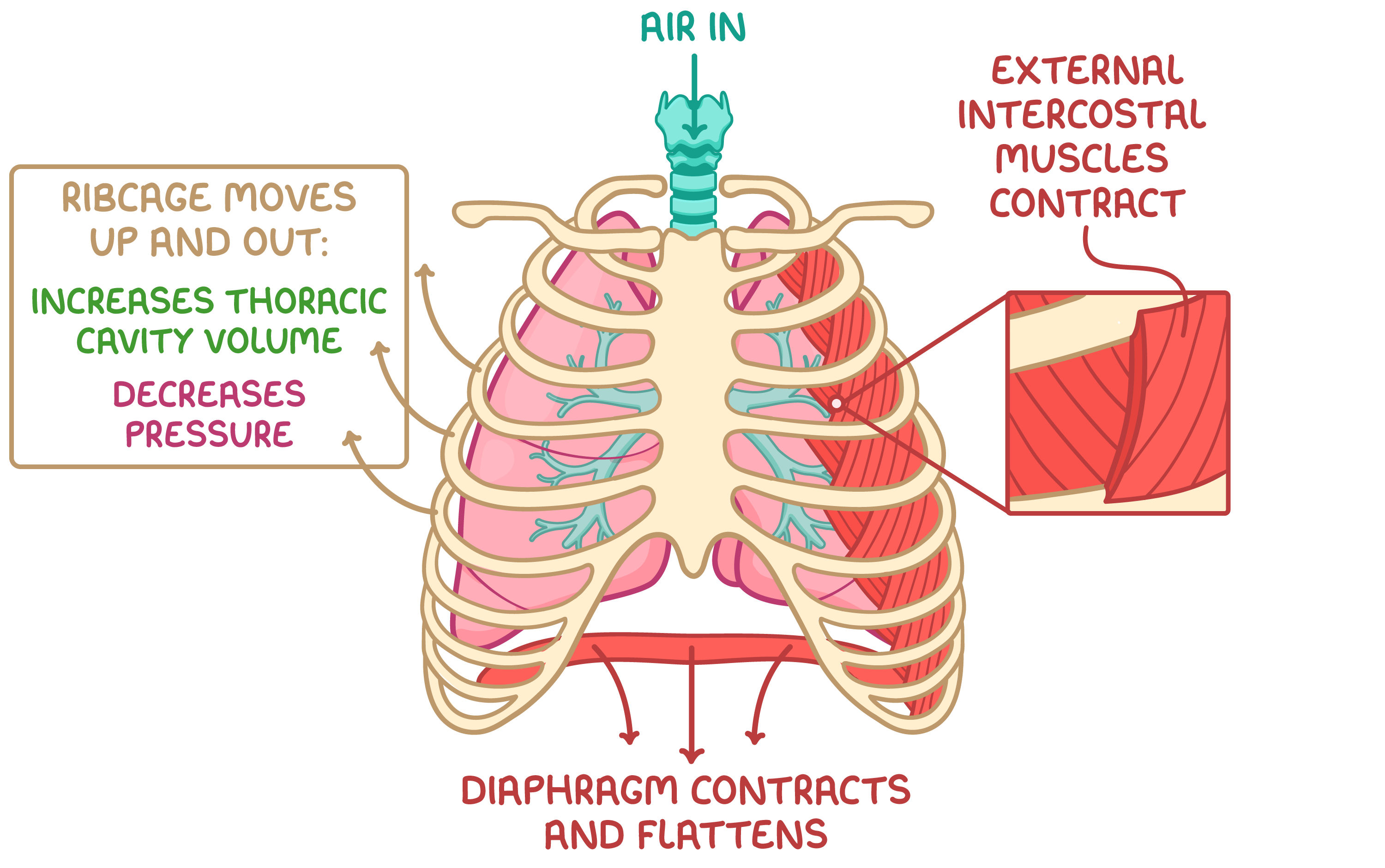
What is inspiration?
It is breathing in and it is an active process requiring energy for muscle contraction
What happens during inspiration?
The external intercostal muscles contract while the internal intercostal muscles relax, moving the ribcage up and out.
The volume of the thoracic cavity increases.
The diaphragm contracts and flattens, further increasing the volume of the thoracic cavity.
The lung pressure decreases below atmospheric pressure.
Air flows into the lungs down the pressure gradient.
What is expiration?
It is breathing out and it is at rest is a passive process so it does not require energy
What is the process of expiration?
The external intercostal muscles relax, moving the ribcage down and in.
The volume of the thoracic cavity decreases.
The diaphragm relaxes and unflattens, further decreasing the volume of the thoracic cavity.
The lung pressure increases above atmospheric pressure.
Air is forced out of the lungs down the pressure gradient.
What is the tool used to measure ventilation?
Spirometer
What can a spirometer measure?
Breathing rate
Title volume
Vital capacity
Inspiratory reserve volume
Expiratory reserve volume
Residual volume
Total lung capacity
How to measure breathing rate?
Counting the number of peaks per minute on a spirometer
What is tidal volume?
The volume of air breathes in or out in an average breath during rest measured from the height of each peak at rest.
What is vital capacity?
The maximum volume of air that can be inhaled or exhaled in one deep breath
What is the inspiratory reserve volume?
The max volume of air that can be inhaled above a normal inhalation.
What is the expiratory reserve volume?
The max volume of air that can be exhaled above a normal exhalation
What is the residual volume?
The volume of air that remains in the lungs after the longest possible exhalation
What is the total lung capacity?
The vital capacity added to the residue volume
Why do insects need gas exchange?
To deliver oxygen to cells
To remove CO2 from cell
What structures are in an insect gas exchange system?
Tracheae
Tracheoles
Spiracles
What are tracheae and their adaptations?
Air filled tubes branching through the body
Reinforce with spirals of chitin to prevent collapsing
Multiple tracheae to increase surface area
What are tracheoles and their adaptations?
Find branches of tracheae that deliver gases to cells
Penetrate directly into tissues which reduces gas diffusion
Thin walls which reduces gas diffusion distance
Highly branched which maximise the surface area
Not reinforced with chitin which allows gas exchange to occur
Tracheal fluid at the end of the tracheoles allows oxygen to dissolve to aid diffusion and reduce water loss
What are spiracles and their adaptations?
External openings of the tracheal system on the exoskeleton along the abdomen and thorax
The open and close which allows control gas exchange within the atmosphere and minimise water loss
How does gas exchange work in insects?
Air enters the tracheal system through open spiricals
Air moves into larger tracheae and diffuses into smaller tracheoles
Oxygen dissolves in water and tracheal fluid and diffuses down its concentration gradient from tracheoles into body cells
Carbon dioxide diffusers down its concentration gradient out of the body cells and into tracheoles
This carbon dioxide is carried back through the spiracles via the tracheoles and out of the body
What are the challenges of gas exchange in Bony fish?
Water is denser and more viscous than air resulting in slower diffusion of oxygen
Water has less oxygen than air
Bony fish are very active so have high oxygen demands
Why is the structure of gills in bony fish?
Covered by an a operculum flap
Consist of stack filaments containing gill lamellae
Surrounded by extensive blood vessels
Why are the adaptations of the gills and bony fish?
The lamellae provide a large surface area
The lamellae membranes are thin to minimise diffusion distance
The girls have a rich blood supply to maintain steep diffusion gradient
The counter current flow of blood and water creates an even steeper concentration gradient
Overlapping filaments tips increases resistance, slowing water flow over gills and allowing more time for gas exchange
What is the system of countercurrent flow?
Blood and water flow over the lamellae in opposite directions
This means oxygen rich blood makes water that is at It’s most oxygen rich when it first moves across the gills this maximise diffusion of oxygen into blood
Oxygen Poor blood returning from body tissues meet oxygen poor water still allowing diffusion of oxygen into the blood
This maintains a steep concentration gradient
How does ventilation in bony fish work?
When fish open their mouth, it increases the volume of the buccal cavity
This increases the pressure which pulls water into the buccal cavity
Water flows over the gills and the opperculum