Locomotor control
1/29
There's no tags or description
Looks like no tags are added yet.
Name | Mastery | Learn | Test | Matching | Spaced |
|---|
No study sessions yet.
30 Terms
What is the somatic motor system
Composed of:
Joints
Skeletal muscles
Spinal motor neurons and interneurons
Controls voluntary movement through interactions between motor neurons and muscle fibers.
Hierarchical organisation of motor control
Level | Key Structures | Functions | Examples |
1. Association Cortex | Prefrontal Cortex, Parietal Cortex | Planning, intention, decision-making; integrates sensory info | Choosing to pick up a cup |
2. Motor Cortex | Primary Motor Cortex (M1), Premotor Cortex | Initiation and direction of voluntary movement | Sending signal to hand muscles to reach |
3. Basal Ganglia & Cerebellum | Striatum, Globus Pallidus, Substantia Nigra, Cerebellar cortex | Basal Ganglia: Initiation/suppression of movementCerebellum: Coordination, error correction, learning | Correcting movement trajectory in real time |
4. Brainstem | Vestibular nuclei, Reticular formation, Red nucleus | Posture, balance, gaze control; relays motor commands | Keeping balance while walking |
5. Spinal Cord | Motor neurons, interneurons, Central Pattern Generators (CPGs) | Execution of movement; reflex control; rhythmic patterns | Walking, knee-jerk reflex |
6. Peripheral Effectors | Skeletal muscles, joints, sensory receptors | Actual movement and proprioceptive feedback | Muscle contraction and feedback to spinal cord/brain |
Somatic motor system
Somatic motor system: the joints, skeletal muscles, and spinal motor neurons and interneurons and how they communicate with each other.
The muscle spindle: proprioception
The muscle spindle is a sensory receptor embedded in the muscle
Intrafusal muscle fibers arranged in parallel with extrafusal fibers
Alpha α motor neurons excite the extrafusal fibers to generate muscle contraction
During muscle contraction, gamma γ motor neurons are also activated and contract intrafusal fibres
Sensory afferent axons type I (Ia axons) respond rapidly to stretch and mediate reflex adjustments when the axon is stretched
Afferent axons form excitatory connections with the α motor neuron innervating the same muscle and inhibitory connections with the α motor neuron innervating the antagonistic muscle
The muscle spindle: proprioception Table
Step | Component / Event | Function / Role |
1 | Muscle spindle is a sensory receptor embedded in the muscle | Detects changes in muscle length and contributes to proprioception |
2 | Intrafusal fibers run parallel to extrafusal fibers | Allow the muscle spindle to detect stretch while maintaining alignment with the contracting muscle |
3 | Alpha (α) motor neurons excite extrafusal fibers | Cause muscle contraction and generate movement |
4 | Gamma (γ) motor neurons contract intrafusal fibers during contraction | Maintain sensitivity of the spindle during shortening of the muscle |
5 | Ia sensory afferents respond to stretch | Send rapid feedback to the spinal cord for reflex adjustment |
6 | Afferent axons synapse with motor neurons: | - Excitatory to α motor neurons of the same muscle- Inhibitory to α motor neurons of the antagonistic muscle (reciprocal inhibition) |
Relationship of muscles and motor neurons
Muscles are organised as functional antagonists
Reciprocal inhibition of flexors and extensors at the same joint
Muscle spindle: a specialized structure involved in proprioception (body sense)
What is a motor unit
Motor unit: An α motor neuron and all the muscle fibers it innervates.
What is Somatotopic organization
Somatotopic organization: motor neurons innervating muscles are spatially arranged in the spinal cord.
What are the 3 types of motor units
3 Types of Motor Units:
Slow (posture): Small red fibers, fatigue-resistant.
Fast-fatigable: Pale, large fibers, high force, quick fatigue (e.g., jumping).
Fast fatigue-resistant: Intermediate properties.
What are the 3 types of motor units
Slow motor units: smaller motor unit, consist of a smaller α motor neuron innervating small red muscle fibres. Slow contraction, small
forces generated. Resistant to fatigue. E.g.Maintaining upright posture.
Fast, fatigable motor units: larger α motor neuron innervating larger, pale muscle fibres,generate more force, sparse mitochondria therefore easily fatigued. E.g. brief exertions requiring large forces, running or jumping.
Fast, fatigue-resistant motor units
Regulation of muscle force: the size principle
• The number of motor units active at any one time determines the amount of force produced by a muscle
• Gradual increase in tension results from the recruitment of motor units in a fixed order according to their sizE
What is the size principle
Size Principle: Motor units are recruited from smallest to largest to control force output smoothly.
Reflexes (Muscle spindle,reflex arc and knee jerk reflex)
Muscle spindle: Detects stretch, consists of intrafusal fibers parallel to extrafusal fibers.
Reflex arc:
Ia afferents excite α motor neurons (same muscle) and inhibit antagonists.
Gamma motor neurons maintain spindle sensitivity during contraction.
Knee-jerk reflex is an example of this stretch reflex system.
Locomotion is characterised by 2 highly stereotyped pattern of movements
• a swing phase, during which the leg (having been extended to the rear) is flexed, raised off the ground, swung forward, and extended again to contact the ground
• a stance phase, during which the leg is in contact with the ground, moving backward in relation to the direction taken by the body
Spinal Control of Movement
Flexion-crossed extension reflex: When one leg withdraws (flexes), the other extends.
Central Pattern Generators (CPGs):
Neural circuits that generate rhythmic patterns (e.g., walking).
Can function without sensory input or brain signals, though inputs refine the pattern.
Built with pacemaker neurons.
Example in Lampreys: Rhythm generated by NMDA receptor properties.
What are CPGs
neural circuits that generate patterns of neural activity that underlie rhythmic motor behaviours
Examples: walking, swimming, scratching, chewing and feeding
What’s a limit of CPGs
However: CPG merely generates a suitable pattern of output; sensory feedback and inputs from the brain adjust the output to suit particular environmental demands
CPGs in Lampreys
Neurotransmitter | Type | Released by | Main Functions in CPG | Role in Swimming |
Glutamate | Excitatory | Excitatory interneurons | - Activates motor neurons and interneurons - Generates rhythmic bursting activity | Drives contractions on each side; initiates and maintains rhythmic movement |
GABA | Inhibitory | Inhibitory interneurons | - Modulates excitatory signals - Regulates burst timing and frequency | Fine-tunes the rhythm; helps stabilize and shape the swimming pattern |
Glycine | Inhibitory | Commissural inhibitory interneurons | - Inhibits neurons on the opposite side - Enables reciprocal (left-right) inhibition | Coordinates alternating muscle activity for smooth, undulating swimming motion |
What’s the Molecular Basis of CPGs
NMDA Receptors:
Allow Ca²⁺ and Na⁺ influx.
Voltage-dependent block by Mg²⁺ creates oscillatory potential in pacemaker neurons
How does the Generation of rhythmic activity in CPG neurons (7)
Steps in Rhythmic Activity Generation:
Initiation by Brainstem Input
Descending signals from the brainstem (e.g., reticulospinal neurons) activate the spinal CPG.
Excitatory Neuron Activation (Glutamate)
Excitatory interneurons release glutamate to stimulate motor neurons and other interneurons.
This initiates bursting activity in one side of the spinal cord.
Intrinsic Bursting of Neurons
Some neurons have ion channels (Na⁺, Ca²⁺, K⁺) that generate rhythmic bursts even without input.
These properties help maintain the rhythm after it starts.
Reciprocal Inhibition (Glycine)
Commissural inhibitory interneurons release glycine to inhibit the opposite side of the spinal cord.
This prevents both sides from firing at the same time, creating left-right alternation.
Feedback Inhibition (GABA/Glycine)
Other inhibitory interneurons provide feedback to excitatory neurons, limiting burst duration and intensity.
This shapes the timing and stability of the rhythm.
Intersegmental Coordination
Rhythmic activity spreads down the spinal cord from one segment to the next.
This creates a traveling wave of alternating contractions along the body (swimming motion).
Sustained Rhythmic Pattern
The balance of excitation and inhibition keeps the rhythm going as long as needed.
Sensory feedback and brain input can modulate the speed or strength of the rhythm.
Descending pathways (2)
Groups of myelinated nerve fibers that carry motor information from the brain or brainstem to effector's muscles, via the spinal cord.
They can be functionally divided into two groups: Pyramidal (voluntary) and extrapyramidal (involuntary) tracts.
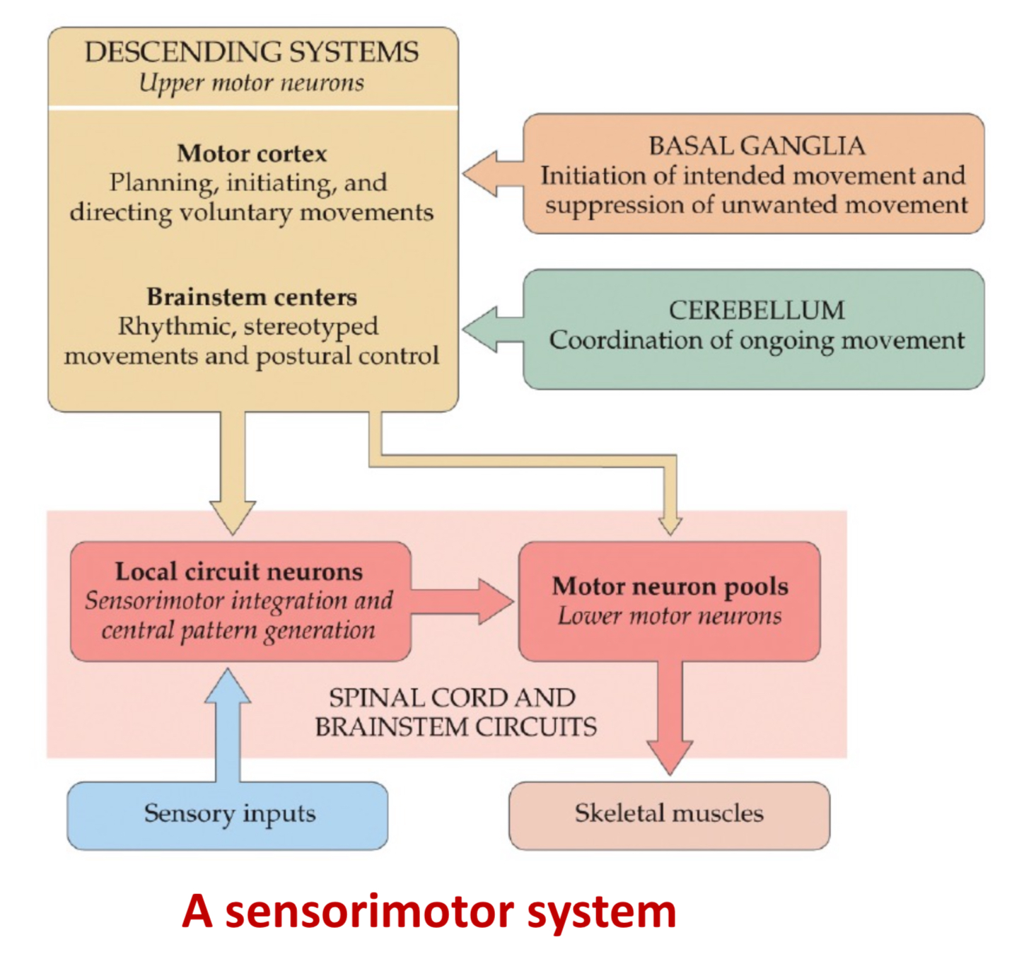
Diagram of A sensorimotor system
What is The Brain Control of Movement
Descending Motor Pathways:
Pyramidal tracts (voluntary): Corticospinal and corticobulbar tracts.
Extrapyramidal tracts (involuntary): Rubrospinal, vestibulospinal, etc.
Motor cortex (M1):
Initiates voluntary movement.
Some axons synapse directly with spinal motor neurons (especially in primates).
Somatotopically organized (motor homunculus).
Premotor cortex: Plans movements; activates before movement occurs.
Contains mirror neurons (respond during action observation).
How does Planning and directing voluntary movements in relation to premotor cortex,primary motor cortex and the somatosensory cortex
1. Premotor Cortex – Planning & Preparation
Function: Plans and organizes movements before they happen.
Role:
Selects the correct movement based on external cues (like seeing an object).
Coordinates with the prefrontal cortex for decision-making and intention.
Sends information to the primary motor cortex to execute the plan.
2. Primary Motor Cortex (M1) – Execution
Function: Executes voluntary movements by sending motor commands to muscles.
Role:
Receives the movement plan from the premotor cortex.
Activates specific muscles through descending motor pathways (e.g., corticospinal tract).
Controls the force, direction, and speed of movements.
3. Somatosensory Cortex – Feedback and Adjustment
Function: Processes sensory input from the body (touch, pressure, proprioception).
Role:
Provides real-time feedback about body position and movement.
Communicates with the motor cortices to adjust movements during execution.
Ensures movements are precise and accurate by detecting errors (e.g., if you’re off-target).
What is The Basal Ganglia Function
Regulates initiation and termination of movement by influencing upper motor neurons.
Works via disinhibition:
Direct Pathway: Facilitates movement via D1 dopamine receptors.
Indirect Pathway: Inhibits competing movements via D2 dopamine receptors.
How does dopamine work
Dopamine activates intended motor programs through the direct pathway and suppresses competing motor programs through the indirect pathway
Basal Ganglia Disorders (2)
Parkinson’s Disease:
Loss of dopaminergic neurons.
Overactive indirect pathway → movement suppression.
Symptoms: bradykinesia, tremors, shuffling gait.
Huntington’s Disease:
Loss of medium spiny neurons in the indirect pathway.
Overactive direct pathway → excess involuntary movement.
Why is C. elegans used as a Model
Simple nervous system: 302 neurons, 118 types, full connectome mapped.
Advantages:
Transparent, genetically tractable, small.
Suited for optogenetics and calcium imaging.
Dopaminergic system modulates locomotion and learning.
Major neurotransmitter pathways present
How does Whole-Brain Imaging in C. elegans Work
Fluorescent calcium indicators allow real-time tracking of single-cell activity.
NeuroPAL: multicolor neuron atlas for brain-wide mapping.
Useful for studying sensorimotor integration:
Stimulus → neural activity → behavior.
Knee Jerk Reflex steps (6)
Tap on the Patellar Tendon = A light tap just below the kneecap (patella) stretches the quadriceps muscle in the thigh.
Stretch of the Muscle Spindle = This stretch activates muscle spindles, which are proprioceptive sensory receptors located within the muscle. They detect the rapid lengthening of the quadriceps.
Activation of Ia Sensory Afferents = The Ia afferent fibers (sensory neurons) from the muscle spindle rapidly carry the signal to the spinal cord (lumbar region).
Monosynaptic Excitation of Alpha Motor Neurons = In the spinal cord, the Ia afferent fibers make a direct excitatory synapse onto the alpha (α) motor neurons that innervate the same quadriceps muscle.
Muscle Contraction = The activated α motor neurons stimulate the quadriceps to contract, producing the knee extension (the leg kicks forward).
Reciprocal Inhibition of Antagonist Muscle = Simultaneously, the Ia afferents also activate interneurons that inhibit the α motor neurons of the antagonistic muscle (the hamstrings). This ensures the hamstrings relax, allowing smooth extension of the knee.