Human & Anatomy: Lab Practical #1 (1-5)
1/218
Earn XP
Description and Tags
1. Intro to Lab; Basic Organization of the Human Body, 2. Histology I: Epithelial Tissues, Connective Tissue, and Integument, 3. Histology II: Cartilage, Bone, Muscle, and Neurons, 4. Axial Skeleton and Muscles, 5. Appendicular Skeleton and Joints
Name | Mastery | Learn | Test | Matching | Spaced | Call with Kai |
|---|
No analytics yet
Send a link to your students to track their progress
219 Terms
Where does the midsagittal (median) plane divide?
Vertically down the midline into equal left and right halves
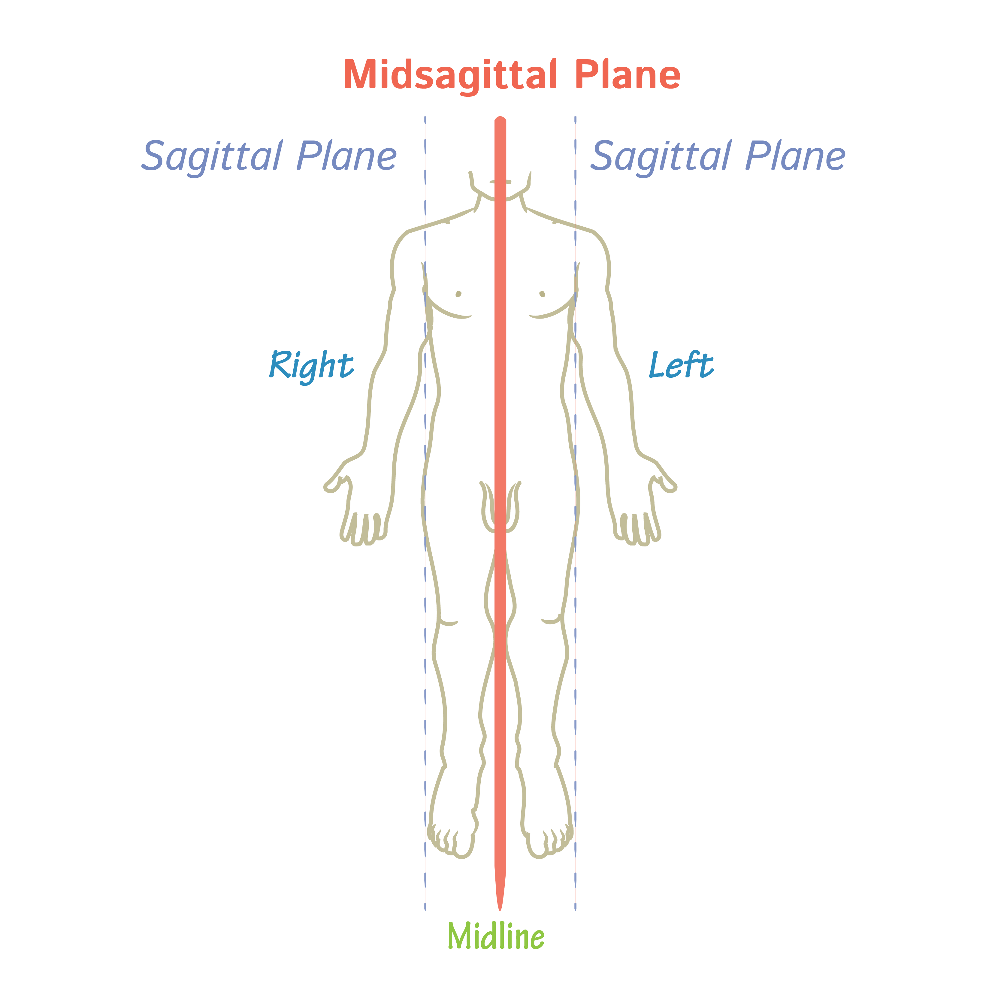
Where does a sagittal (parasagittal) plane divide?
Vertically into left and right halves

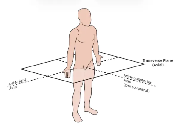
Where does a transverse (horizontal/cross-sectional) plane divide?
Horizontally into superior and inferior parts
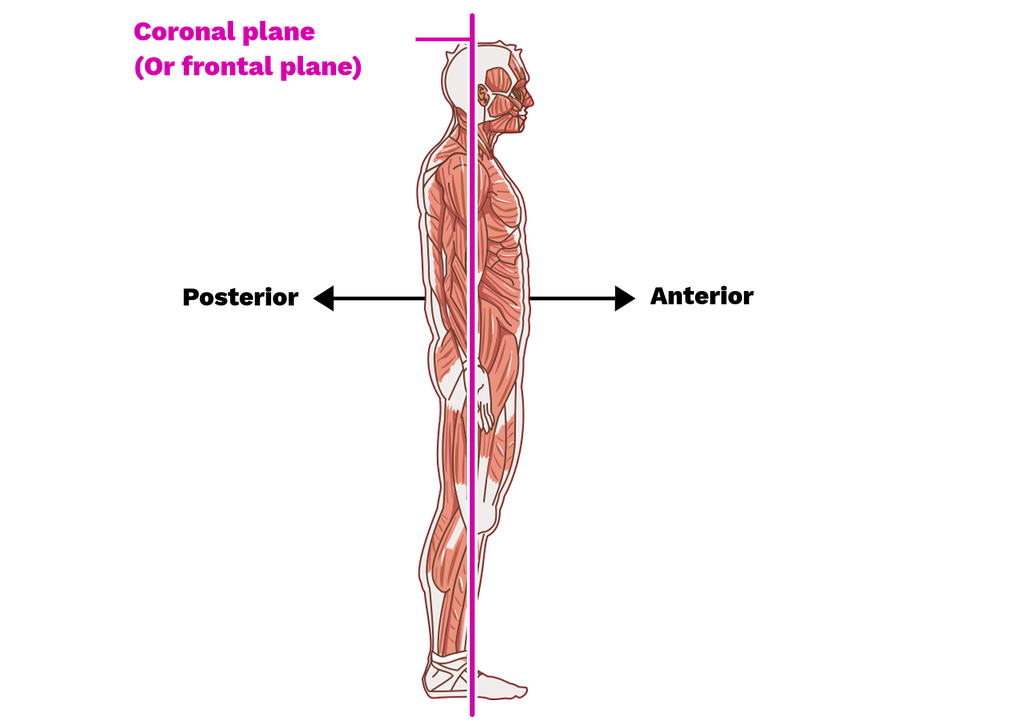
Where does a coronal (frontal) plane divide?
Vertically into anterior and posterior parts (or ventral and dorsal parts)
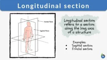
What is a longitudinal section?
Vertical cut through the longest axis of an object from its furthest points (from end to end) that shows its internal structure
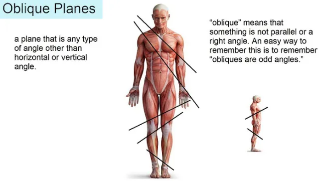
What is an Oblique section?
A cut that is nerther a cross/longitudinal section.
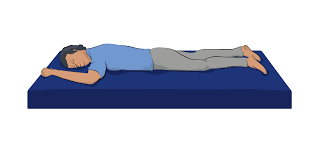
What is a body in a prone position?i
Lying flat, face down with their back/dorsal side upwards
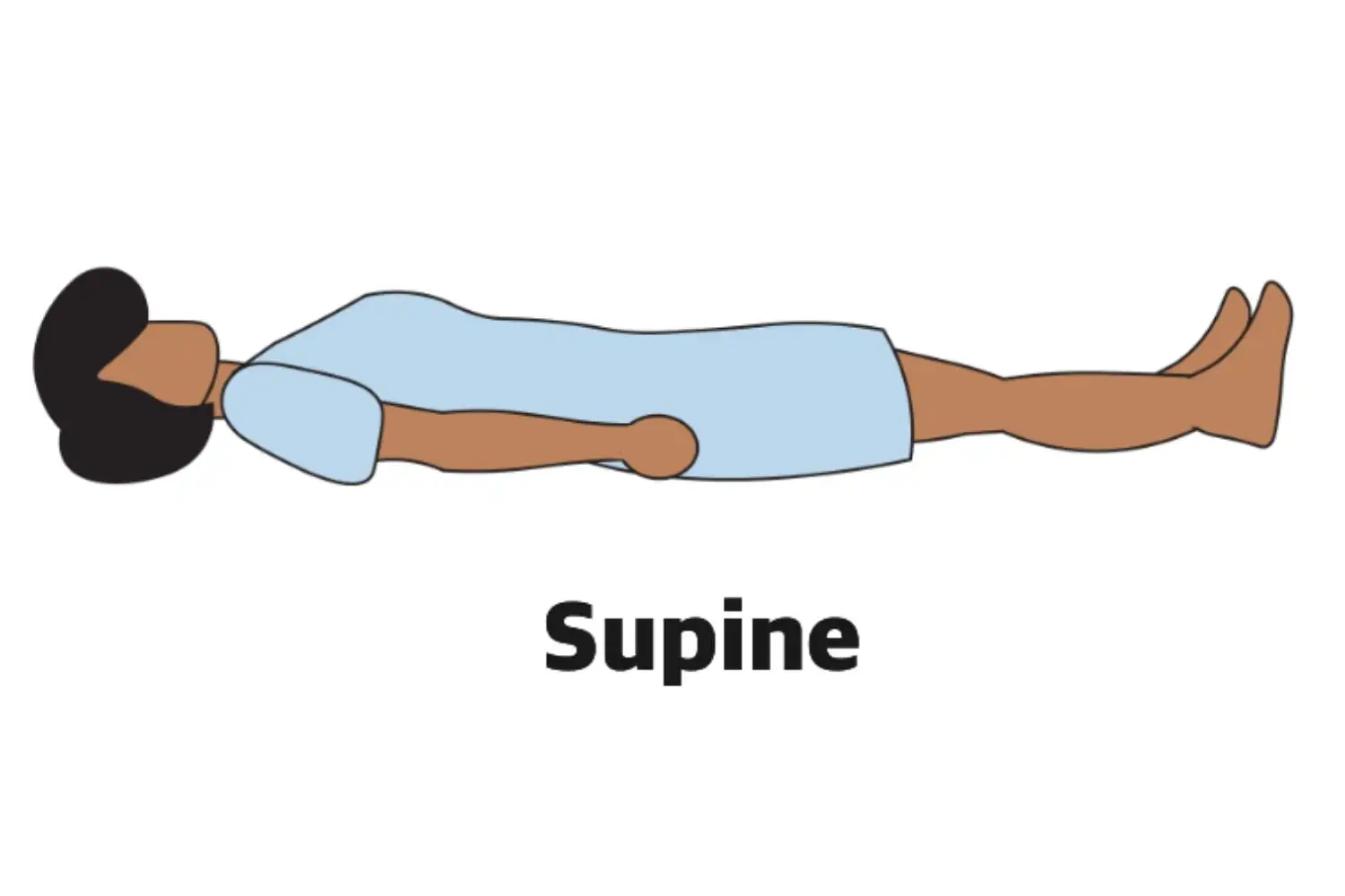
What is a body in a supine position?
Lying flat on their back, facing upwards
What is the directional term “Superior” (cranial) referring to?
Towards the head end of the body or Above/Higher
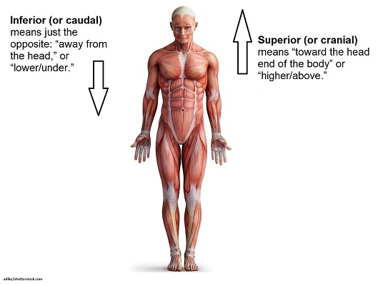
What is the directional term “Inferior” (caudal) referring to?
Away from the head or Lower/Under

What does the directional term “Anterior” (ventral) mean?
Front side of the body or belly-side
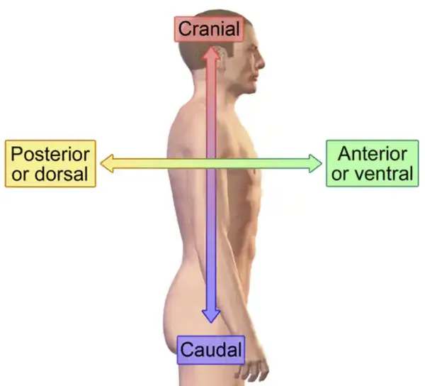
What does the directional term “Posterior” (dorsal) mean?
Back side of the body or the rear side

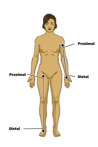
What does the directional term “Proximal” mean?
Closer to the point of attachment (of a limb)

What does the directional term “Distal” mean?
Further from the point of attachment
What does the directional term “Lateral” mean?
Away from the midline wW
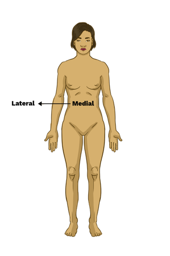
What does the directional term “Medial” mean?
Towards the midline

What does the directional term “Superficial” mean?
Towards the surface
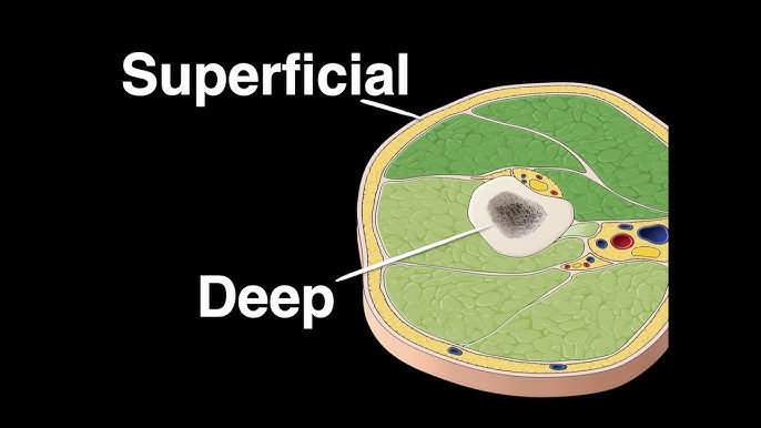
What does the directional term “Deep” mean?
Away from the surface

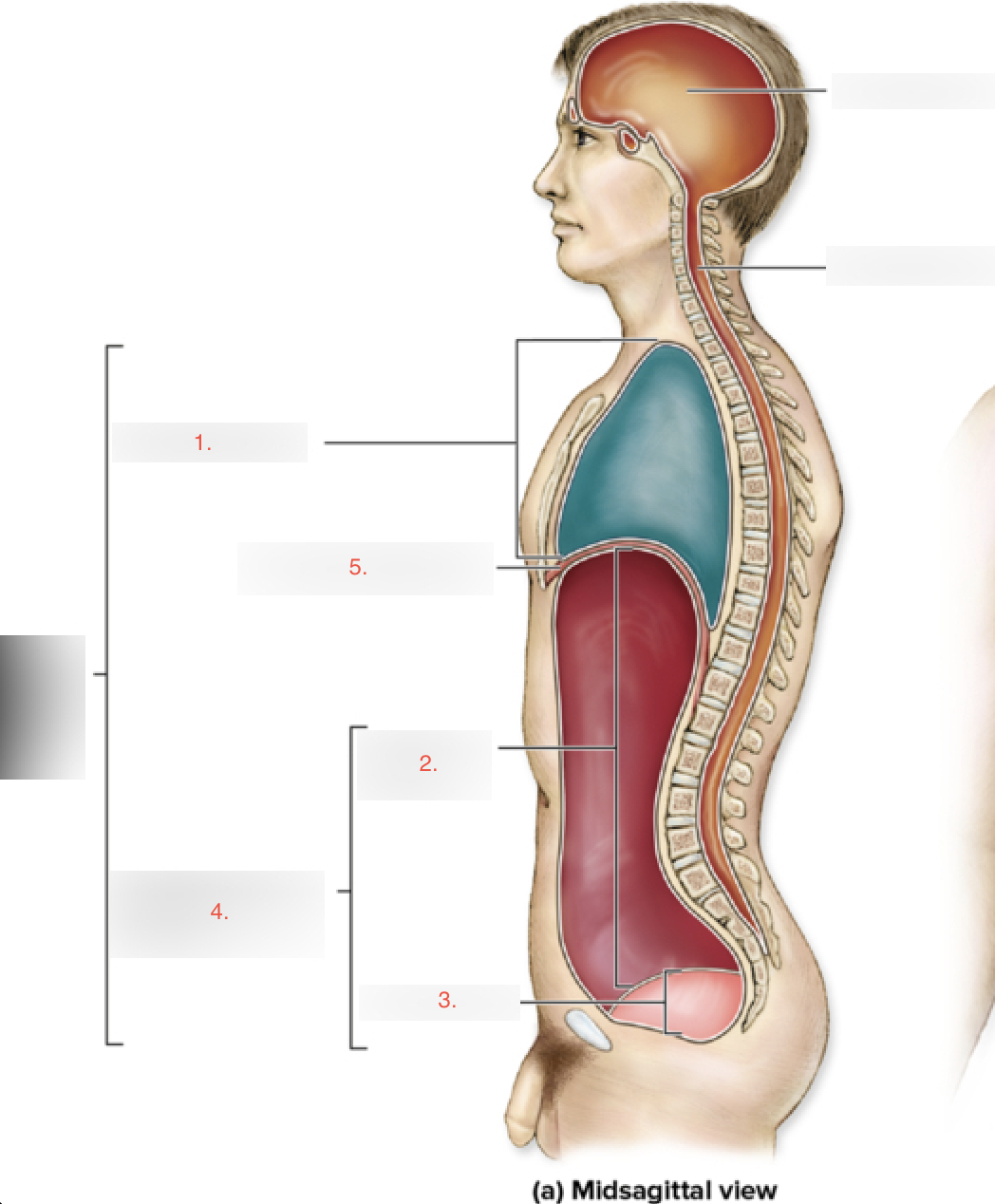
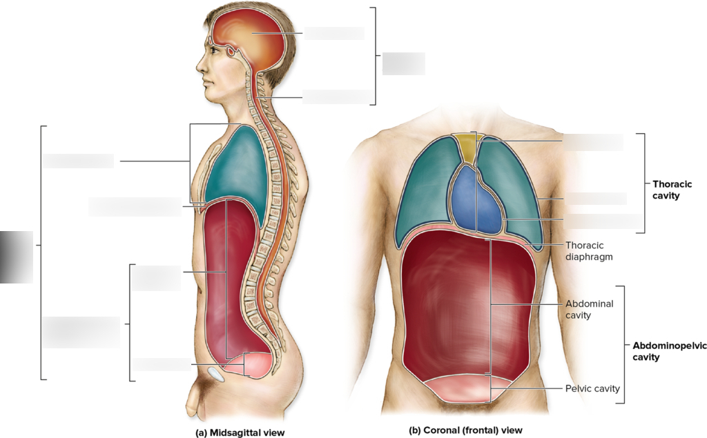
What are the 4 Body Cavities listed here along with the organ listed?
Thoracic Cavity
Abdominal Cavity
Pelvic Cavity
Abdominopelvic Cavity
Diaphragm
What is the mediastinum?
The median space in the thoracic cavity that contains the heart, thymus, esophagus, trachea, and major blood vessels.

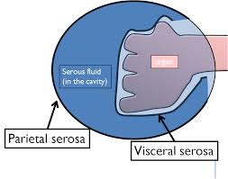
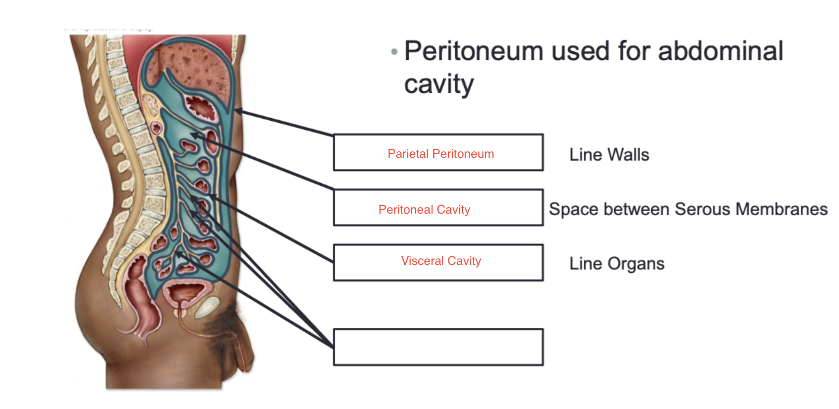
What are Visceral Serous Membranes and the 3 types?
Membranes covering the surface of an organ
Visceral pericardium
Visceral peritoneum
Visceral pleura

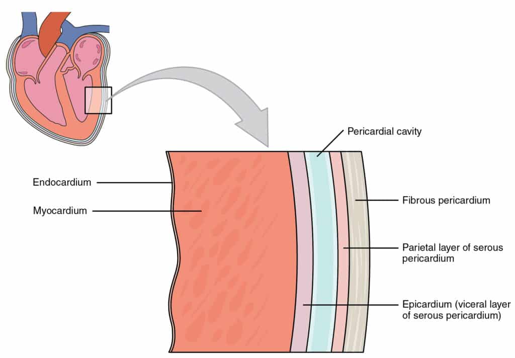
What is the Visceral Pericardium?
Serous membrane that covers the surface of the heart
What is the Visceral Peritoneum?
Serous membrane that covers the surface of organs within the abdominopelvic cavity. ex. stomach, liver, and intestines

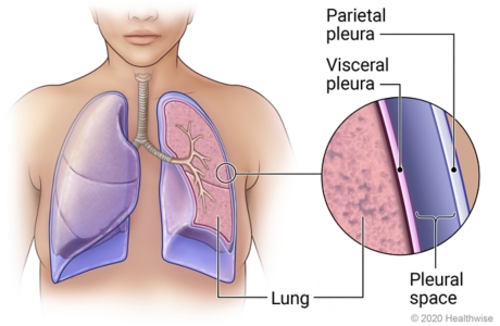
What is the Visceral Pleura?
Serous membrane that covers the surface of the lungs

What are Parietal Serous Membranes and its 3 types?
Serous membranes that line the cavity containing an organ(s)
Parietal pericardium
Parietal peritoneum
Parietal pleura
What is the Parietal Pericardium?
Serous membrane that lines the pericardial cavity

What is the Parietal Peritoneum?
Serous membrane that lines the peritoneal cavity

What is the Parietal Pleura?
Serous membrane that lines the pleural cavity

What forms the Pericardial Cavity and what is it?
Formed by the serous membranes (parietal pericardium & visceral pericardium) with serous fluid inside of it. Cavity surrounding the heart.

What forms the Pleural Cavity and what is it?
Formed by the serous membranes (visceral pleura & parietal pleura) and filled with serous fluid.
Cavity surrounding both lungs
What forms the Peritoneal Cavity and what is it?
Formed by the serous membranes (visceral peritoneum & parietal peritoneum) and filled with serous fluid.
Cavity surrounding organs like the liver, stomach, and intestines.

What is Mesentery?
A double layer (fold) of visceral peritoneum that anchors things in place for ex. small intestines, stomach, pancreas, and other organs to the abdominal wall
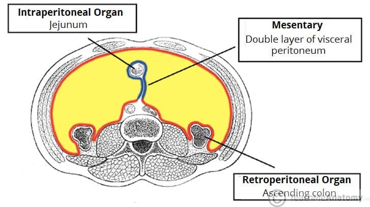
What does Retroperitoneal mean?
Refers to the location of organs and is not a membrane/cavity
Organs behind the peritoneal cavity that are partially “covered” by parietal peritoneum
Ex. kidneys, pancreas, & ureters
Body Regions and Parts: Cephalic
Head

Body Regions and Parts: Orbital
Eye
Body Regions and Parts: Nasal
Nose
Body Regions and Parts: Oral
Mouth
Body Regions and Parts: Auricular
Ear
Body Regions and Parts: Buccal
Cheek
Body Regions and Parts: Cervical
Neck
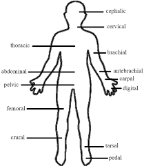
Body Regions and Parts: Thoracic
Thorax

Body Regions and Parts: Abdominal
Abdomen

Body Regions and Parts: Pelvic
Pelvis

Body Regions and Parts: Dorsal
Back
Body Regions and Parts: Deltoid
Shoulder
Body Regions and Parts: Vertebral
Spinal column
Body Regions and Parts: Gluteal
Buttock
Body Regions and Parts: Axillary
Armpit
Body Regions and Parts: Brachial
Arm

Body Regions and Parts: Antebrachial
Forearm

Body Regions and Parts: Manus
Hand
Body Regions and Parts: Carpal
Wrist

Body Regions and Parts: Digital
Fingers/Toes

Body Regions and Parts: Coral
Hip
Body Regions and Parts: Femoral
Thigh

Body Regions and Parts: Patellar
Kneecap
Body Regions and Parts: Popliteal
Behind the knee
Body Regions and Parts: Crural
Leg

Body Regions and Parts: Pet
Foot
Body Regions and Parts: Tarsal
Ankle

Body Regions and Parts: Calcaneal
Heel of foot
What is the lowest power objective lens and its magnification?
Scanning Objective
4X magnification
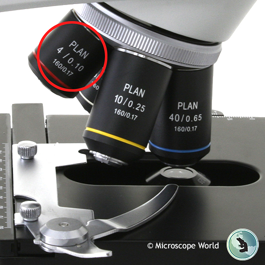
What is the middle power objective lens and its magnification?
Low Power Objective
10X Magnification
What is the highest power objective lens and its magnification?
High Power Objective
40X magnification
What does the Ocular Lens magnify by?
10X
How can I calculate Total Magnification?
The objective lens X ocular lens
What is resolution (resolving power)?
The ability of the microscope to tell apart two objects that are close together.
Depends on the wavelength of light, more resolution with less wavelength
What is Depth of Field?
Focusing in a compound microscope is limited to this particular depth
Decreasese with more high powered lens
What is Field of View?
The amount of the object that you can see through the microscope
Decreases with higher objectives
What is Field Diameter & what are they for each objective lens?
The distance across the field of view
4X: 5.0mm
10X: 2.0mm
40X: 0.5mm
What are the labelled components of a Compound Light Microscope? Pt.1
Arm
Light Switch
Control knob
Coarse focus knob
Fine focus knob
Mechanical stage knob
Aperture Iris diaphragm lever
Condenser
Field iris diaphragm ring
Base
What are the labelled components of a Compound Light Microscope? Pt. 2
Ocular lenses
Rotating nosepiece
Objective lens
Stage
Condenser lens
Stage clip
Condenser lens adjustment knob
What is Histology?
microscopic study of tissues
How is epithelial tissue classified?
By # of cell layers and shape of the cells
What are the four epithelial tissue?
Simple Cuboidal Epithelium
Simple Columnar Epithelium
Stratified Squamous Epithelium (keratinized form)
Pseudostratified Columnar Epithelium
What are the 5 connective tissues?
Mesenchyme
Areolar Connective Tissue
Dense Regular Connective Tissue
Dense Irregular Connective Tissue (dermis of the skin)
Adipose Connective Tissue
How can we distinguish types of connective tissue?
By looking at their types of cells, form of the extracellular matrix, relative abundance of protein fibers
What are the two layers of the Integument?
Epidermis & Dermis
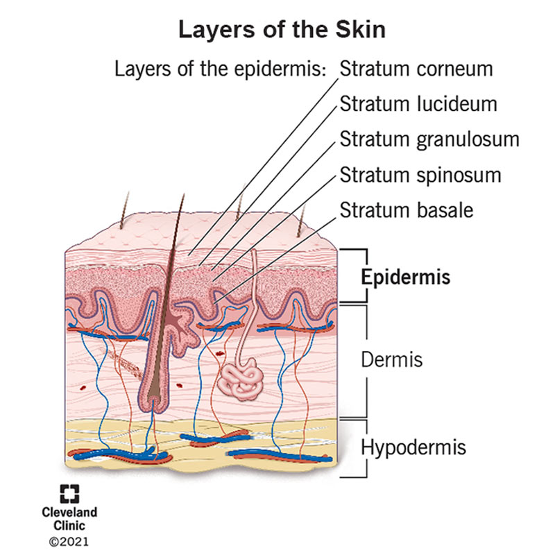
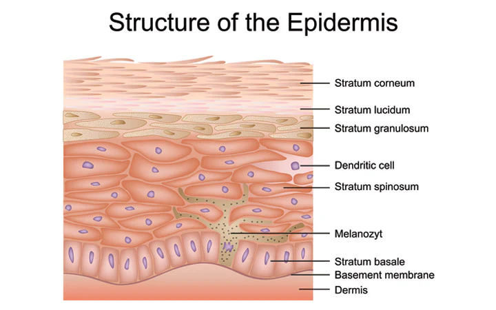
What are the 5 layers of the Epidermis? (order from superficial to deep)
What stratum corneum
stratum lucidum
stratum granulosum
stratum spinosum
stratum basale
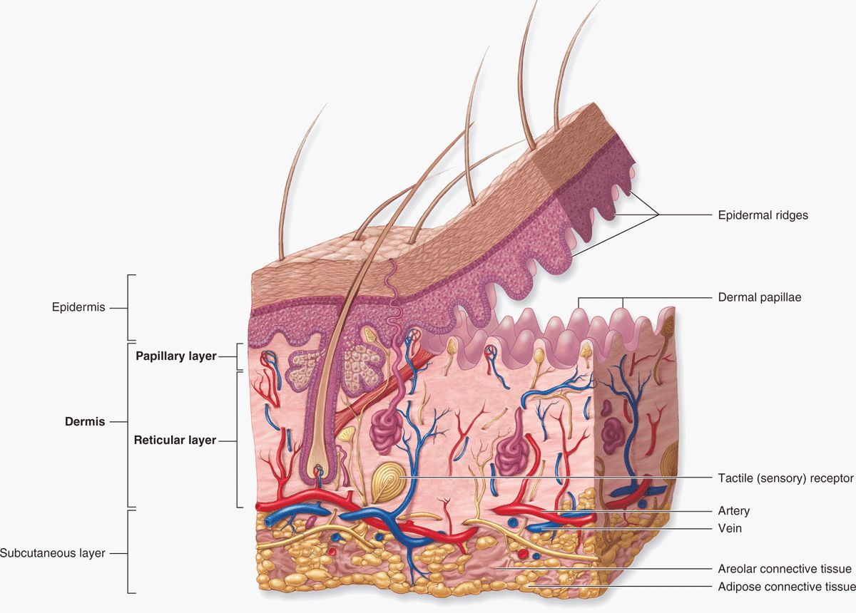
What does the Dermis consist of?
Dermal papilla(e)

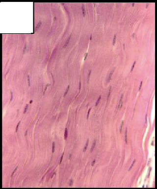
What kind of tissue is a. ?
Dense Regular Connective Tissue
Where is dense regular connective tissue found?
Tendons & Ligaments
What is the predominant cell in dense regular connective tissue?
Fibroblasts
What types of protein fibers are present in dense regular connective tissue and how are they arranged?
Collagen fibers arranged in parallel bundles

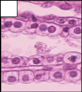
What kind of tissue is b. ?
Simple Cuboidal Epithelium
What is simple cuboidal epithelium?
ONE layer (simple) of cube shaped cells (cuboidal)

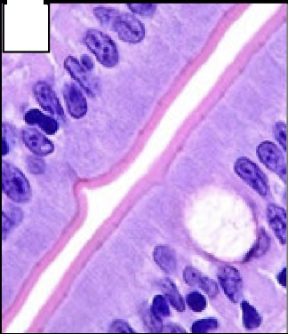
What kind of tissue is c. ?
Simple Columnar Epithelium
What is Simple Columnar Epithelium and what cells are in it?
ONE layer (simple) of column shaped cells (columnar)
Goblet Cells

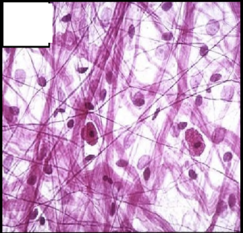
What kind of tissue is d. ?
Areolar Connective Tissue
What are the most prevalent cells in areolar connective tissue?
fibroblasts
What types of protein fibers are present in areolar connective tissue?
Collagen (pale pink & thick), elastic (thin and dark), and reticular

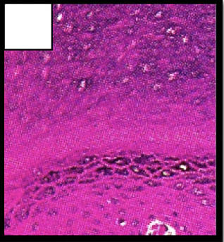
What kind of tissue is e. ?
Stratified Squamous Epithelium
What is Stratified Squamous Epithelium?
MANY layers (stratified) of flat-shaped (squamous) cells in THICK SKIN
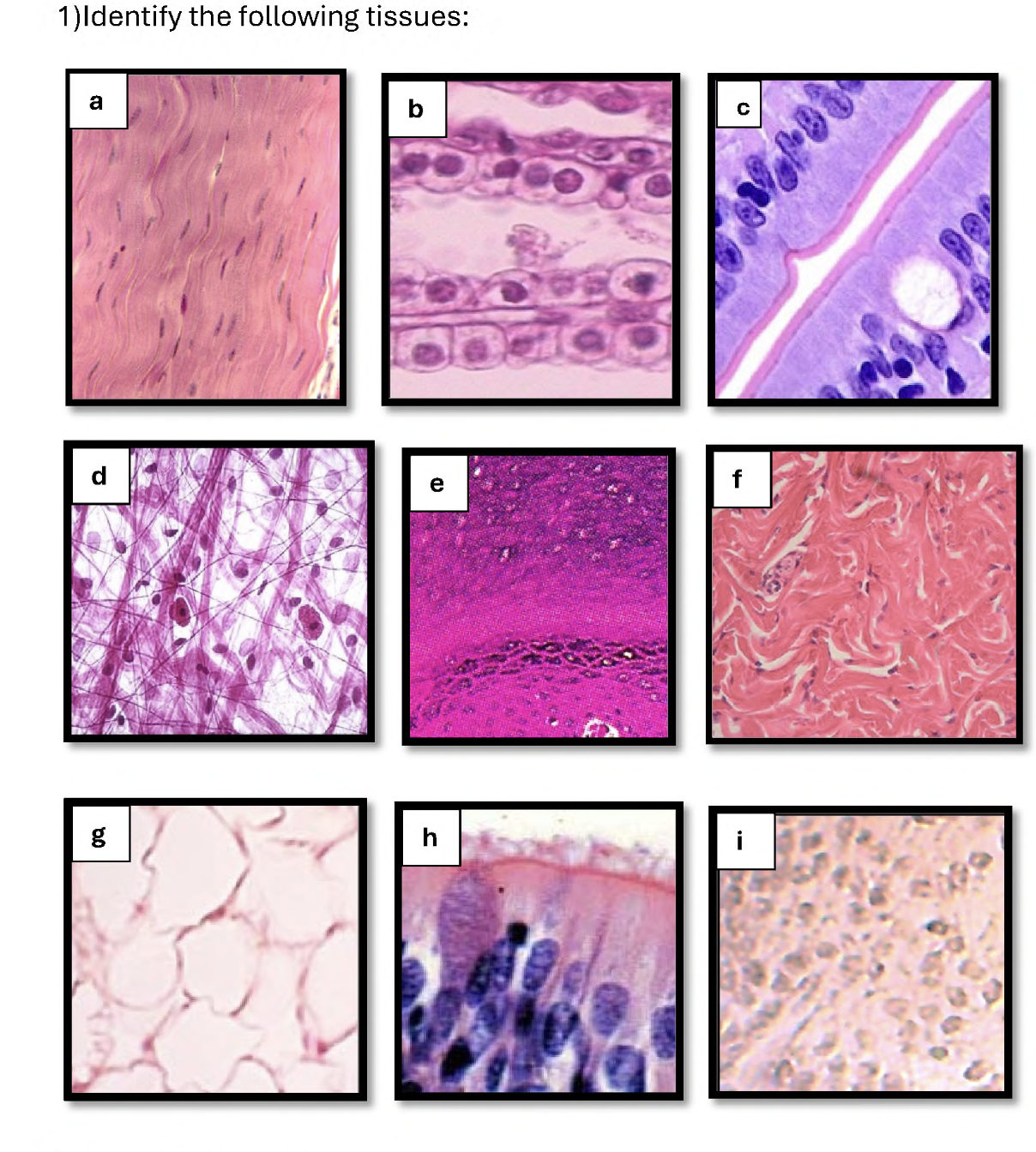
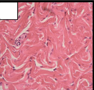
What type of tissue is f. ?
Dense Irregular Connective Tissue
What dominant types of protein fibers are present in Dense Irregular Connective Tissue and how could you describe their arrangement?
Collagen
Swirly (random directions) and deep pink

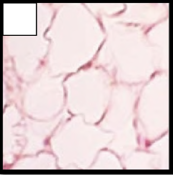
What kind of tissue is g. ?
Adipose Connective Tissue
What is the cell in Adipose Connective Tissue and what do they look like?
Adipocytes (lipid interior)
White blobs

What kind of tissue is h. ?
Pseudostratified Columnar Epithelium
Where is dense irregular connective tissue found?
In the dermis of the skin (thick skin)