han 202 - pregnancy n human development (ch 28+29)
1/74
There's no tags or description
Looks like no tags are added yet.
Name | Mastery | Learn | Test | Matching | Spaced | Call with Kai |
|---|
No analytics yet
Send a link to your students to track their progress
75 Terms
pregnancy
event that occur from fertilization until birth
divided into 3-month trimesters
gestation period
time from last menstrual period until birth (abt 280 days)
avg 266 days from conception to childbirth
conceptus
products of conception
pre-embryonic stage - from fertilization thru first 16 days
embryo - from day 16 to week 8
fetus - week 9 till birth
oocyte viability
for 12-24 hrs
sperm viability
for 24-48 hrs after ejaculation
fertilization
when sperm’s chromosomes combine with secondary oocyte (immature egg) to form fertilized egg (zygote)
for fertilization to occur
coitus (sex) must occur no more than
2 days before ovulation
24 hrs after ovulation
sperm propelled by
whiplike tail movement of flagella
endometrial cilia n forceful uterine contractions which disperse them throughout uterine cavity
sperm ejaculation
leak out vagina immediately after deposition
destroyed by acidic vaginal envi
fail to make it thru cervical mucus
dispersed in uterine cavity or destroyed by phagocytes
few (100 - few thousand) reach uterine tubes
total trip = 5 inches
sperm reach oocyte
need to pass 2 layers
corona radiata
zona pellucida
sperm must be capacitated before penetrating oocyte
secretions of female tract weaken acrosome membrane
capacitation
sperm membrane become fragile so hydrolytic enzyme can be released
make sperm membrane more permeable to calcium
activate receptors for chemical attractants
sperm penetration
acrosomal process forms n binds to receptors on oocyte’s plasma membrane
sperm n oocyte membranes fuse
nucleus pulled into oocyte cytoplasm
only 1 sperm allowed to penetrate oocyte (monospermy)
when sperm enters oocyte
waves of ca2+ released into oocyte cytoplasm which activates
oocyte to prep for 2nd meiotic division
cortical reaction
cortical reaction
zonal inhibiting proteins r released
blocks other sperm from entering
polyspermy
not desired
results in non-viable embryo
pre-embryonic stage
first 16 days of development, cumulates in an embryo
3 major processes
cleavage
implantation
embryogenesis
cleavage
rapid mitotic division of zygote without increase in size
increases SA n # of cells
easier for uptake in nutrients, o2, waste removal
zygote formation
blastomere (36 hrs) - 2-8 cells
morula (72 hrs) - 16+ cells
blastocyte (4-5 days) - fluid filled hollow sphere ; reaches uterus
identical twins
monozygotic - 1 placenta
one egg/sperm = one zygote
embryoblast divide into 2 within 2 weeks of fertilization
fraternal twins
dizygotic - 2 placenta
2 eggs/sperm = 2 zygote
blastocyte
trophoblast (outer layer) cells - single layer of flat cells
participate in placenta formation
inner cell mass
becomes embryonic disc
ectopic pregnancy
when egg implants itself outside uterus
most common is implanting itself in fallopian tube
blastocyst
floats for 2-3 days
nourished by uterine secretions
aborted if implantation fails
implantation
begins 6-7 days after ovulation
trophoblast adheres to endometrium
secretes enzymes to irritate endometrium
human chorionic gonadotropin (hCG)
secreted by trophoblast cells, later becomes chorion
prompts corpus luteum to continue secretion of progesterone n estrogen
hCG levels rise until the end of second month, then declines as placenta begin secrete progesterone n estrogen
hCG levels r used in pregnancy tests
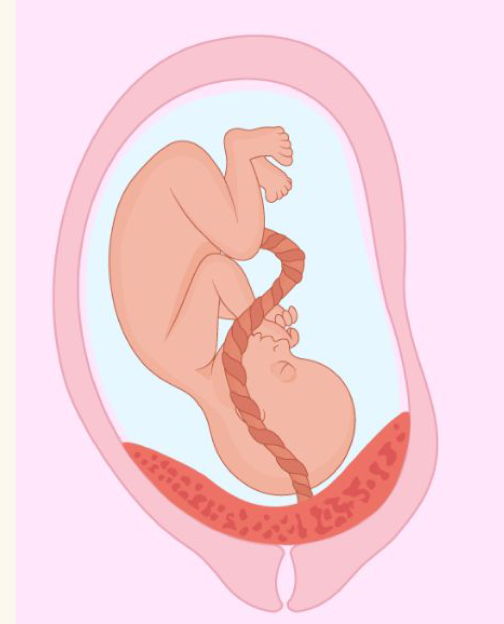
placenta formation
from embryonic n maternal tissues
embryonic tissue - chorion (develop from inner cell mass)
maternal tissue - decidua basalis
mom + baby blood supply
lie close but don’t intermix
development of circulation
first blood cells arise in yolk sac
end of third week
embryo has system of paired vessels
can hear baby heartbeat
what carries o2 from placenta to baby
blood vessel carrying o2 from placenta to baby is umbilical vein
unique vascular modifications
umbilical arteries carry deoxygenated blood
umbilical veins carry oxygenated vein
teratogens
harmful substances that can cross placental barriers
enters fetal blood n cause congenital abnormalities or death
extraembryonic membranes
amnion
yolk sac
allantois
chorion
all formed within first 2-3 weeks of development
amnion
forms amniotic sac
yolk sac
forms part of digestive tube
allantois
umbilical cord
chorion
helps form placenta
embryogenesis
gastrula → fetus = gastrulation
implantation: blastocyte convert to gastrula
inner cell mass become embryonic disc
3 primary germ layers n extraembryonic membranes develop (week 3)
embryonic disc subdivision
into epiblast n hypoblast
gastrulation
embryonic disc (2 layers) become 3-layered embryo
ectoderm, mesoderm, endoderm
primitive streak (dorsal groove) appears
notochord
mesodermal cells
form axial support
ectoderm germ layer
external layer
NS n skin epidermis
ectoderm specialization
neurulation
first major event of organogenesis
give rise to brain n spinal cord
neural plates fold inward as neural groove n fuse into neural tube
neural crest cells
neural crest cells
cranial
spinal
sympathetic ganglia
adrenal medulla
endoderm
epithelial linings of digestive, respiratory, urogenital systems
epithelia
endoderm + ectoderm
mesoderm
forms
notochord
somite
intermediate mesoderm
lateral plate mesoderm
somatic mesoderm
splanchnic mesoderm
notochord
nucleus pulposus of intervertebral disc
somite
sclerotome - vertebrae n ribs
dermatome - dermis of dorsal body region
myotome - trunk n limb musculature
intermediate mesoderm
kidneys
gonads
lateral plate mesoderm - somatic mesoderm
parietal serosa
dermis of ventral body region
CT of limbs (bones, joints, ligaments)
lateral plate mesoderm - splanchnic mesoderm
wall of digestive n respiratory tracts (except epithelial lining)
visceral serosa
heart
blood vessels
anencephaly
no brain produced
nonviable for life
organogenesis
formation of body organs n systems
week 8:
all organ systems r present, but not fully functional
end of embryonic period - embryo become fetus
fetal development
time of rapid growth of body structures established in the embryo
occurs from week 9 thru birth
adjustments to pregnancy
relaxin (placenta) causes pelvic ligaments n pubic symphysis to relax to ease birth passage
reproductive organs become engorged with blood
increase in lordosis
uterus expands, occupying most of abdominal cavity
adjustments to pregnancy - GI tract
morning sickness due to elevated estrogen n progesterone levels
adjustments to pregnancy - urinary system
more urine made due to increased metabolism n fetal wastes
stress incontinence
adjustments to pregnancy - respiratory system
tidal volume increase
dyspnea may occur later in pregnancy
adjustments to pregnancy - CV system
blood volume increase 25-40%
BP n pulse rise
venous return from lower limbs may be impaired
initiation of labor
last few weeks of preg
fetal secretion of cortisol stimulates placenta to secrete more estrogen
surfactant protein A
from fetal lungs, causes cervix softening
fetal oxytocin
placenta makes prostaglandins
maternal emotional n physical stress
activates hypothalamus, cause oxytocin release → powerful uterine contraction
pos feedback mechanism
more estrogen in preg
causes production of oxytocin receptors
assists w smooth birth
antagonizes calming effects of progesterone
braxton hicks contractions (weak irregular contractions) in uterus - false labor
parturition
act of giving birth
expel infant from uterus
stages of labor
dilation - longest stage (6-12+ hrs)
expulsion - 30 mins
placental - afterbirth - 30 mins later
dilation stage
initial weak contractions
15-30 mins apart, 10-30 secs long
gets more vigorous n rapid
cervix dilates to 10cm
amnion ruptures, releasing amniotic fluid - water breaking
engagement occurs
head enters true pelvis
pelvis
true pelvis - where baby exits
false pelvis - made by iliac crest
baby go from false to true pelvis
early dilation
baby head sideways to pass thru true pelvis
late dilation
baby head rotate posteriorly once past true pelvis
expulsion stage
contractions every 2-3 mins, 1 min long
want to push (absence of local anesthesia)
crowning occurs when largest dimension of head distends vulva
30-50 mins
end: infant delivered
placental stage
more strong contractions → placenta detaches
delivery of afterbirth (placenta n membranes) occurs 30 mins after birth
all placenta fragments must be removed to prevent postpartum bleeding
placenta previa
placenta form low in uterus, adjacent to and/or covering cervix
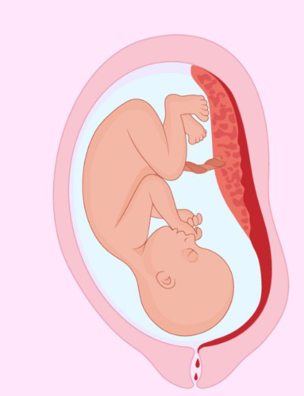
placental abruption
placenta separates from uterus wall before birth
first breath
increase co2 → central acidosis → stimulates respiratory control centers to trigger first inspiration
surfactant in alveolar fluids help reduce surface tension
respiratory rate - abt 45 breaths per min for first 2 weeks, then declines
premies usually put on respirators, lungs immature
neonatal period
4 week period immediately after birth
newborn’s physical status
assessed 1 n 5 mins after birth using APGAR score
appearance - skin color
pulse rate
grimace - reflex response
activity - mm tone
respiration
APGAR score
0-3 - critically low
4-6 - fairly low
7-10 - normal/healthy
lactation
make milk thru mammary glands
anterior pituitary release prolactin
oxytocin causes letdown reflex → actual ejection of milk from mammary glands
colostrum
yellowish secretion rich in vit A, protein, minerals, IgA antibodies
released first 2-3 days