Intro to brain structure and function
1/106
There's no tags or description
Looks like no tags are added yet.
Name | Mastery | Learn | Test | Matching | Spaced |
|---|
No study sessions yet.
107 Terms
what are the layers of the brain (superficial-innermost)
scalp + loose connective tissue, periosteum
bone
dura mater
arachnoid mater
pia mater
where can spread of infection occur between in brain
from scalp to cranial cavity or brain via emissary veins
what are the 3 meninges
dura mater (thick, tough)
arachnoid mater (blood vessels beneath, more flexible)
pia mater (closely applied to brain matter→ follows contours of brain = very thin)
what are the 3 dural reflections/folds of the dura mater
falx cerebri (related to cerebral cortex)
tentorium cerebelli
falx cerebelli (related to cerebellum)
what does rostral mean
anterior
what does caudal mean
posterior
what is parietal formaina
small crevices in bone of brain→ contain emissary veins
what is the string-like structure in arachnoid mater
trabeculi = connective tissue
what is sub-arachnoid space used for
harbouring CSF
what direction does falx cerebri run
sagittally and vertically
what shape is falx cerebri
sickle-shaped
what direction does tentorium cerebelli run
horizontally
which direction does falx cerebelli run
sagittally
what partially separates the two cerebral hemispheres
by falx cerebri
what separates cerebella hemispheres
falx cerebelli
what does tentorium cerebelli separate
separates occipital lobe and back of cerebral hemispheres
and occipital lobe from cerebellum
what structures are found at margins of falx cerebri
superior sagittal sinus
inferior sagittal sinus
what is a structural feature of arachnoid mater
arachnoid granulations
what occurs in arachnoid granulations
reabsorption of CSF occurs- keeps it in circulation
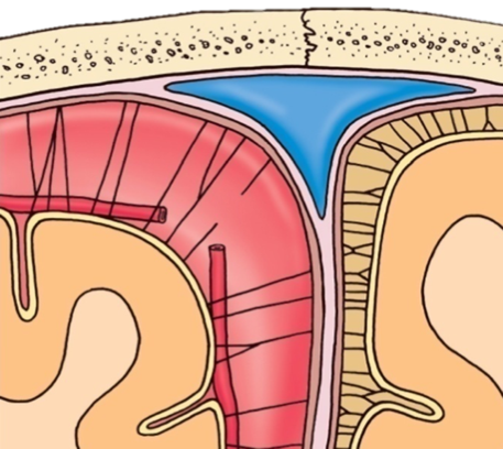
what type of haematoma is shown here
subarachnoid heaematoma
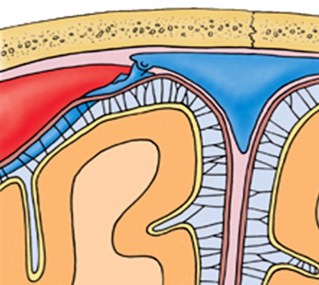
what type of haematoma is shown here
subdural haematoma
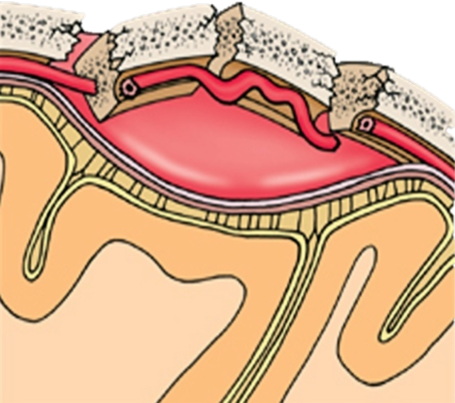
what type of haematoma is shown here
epidural haematoma
what does arachnoid mater contain
blood vessels
CSF
granulations
what creates foldings of brain
cortical sulci and cortical gyri
what sulcus is in front of central sulcus
precentral sulcus
what sulcus is behind central sulcus
post central sulcus
what are coritcal gyri
elevated parts/ridges of brain
what gyrus is found in front of central sulcus
precentral gyrus= primary motor cortex
what gyrus is found behind centra sulcus
post central gyrus= primary (somato)sensory cortex
what are the 4 lobes of the brain
frontal
parietal
temporal
occipital
what is frontal lobe responsible for
personality
attention
motivation
planning movement
what is parietal lobe responsible for
integrating sensory info
language processing
what is temporal lobe responsible for
memory
sensory processing
language comprehession
what is occipital lobe responsible for
vision
what are the 3 structural parts of brain derived from embryological structure
hindbrain (rhombencephalon)
midbrain (mesencephalon)
forebrain (prosencephalon)
what 2 structures does hindbrain differentiate into
metencephalon
mylencephalon
what structures are derived from metencephalon
pons
cerebellum
what structure is derived from mylencephalon
medulla oblongata
what structure does midbrain differentiate into
mesencephalon
what structures are derived from mesencephalon
tectum (colliculi)
tegmentum
peduncles
what structures does forebrain differentiate into
diencephalon
telencephalon
what structures are derived from diencephalon
thalamus
hypothalamus
what structures are derived from telencephalon
basal ganglia and cortex
what are the 3 aspects of brain stem (superior- inferior)
midbrain
pons
medulla oblongata
which cranial nerve branches off superior from cerebral peduncles
oculomotor nerve (3rd cranial nerve supplying eye)
which cranial nerve lies beneath cerebral peduncles
trochlear nerve
why is the red nucleus red
enriched with iron
what function is cerebllum responsible for
balance
coordination
synchronisation of muscles
what is found in ventricles
CSF
what volume of CSF is exchanged 3 times/day
150ml
how much CSF is produced approx. each day
500 ml
function of CSF
assists in circulating substances
provides cushioning
absorbs shock
where is CSF produced
choroid plexus (cells that line ventricles)
what is CSF exchanged between
ECF and blood stream
accumulation of CSF in ventricles causes what
hydrocephalus
what septum covers lateral ventricle
septum pellucida
which part of the brain contains the cerebral aqueduct
midbrain
what vertebral level in an adult can you take CSF samples
L3-L4
L4-L5
what vertebral level in infants is it safe to take CSF samples
at or below L4-L5
what are the ventricles
inter-connected, fluid filled cavities
cushion brain
bathe it in CSF
components of ventricles
lateral ventricles
third ventricles
cerebral aqueduct
fourth ventricle
what is the arterial supply of brain
internal carotid artery
vertebral arteries
terminal branches of internal carotid artery
anterior cerebral aa
middle cerebral aa
what do vertebral arteries join to form
basilary artery
what does basilar artery give rise to
posterior cerebral arteries
what is circle of willis
formed from cerebral blood supply
major anastomosis for brain
what arteries form subcortical blood supply
anterior cerebral artery
lenticulostriate arteries
middle cerebral artery
which sinuses form surface venous drainage
superior sagittal sinus
confluence of sinuses
transverse sinuses
sigmoid sinus
what sinuses and veins form medial venous drainage
inferior sagittal sinus
straight sinus
internal cerebral veins
what structures from blood brain barrier
endothelial tight junctions
basement membrane
pericytes
astrocytes
what is blood brain barrier
ensures circulatory system (blood) is kept separate from extracellular fluid
basement membrane surrounds what cells
endothelial ccells
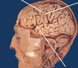
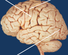
label the cortical sulci
left= precentral sulcus
right= central sulcus
bottom= lateral sulcus
label the cortical sulci
left= central sulcus
right= precentral sulcus
bottom= parieto-occipital sulcus
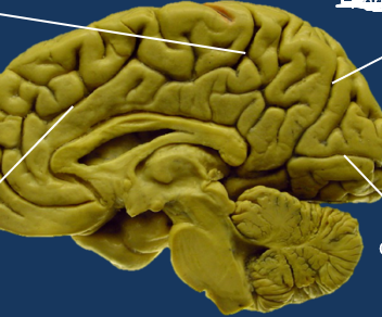
label the sulci
top left= marginal sulcus
top right= parieto-occipital sulcus
lower left= cingulate sulcus
lower right= calcarine sulcus
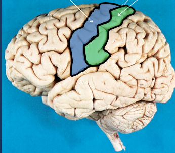
label the gyri
left= precentral gyrus
right= postcentral gyrus
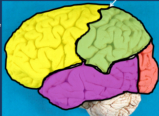
label the lobes
top left= frontal
top right= parietal
bottom right= occipital
bottom left= temporal lobe
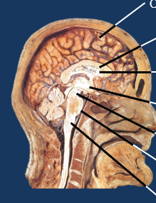
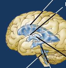
label from top to bottom
cerebral hemisphere
corpus callosum
ventricle
diencephalon
midbrain
pons
medulla
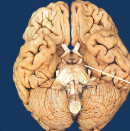
inferior view of brain ventral view of midbrain
cerebral peduncles
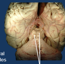
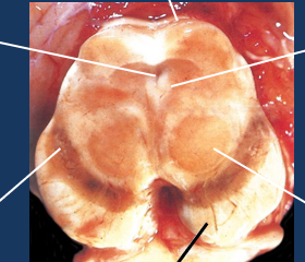
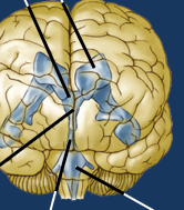
posterior view of brain with dorsal view of midbrain
tectum: superior and inferior colliculi

label top labels of transverse midbrain
top left→ cerebral aqueduct
middle top→ tectum
top right→ periaqueductal grey
label bottom labels of transverse midbrain
bottom left→ substantia nigra
bottom middle→ cerebral peduncles
bottom right→ red nucleus
what is cerebellum responsible for
balance
coordination
synchronisation of muscles
components of midbrain
cerebral peduncles
substantia nigra
red nucleus
tectum
reticular formation
what are cerebral peduncles responsible for
motor tracts
what is the tectum responsible for
vision and hearing
what is reticular formation responsible for
consciousness
role of pons
some direct connections with cortex
what is medulla responsible for
respiration
heart rate
vomiting
sneezing
role of thalamus
major relay station for sensory information from body
role of hippocampus
memory
spatial navigation
role of hypothalamus
hormone synthesis
temperature
hunger
thirst
sleep
what is role of caudate nucleus and putamen (basal ganglia)
planning movement
cognition
emotion
label ventricular system
top= lateral ventricles
middle side= interventricular foramen
bottom right= third ventricle
middle bottom= cerebral aqueduct
bottom left= fourth ventricle
label ventricular system
top right= lateral ventricle
top left= interventricular foramen
middle= third ventricle
left bottom= cerebral aqueduct
right bottom= fourth ventricle

Label left side (top to bottom)
superior sagittal sinus
choroid plexus
interventricular foramen
third ventricle
interpeduncular cistern
cisterna pontis
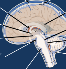
label right side top to bottom
arachnoid granulation
lateral ventricle
cerebral aqueduct
fourth ventricle
cisterna magna
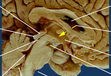
label left side top to bottom
midbrain
colliculi
cerebral aqueduct
fourth ventricle
cisterna magna
cisterna pontis

label right side top to bottom
lateral ventricle
inter-ventricular foramen
third ventricle
interpeduncular cistern
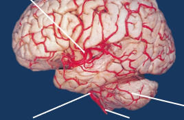
label the arteries
top= middle cerebral artery
bottom left =basilar artery
bottom middle= vertebral artery
bottom right= superior cerebellar artery