Week 2 - Sensory Neuroscience (Part 1 - Visual Neuroscience)
1/17
There's no tags or description
Looks like no tags are added yet.
Name | Mastery | Learn | Test | Matching | Spaced | Call with Kai |
|---|
No analytics yet
Send a link to your students to track their progress
18 Terms
The eye (image)
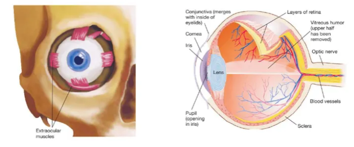
What is the retina?
Light-sensitive tissue lining at the back of the eye.
It acts like the sensor in a camera by capturing focused light, converting it into electrical signals, and sending them via the optic nerve to the brain, which interprets them as vision.
It is composed of specialised cells (photoreceptors like rods and cones, neurons, and glial cells) that detect light, colour, and movement.
The retina is crucial for sight, translating visual input into nerve impulses for the brain to form images.
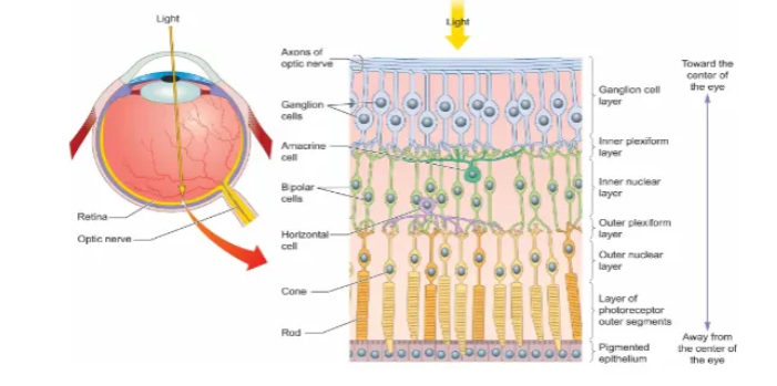
Structure of the retina (image)
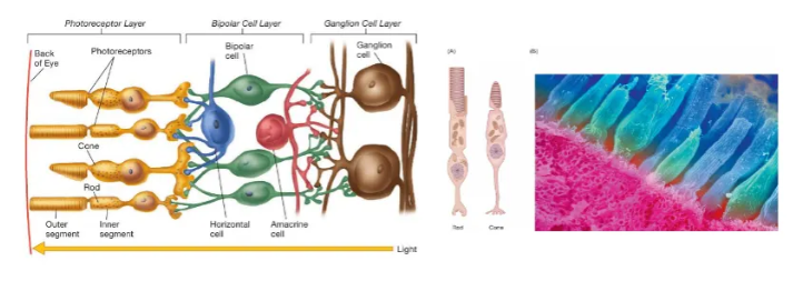
Retina photoreceptors (image)
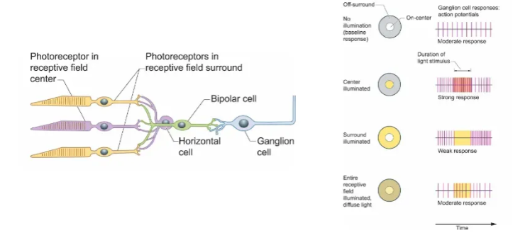
What are ganglion cells?
Specialised neurons known as the final output neurons of the retina.
They collect processed visual signals from bipolar and amacrine cells and send them as electrical impulses (action potentials) through the optic nerve to the brain for vision.
Ganglion cell axons extend towards the brain and form the optic nerve.
Only a few cones (vs. many rods) feed into a single ganglion cell.
There are two main types: M cells (magnocellular = large) and P cells (parvocellular = small).
M cells are responsive to coarse pattern and detect rapid motion.
P cells preserve colour information.
Each send their input to different destinations in the brain.
More broadly, they are clusters of neuron cell bodies in the peripheral nervous system (such as in dorsal root or autonomic ganglia) that relay signals, but in vision they are crucial for transmitting ‘digitised’ image information from eye to brain.
What are cones?
Specialised photoreceptor cells crucial for colour vision and sharp detail (visual acuity) in bright light.
Humans have three types, each sensitive to different wavelengths (red, green, blue), allowing the brain to interpret a full spectrum of colours.
They are concentrated in the fovea (center of the retina) for focused, detailed sight and work alongside rods to provide comprehensive sight.
What are rods?
Light-senstive cells in the retina responsible for vision in low light (night vision, also known as scotopic vision) and peripheral vision.
They don’t detect colour, producing black-and-white images.
They are cylindrical photoreceptors that contain rhodopsin, a pigment that breaks down with light, triggering neural signals sent to the brain, allowing us to see in dim conditions.
Receptive fields of ganglions (image)
Anatomical pathways from eye to cortex (image)
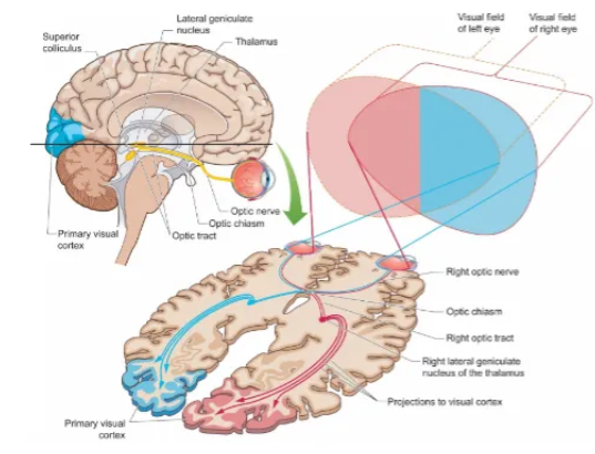
What is the Tectopulvinar Pathway?
A fast-acting, unconscious visual route bypassing the main visual cortex, starting from the retina to the superior colliculus (midbrain), then the pulvinar nucleus (thalamus), and projecting to visual/parietal/striatal areas, crucial for rapidly detecting motion and guiding eye/head movements (orientation) towards peripheral stimuli, explaining residual vision in blindsight patients.
What is the Geniculostriate Pathway?
The main visual route in the brain, carrying retinal information from the Lateral Geniculate Nucleus (LGN) in the thalamus to the primary visual cortex (V1) in the occipital lobe.
It is crucial for conscious vision, processing details like shapes, colours, and patterns, and is known as the optic radiation or geniculocalcarine tract.
90% optic nerve fibres project here
Axons terminate in the relay station LGN
Information then continues to V1
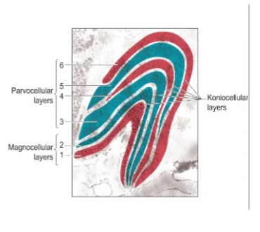
What is the lateral geniculate nucleus (LGN)?
A cricual part of the thalamus, acting as the main relay station for visual information from the retina to the visual cortex.
Six main layers stacked on top of one another, then folded into a knee-like shape.
Each layer receives input from only one eye, but all layers receive information from the contralateral visual field.
Retinotopically organised.
It organises signals before sending them to the occipital lobe for higher processing, controlling the flow of visual data, and refining visual perception.
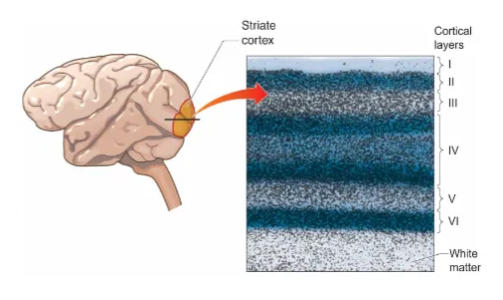
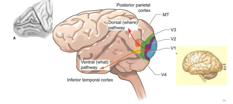
What is the primary visual cortex (V1 / striate cortex)?
Located in the occipital lobe, it is responsible for receiving raw data from the eyes (via the thalamus) and extracting basic features like edges, orientation, and movement, forming the foundation for seeing the world.
It's organized into specialized columns of neurons that analyze specific visual properties and sends this fundamental data to other visual areas for complex recognition and interpretation.
Projections from the LGN to V1 maintain their spatial organisation.
Cortical magnification factor describes the milimeters of cortical surface that are devoted to one degree of angle in the visual world.
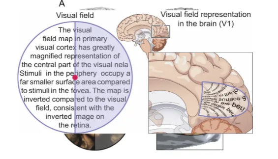
What is retinotopic organisation of the V1?
The V1 contains a distorted but orderly map of the visual field, where adjacent neurons process information from nearby locations on the retina, preserving spatial relationships.
This mapping is like a flipped, compressed map: signals from the retina's center (fovea) get disproportionately large cortical representation, while the periphery gets compressed, allowing the brain to efficiently process detailed central vision and broader peripheral context.
What is the calcarine fissure (or sulcus)?
A prominent groove on the medial surface of the occipital lobe of the brain, crucial for vision as it houses the primary visual cortex (V1), separating the cuneus above from the lingual gyrus below, with damage potentially causing blindness in half the visual field (hemianopia).
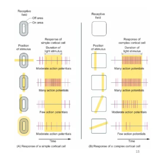
What is the difference between the receptive fields of simple and complex cells?
Simple cells in the primary visual cortex have distinct ON/OFF regions, responding best to bars of light/dark at specific locations.
Complex cells have overlapping ON/OFF areas and respond to oriented edges or bars of a specific orientation anywhere within their larger receptive field, showing phase invariance, with complex cells often built from inputs from several simple cells.
This hierarchical structure (simple -> complex) allows for processing features like orientation and movement robustly across different positions in the visual field.
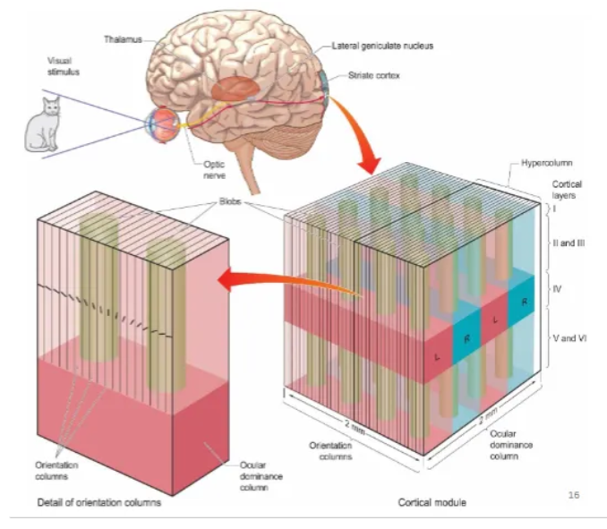
What is a hypercolumn in the V1?
A fundamental organizational unit, roughly 1x1 mm, that processes visual information for a specific point in space by containing a complete set of orientation columns (for all line angles) and ocular dominance columns (for input from both eyes).
Think of it as a miniature, functional module that captures all the basic features (orientation, eye preference) needed to analyze a small patch of the visual world.
Explain binocular integration in the V1.
The brain's crucial process of combining the slightly different images from your two eyes to create a single, unified 3D perception of the world, forming depth (stereopsis) and recognizing objects.
Neurons in V1 receive input from both eyes, creating specialized responses to specific visual cues like depth (binocular disparity) and matching orientations, allowing for sophisticated depth perception and object recognition, with complex interactions happening within V1 itself, not just simple convergence.
In V1, information from the two eyes is integrated (unlike LGN).
Binocular disparity - the image that falls on each retina is different as the eyes are positioned in different locations.
The brain uses this information to determine depth
Some cells in striate cortex are especially tuned to certain amounts of binocular disparity