Abdomen
1/29
There's no tags or description
Looks like no tags are added yet.
Name | Mastery | Learn | Test | Matching | Spaced |
|---|
No study sessions yet.
30 Terms
For which two of the following projections of the abdomen is the x-ray beam placed in the horizontal position?
PA, upright
AP, lateral decubitus
What is the respiration phase for an AP or PA abdominal image obtained with the patient in the upright position?
Expiration
All of the following organs lie in the abdominal cavity, except:
urinary bladder
Where is the center of the IR positioned for an AP abdominal image performed with the patient in the supine position?
Iliac crests
The space between the two layers of peritoneum is called the:
peritoneal cavity
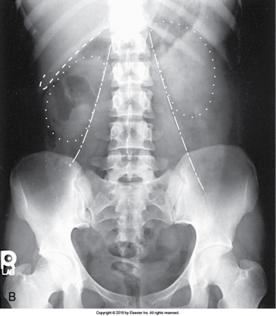
Which structures are outlined with dots in this AP image of the abdomen?
Kidneys
The serous membrane that lines the abdominopelvic walls is called the:
peritoneum
Where is the center of the IR positioned for an AP abdominal image obtained in the left lateral decubitus position?
2 inches above the iliac crests
Where is the center of the IR positioned for an upright PA abdominal image?
2 inches above the iliac crests
What is the respiration phase for an AP abdominal image obtained with the patient in the left lateral decubitus position?
Expiration
All of the following are used to evaluate rotation on a KUB image, except:
superimposed posterior ribs
The pathologic accumulation of fluid in the peritoneal cavity is termed:
ascites
A properly exposed abdominal image will exhibit all of the following, except the:
pancreas
Which of the following are clearly shown on a lateral abdomen projection performed with the patient in the dorsal decubitus position?
1. Prevertebral space
2. Air-fluid levels
3. Urinary bladder
1 and 2
What is the cavity posterior to the peritoneum?
retroperitoneum
Which of the following is placed perpendicular to the long axis of the grid for a lateral projection of the abdomen?
Midcoronal plane
Where is the center of the IR positioned for an AP abdominal image performed with the patient in the upright position?
2 inches above the iliac crests
Demonstrating small amounts of intraperitoneal gas in patients with an acute abdomen is often necessary. How long should the patient lie in the left lateral position before the exposure is made?
5 minutes
What is the respiration phase for an AP abdominal image obtained with the patient in the supine position?
Expiration
The folds of peritoneum that support the abdominal organs are called the:
1. omenta.
2. mesentery.
3. pleura.
1 and 2
A three-way abdominal series may be ordered to rule out all of the following except:
tumor mass
Which two of the following organs lie in the pelvic cavity?
Urinary bladder
Rectum
Which of the following are prime considerations in producing an optimal image of the abdomen?
1. Apply compression.
2. Explain the breathing procedure to the patient.
3. Do not start the exposure for 1 to 2 seconds after suspension of respiration.
2 and 3
The central-ray angulation for an AP abdominal image is:
0 degrees
The most commonly performed abdominal examination is referred to as a(n):
KUB
The inner portion of the sac that covers the abdominal organs is termed the:
visceral peritoneum
The outer portion of the sac that lines the abdominopelvic cavity is termed the:
parietal peritoneum
If a patient is unable to stand for an upright AP abdominal image, which position should be used?
Left lateral decubitus
One of the primary reasons a left lateral decubitus abdominal image is performed is to demonstrate:
air-filled levels
Where is the center of the IR positioned for a lateral projection of the abdomen performed with the patient in the dorsal decubitus position?
2 inches above the iliac crests