RESPIRATORY SYSTEM
The Anatomy and Physiology of the Respiratory System
WHAT IS HUMAN RESPIRATION?
- Human respiratory system allows one to obtain oxygen, eliminate carbon dioxide
- Breathing consists of two phase
- Inspiration - the process of taking in air
- Expiration - the process of blowing out air
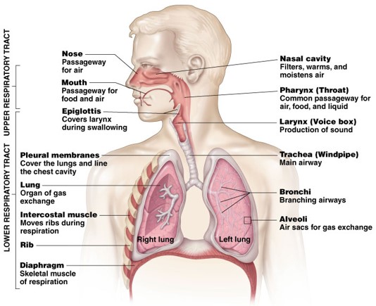
Upper respiratory tract function
- Passageway for respiration
- Receptors for smell
- Filters incoming air to filter larger foreign material
- Moistens and warms incoming air
- Resonating chamber for voice
Lower respiratory tract functions
- Larynx - maintains an open airway, routes food and air appropriately, assists in sound production
- Trachea - transports air to and from lungs
- Bronchi - branch into lungs
- Lungs - transport air to alveoli for gas exchange
FOUR RESPIRATION PROCESS
- Breathing (ventilation) - air into and out of lungs
- External respiration - gas exchange between air and blood
- Internal respiration - gas exchange between blood and tissues
- Cellular respiration - oxygen use to produce ATP, carbon dioxide as waste
Functions of the Respiratory System
- Air Distributor
- Gas exchanger
- Filters, warms, and humidifies air
- Influences speech
- Allows for sense of smell
Divisions of the Respiratory System
Upper respiratory tract (outside thorax)
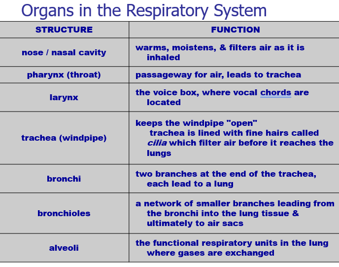
- Nose
- Nasal Cavity
- Sinuses
- Pharynx Larynx
Divisions of the Respiratory System
Lower respiratory tract (within thorax)
- Trachea
- Bronchial Tree
- Lungs
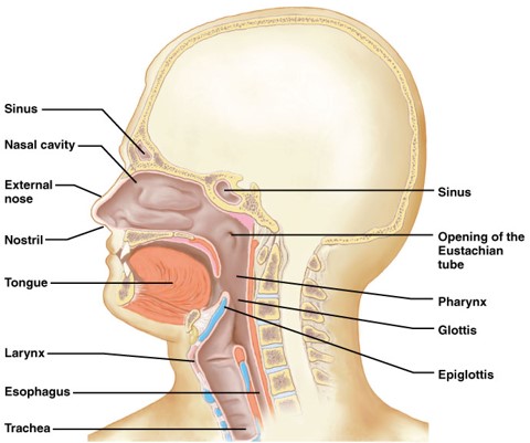
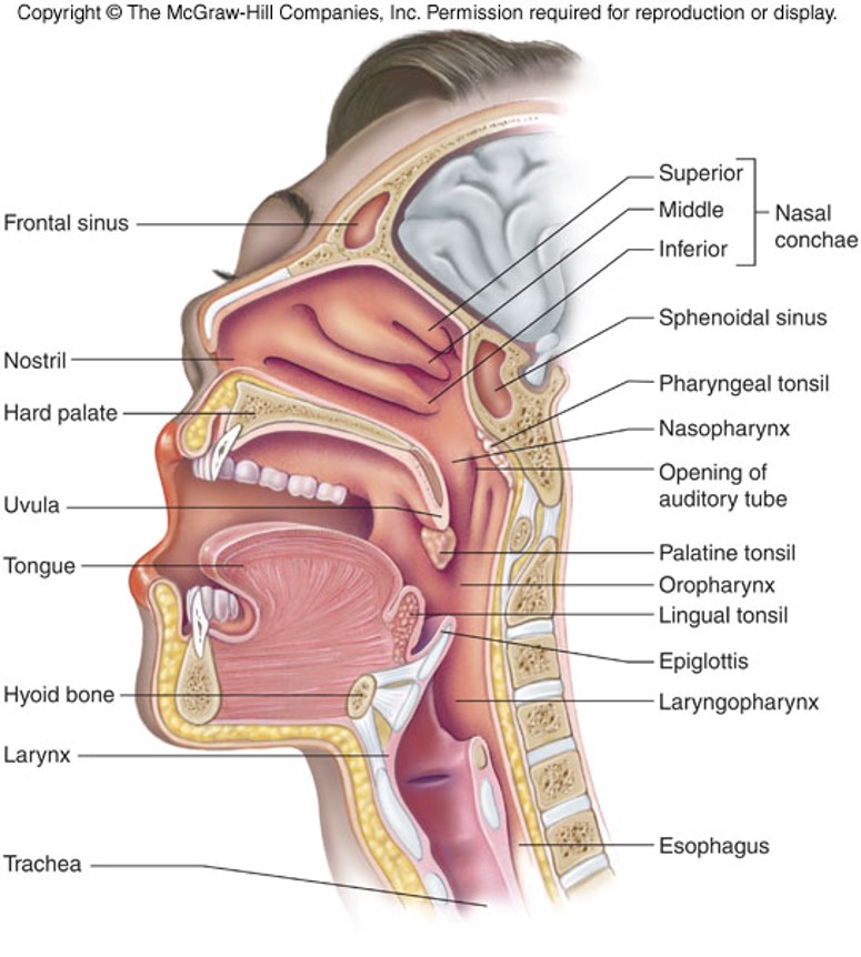
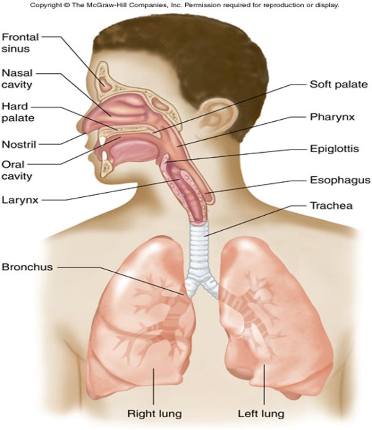
Structures of the Upper Respiratory Tract
Nose - warms and moistens air
- Palatine bone separates nasal cavity from mouth
- Cleft palate - Palatine bone does not form correctly, difficulty in swallowing and speaking.
- Septum - separates right and left nostrils
- rich blood supply = nose bleeds
- Sinuses - 4 air containing spaces – open or drain into nose - (lowers weight of skull).
Pharynx (throat)
- Base of skull to esophagus
3 divisions
- Nasopharynx - behind nose to soft palate.
- Adenoids swell and block.
- Oropharynx - behind mouth, soft palate to hyoid bone.
- tonsils
- Laryngopharynx - hyoid bone to esophagus.
• Changes shape to allow for vowel sounds = phonation.
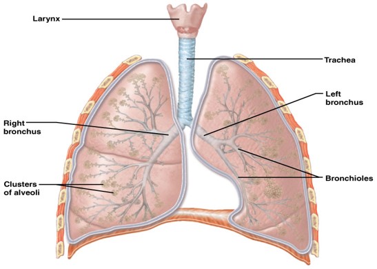
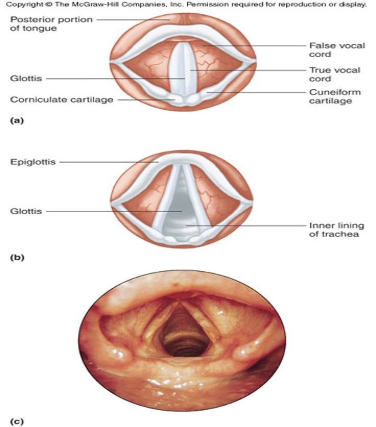
Larynx
- voice box
- Root of tongue to upper end of trachea.
- Made of cartilage
2 pairs of folds
• Vestibular - false vocal cords
• True vocal cords
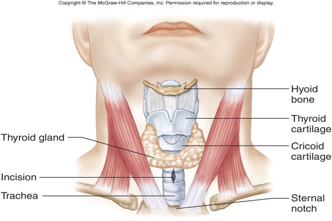
Thyroid cartilage
- Adam’s apple
- larger in males due to testosterone.
Epiglottis
- flap of skin (hatch) on trachea, moves when swallowing and speaking and closes off trachea when swallowing food
Trachea (windpipe)
- Larynx to bronchi
- Consists of smooth cartilage and C shaped rings of cartilage.
- Tracheostomy - cutting of an opening in trachea to allow breathing.
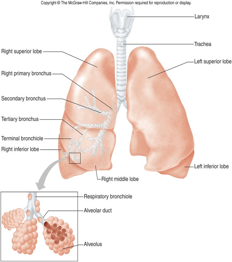
 Bronchi
- Tubes that branch off trachea and enter into lungs
- Ciliated– WHY?
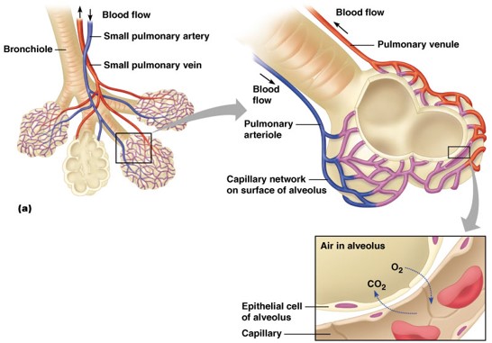
- Branches: Primary bronchi — secondary bronchi — tertiary bronchi — bronchioles – Bronchioles branch into microscopic alveolar ducts. Terminate into alveolar sacs – Gas exchange with blood occurs in sacs.
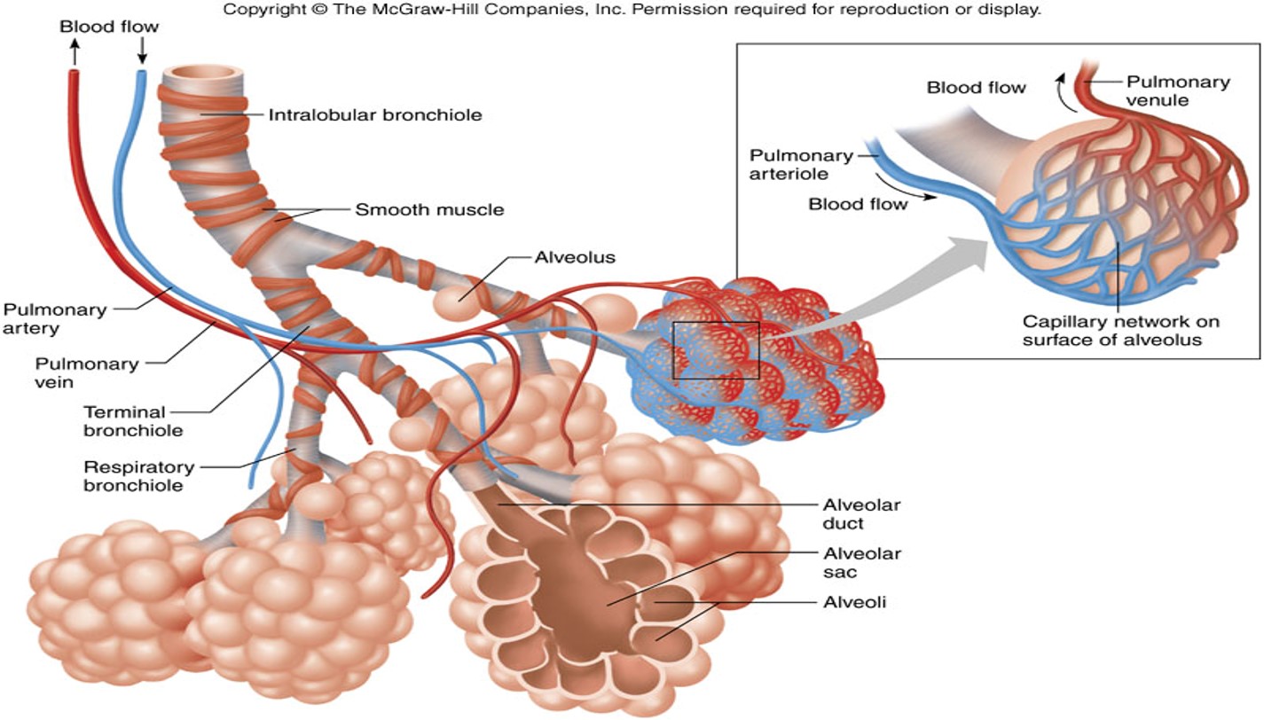 Lungs
Lungs
- Extend from diaphragm to clavicles
- Divided into lobes by fissures.
- Visceral pleura adheres to the lungs
- Pleurisy = inflammation of the pleural lining (Plural – bronchi)
Respiratory Physiology
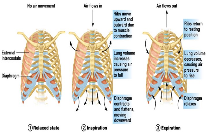
- Pulmonary Ventilation = breathing
- Mechanism
- Movement of gases through a pressure gradient - hi to low.
- Inspiration - When atmospheric pressure (760 mmHg) is greater than lung pressure ---- air flows in
- Expiration - When lung pressure is greater than atmospheric pressure ---- air flows out
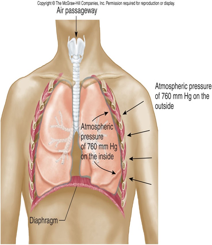
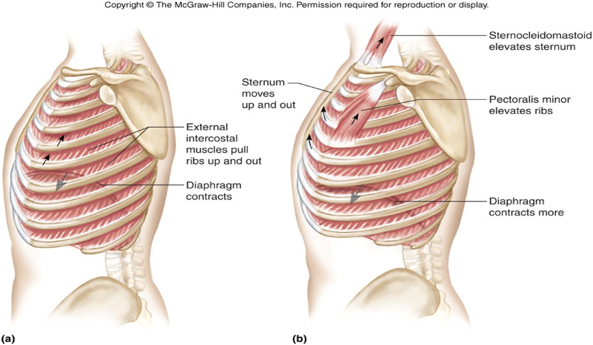
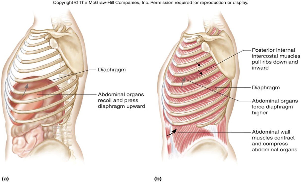
- Pressure gradients are established by changes in thoracic cavity.
- increase size in thorax = a decrease in pressure --- air moves in.
- Decrease size in thorax = increase in pressure --- air moves out.
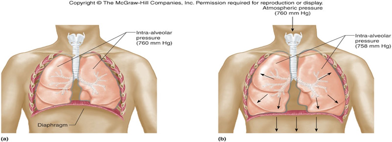
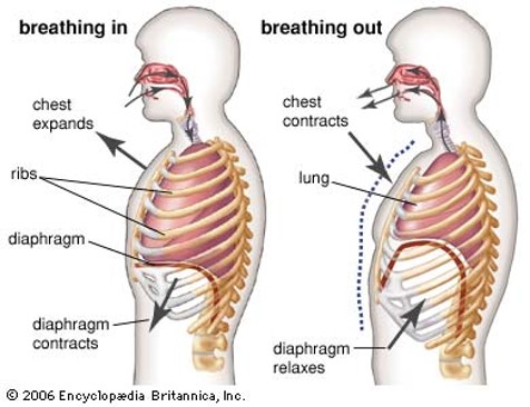
- Inspiration - contraction of diaphragm and intercostal muscles
- Expiration - relaxation of diaphragm and intercostal muscles
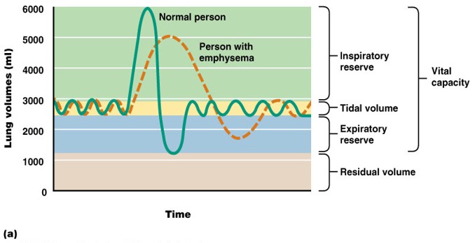
 Volumes of Air Exchange
• Tidal volume
- amount of air exhaled normally after a typical inspiration. (about 500 ml)
• Expiratory Reserve volume
- additional amount of air forcibly expired after tidal expiration (1000 - 1200 ml).
• Inspiratory Reserve volume
- (deep breath) amount of air that can be forcibly inhaled over and above normal.
• Residual volume
- amount of air that stays trapped in the alveoli (about 1.2 liters).
• Vital capacity
- the largest volume of air an individual can move in and out of the lungs.
• Vital capacity = sum of IRV+TV+ERV
Depends of many factors
• size of thoracic cavity
• posture
• volume of blood in lungs
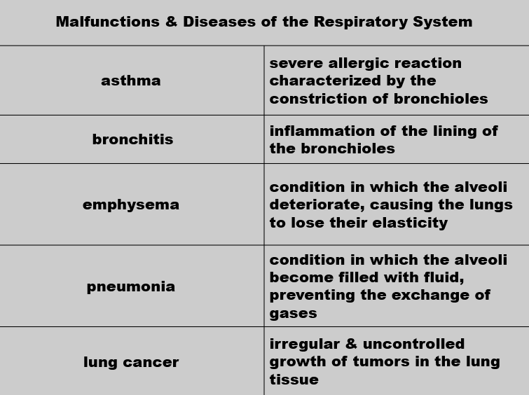
- congestive heart failure, emphysema, disease, etc.
• Eupnea
- normal quiet breathing, 12-17 breaths per minute.
• Hyperpnea
- increase in breathing to meet an increased demand by body for oxygen.
• Hyperventilation
- increase in pulmonary ventilation in excess of the need for oxygen.
Someone hysterical (exertion) → Breathe into paper bag.
• Hypoventilation - decrease in pulmonary ventilation.
• Apnea - temporary cessation of breathing at the end of normal expiration.
Heimlich Maneuver
- Lifesaving technique that is used to open a windpipe that is suddenly obstructed.
- Air already in lungs used to expel object.
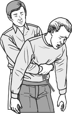
Technique - Conscious victim
– Ask the victim if he/she can talk
– Stand behind victim and wrap your arms around their waist
– Make a fist with one hand and grasp it with the other hand
– Place thumb side of fist below xiphoid process and above navel.
– Thrust your fist in and upward - about 4 times.
- DO NOT press on ribs or sternum
Technique - Unconscious victim
• Catch victim if they begin to fall - place on floor face up.
• Straddle hips
• Place one hand on top of other on the victims abdomen - above navel and below xiphoid process.
• Forceful upward thrusts with heel of hand - several times if necessary.